. 2019 Nov 14:10:1113.
doi: 10.3389/fneur.2019.01113. eCollection 2019.

Development of a Routinely Applicable Imaging Protocol for Fast and Precise Middle Cerebral Artery Occlusion Assessment and Perfusion Deficit Measure in an Ovine Stroke Model: A Case Study

Affiliations

Development of a Routinely Applicable Imaging Protocol for Fast and Precise Middle Cerebral Artery Occlusion Assessment and Perfusion Deficit Measure in an Ovine Stroke Model: A Case Study

Andrea Maria Herrmann et al. Front Neurol. .

Erratum in

Abstract

Temporary middle cerebral artery occlusion (MCAO) in sheep allows modeling of acute large vessel occlusion stroke and subsequent vessel recanalization. However, rapid and precise imaging-based assessment of vessel occlusion and the resulting perfusion deficit during MCAO still represents an experimental challenge. Here, we tested feasibility and suitability of a strategy for MCAO verification and perfusion deficit assessment. We also compared the extent of the initial perfusion deficit and subsequent lesion size for different MCAO durations. The rete mirabile prevents reliable vascular imaging investigation of middle cerebral artery filling status. Hence, computed tomography perfusion imaging was chosen for indirect confirmation of MCAO. Follow-up infarct size evaluation by diffusion-weighted magnetic resonance imaging revealed fluctuating results, with no apparent relationship of lesion size with MCAO at occlusion times below 4 h, potentially related to the variable collateralization of the MCA territory. This underlines the need for intra-ischemic perfusion assessment and future studies focusing on the correlation between perfusion deficit, MCAO duration, and final infarct volume. Temporary MCAO and intra-ischemic perfusion imaging nevertheless has the potential to be applied for the simulation of novel recanalization therapies, particularly those that aim for a fast reperfusion effect in combination with mechanical thrombectomy in a clinically realistic scenario.

Keywords: CT perfusion; DSA; MCAO; brain imaging; cerebral ischemia; reperfusion; sheep stroke model; translational research.


Figures

Figure 1
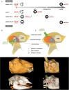
Figure 1
Study design and surgical approaches. (A) Overview on study design with two experimental series. Clip pictograms indicate (transient) MCAO by vessel clipping whereas the forceps indicate MCAO by electrocoagulation. (B,C): Schematic illustration of the surgical approaches (top), 3D-Reconstruction showing the skin incision (middle), 3D-bony-reconstruction with muscle (red), and craniectomy (blue) overlays (bottom). (B) The surgical approach according to Wells et al. (series a). The proximal MCA and terminal ICA were reached easily. Bony CT reconstruction shows the partial removal of the coronoid process. (C) Approach according to Boltze et al. (series b), in which the distal branches of the MCA were followed proximally until the optic nerve and the terminal internal carotid artery (ICA) had been identified. A partial resection of the coronoid process was not necessary (CT reconstruction). Dotted lines: skin incision; blue areas: craniectomy; *: muscle dissection (series a only). The green arrows describes the surgeon's approach and the approximate line of vision.
Figure 2
Figure 2
Results from case 1. (A) MRI images on day 2 after MCAO from case 1. Upper panels show DWI images in coronal view with ischemic lesions of the midbrain tegmental area within the right crus cerebri and right thalamus (white arrows). Lower panels show consecutive edema on T2w images in mid-sagittal views. (B) CCA DSA images before and after clip MCAO. After clip MCAO, no clear cut-off of MCA main trunk was visible with possible faint MCA filling (DSA image in left panel) at the clip level (unsubtracted image in mid panel). After clip removal (DSA image in right panel), filling of the main MCA trunk was visible. However, the distal MCA branch vasculature was not seen due to vessel overlap from rete mirabile and larger extracranial arteries.
Figure 3
Figure 3
Results from cases 2 and 3. MRI images of case 2 (upper panels) and case 3 (lower panels) on day 2 after 2 h of transient MCAO. DWI and T2w images show a large right MCA territory infarct lesion (DWI lesion volume, 25.6 ml) with swelling and mild herniation through the craniectomy site (arrows in upper left and mid panels). MRA (upper right panel) demonstrates adequate MCA recanalization after right temporary MCAO. In case 3, no relevant MCA territory ischemia is seen on DWI images (lower left panel; DWI lesion volume, 0.5 ml), whereas T2w images exhibit vasogenic edema in the area of the craniectomy with mild hemorrhagic foci (arrow in lower middle panel). Again, MRA demonstrates adequate MCA vessel recanalization after right temporary MCAO (lower right panel).
Figure 4
Figure 4
Results from case 4. DSA image (left panel) showing superselective rete mirabile injection (red arrow) after permanent MCAO (animal 4) without clear evidence of MCA main trunk occlusion (red arrow). 3T TOF MRA showing duplicated right MCA main trunk post temporary MCAO (mid panel, arrows) and clear evidence of right MCA occlusion after permanent MCAO (right panel).
Figure 5
Figure 5
Results from case 5. CTP images from case 5. Directly after clip MCAO profound hypoperfusion is visible on Tmax maps (upper panels) without reduction in CBV (lower panels). False color scale indicates Tmax from 0 (purple) to 2.5 s (red) in upper panels and CBV from 0 ml/100 g (purple) to 6 ml/100 g (red) in lower panels, respectively.

Similar articles

Cited by

References

    1. Goyal M, Menon BK, van Zwam WH, Dippel DWJ, Mitchell PJ, Demchuk AM, et al. . Endovascular thrombectomy after large-vessel ischaemic stroke: a meta-analysis of individual patient data from five randomised trials. Lancet. (2016) 387:1723–31. 10.1016/S0140-6736(16)00163-X - DOI - PubMed
    1. Linfante I, Cipolla MJ. Improving reperfusion therapies in the era of mechanical thrombectomy. Transl Stroke Res. (2016) 7:294–302. 10.1007/s12975-016-0469-3 - DOI - PMC - PubMed
    1. Savitz SI, Baron J.-C., Yenari MA, Sanossian N, Fisher M. Reconsidering neuroprotection in the reperfusion era. Stroke. (2017) 48:3413–9. 10.1161/STROKEAHA.117.017283 - DOI - PubMed
    1. Herrmann AM, Meckel S, Gounis MJ, Kringe L, Motschall E, Mülling C, et al. . Large animals in neurointerventional research: a systematic review on models, techniques and their application in endovascular procedures for stroke, aneurysms and vascular malformations. J Cereb Blood Flow Metab. (2019) 39:375–94. 10.1177/0271678X19827446 - DOI - PMC - PubMed
    1. Boltze J, Förschler A, Nitzsche B, Waldmin D, Hoffmann A, Boltze CM, et al. . Permanent middle cerebral artery occlusion in sheep: a novel large animal model of focal cerebral ischemia. J Cereb Blood Flow Metab. (2008) 28:1951–64. 10.1038/jcbfm.2008.89 - DOI - PubMed

LinkOut - more resources