

Development of a Routinely Applicable Imaging Protocol for Fast and Precise Middle Cerebral Artery Occlusion Assessment and Perfusion Deficit Measure in an Ovine Stroke Model: A Case Study
- PMID: 31798511
- PMCID: PMC6868089
- DOI: 10.3389/fneur.2019.01113
Development of a Routinely Applicable Imaging Protocol for Fast and Precise Middle Cerebral Artery Occlusion Assessment and Perfusion Deficit Measure in an Ovine Stroke Model: A Case Study
Erratum in
-
Corrigendum: Development of a Routinely Applicable Imaging Protocol for Fast and Precise Middle Cerebral Artery Occlusion Assessment and Perfusion Deficit Measure in an Ovine Stroke Model: A Case Study.Front Neurol. 2020 Jan 31;11:46. doi: 10.3389/fneur.2020.00046. eCollection 2020. Front Neurol. 2020. PMID: 32082249 Free PMC article.
Abstract
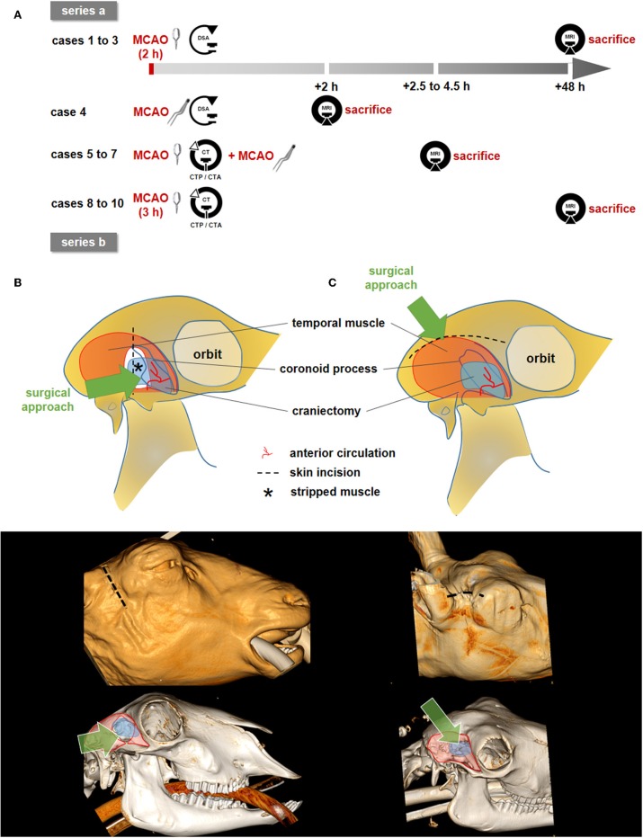
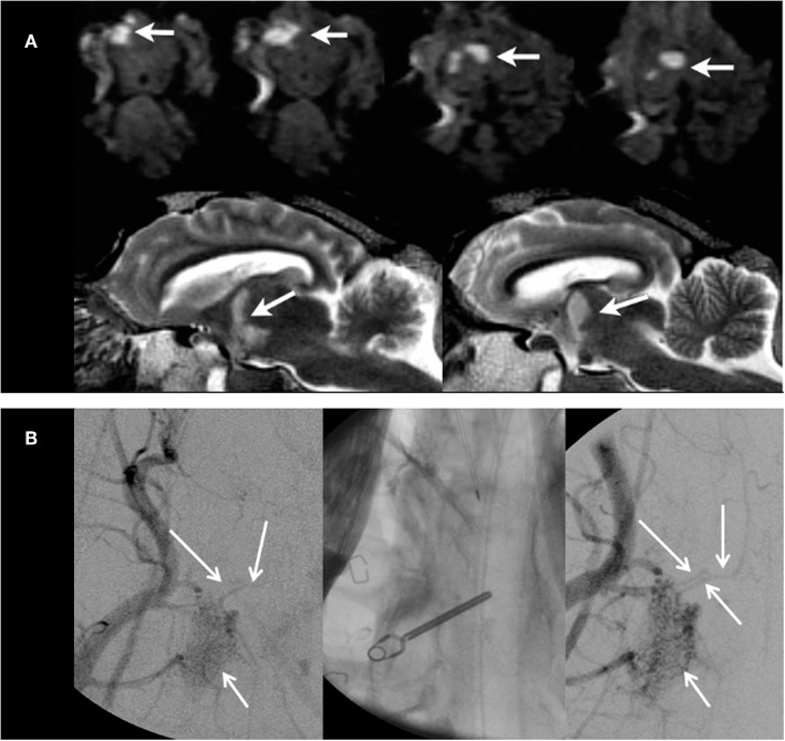
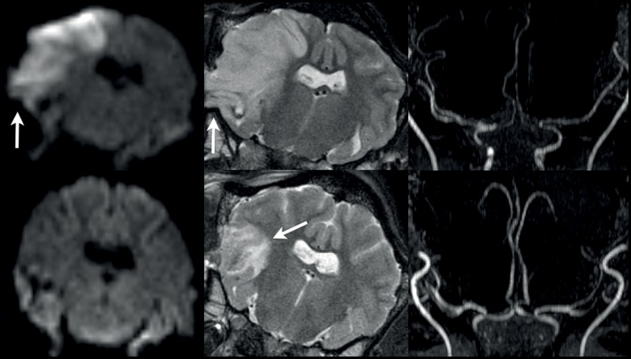
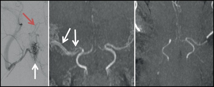
Temporary middle cerebral artery occlusion (MCAO) in sheep allows modeling of acute large vessel occlusion stroke and subsequent vessel recanalization. However, rapid and precise imaging-based assessment of vessel occlusion and the resulting perfusion deficit during MCAO still represents an experimental challenge. Here, we tested feasibility and suitability of a strategy for MCAO verification and perfusion deficit assessment. We also compared the extent of the initial perfusion deficit and subsequent lesion size for different MCAO durations. The rete mirabile prevents reliable vascular imaging investigation of middle cerebral artery filling status. Hence, computed tomography perfusion imaging was chosen for indirect confirmation of MCAO. Follow-up infarct size evaluation by diffusion-weighted magnetic resonance imaging revealed fluctuating results, with no apparent relationship of lesion size with MCAO at occlusion times below 4 h, potentially related to the variable collateralization of the MCA territory. This underlines the need for intra-ischemic perfusion assessment and future studies focusing on the correlation between perfusion deficit, MCAO duration, and final infarct volume. Temporary MCAO and intra-ischemic perfusion imaging nevertheless has the potential to be applied for the simulation of novel recanalization therapies, particularly those that aim for a fast reperfusion effect in combination with mechanical thrombectomy in a clinically realistic scenario.
Keywords: CT perfusion; DSA; MCAO; brain imaging; cerebral ischemia; reperfusion; sheep stroke model; translational research.
Copyright © 2019 Herrmann, Cattaneo, Eiden, Wieser, Kellner, Maurer, Haberstroh, Mülling, Niesen, Urbach, Boltze, Meckel and Shah.
Figures
References
-
- Herrmann AM, Meckel S, Gounis MJ, Kringe L, Motschall E, Mülling C, et al. Large animals in neurointerventional research: a systematic review on models, techniques and their application in endovascular procedures for stroke, aneurysms and vascular malformations. J Cereb Blood Flow Metab. (2019) 39:375–94. 10.1177/0271678X19827446 - DOI - PMC - PubMed
LinkOut - more resources
Full Text Sources