Review
. 2019 Nov 14:10:2657.
doi: 10.3389/fimmu.2019.02657. eCollection 2019.

Mass Cytometry Imaging for the Study of Human Diseases-Applications and Data Analysis Strategies

Affiliations
Review

Mass Cytometry Imaging for the Study of Human Diseases-Applications and Data Analysis Strategies

Heeva Baharlou et al. Front Immunol. .

Abstract

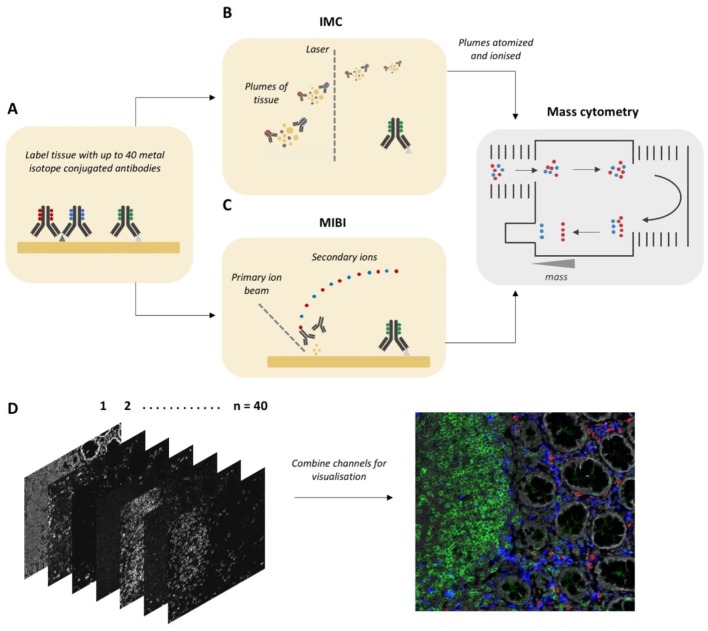
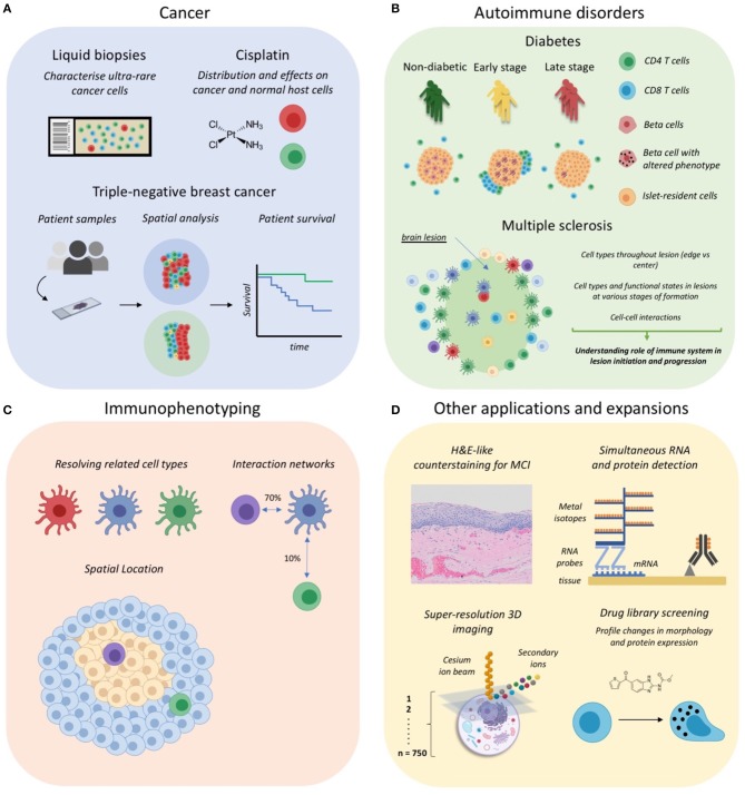
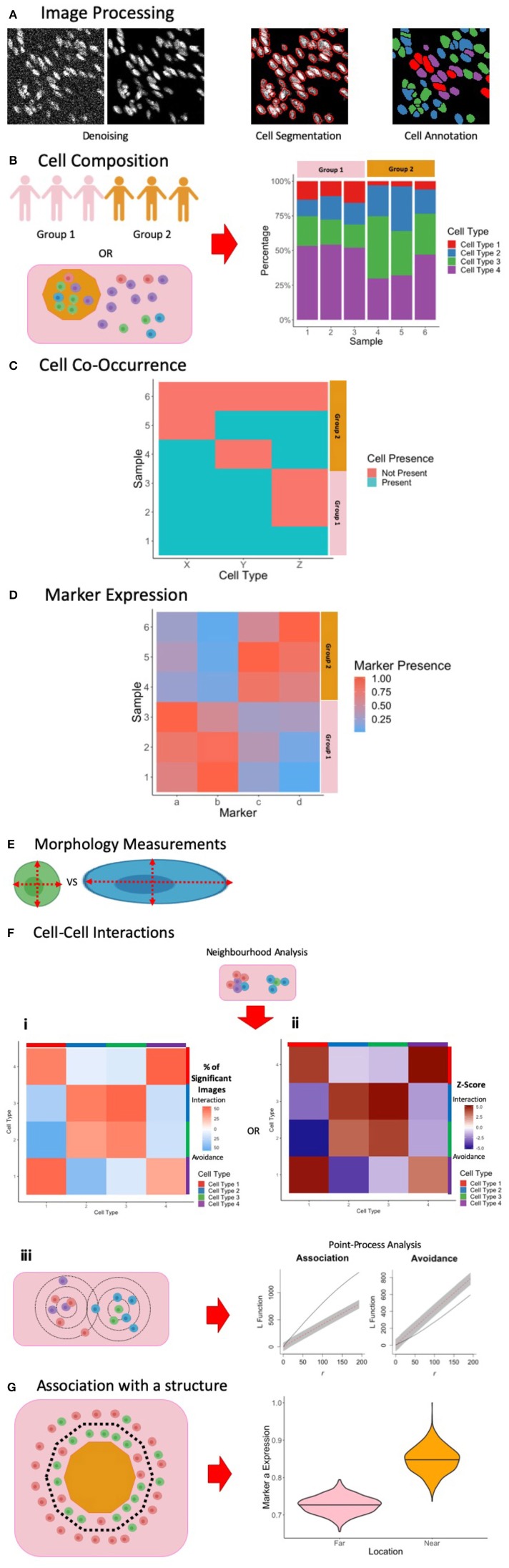
High parameter imaging is an important tool in the life sciences for both discovery and healthcare applications. Imaging Mass Cytometry (IMC) and Multiplexed Ion Beam Imaging (MIBI) are two relatively recent technologies which enable clinical samples to be simultaneously analyzed for up to 40 parameters at subcellular resolution. Importantly, these "Mass Cytometry Imaging" (MCI) modalities are being rapidly adopted for studies of the immune system in both health and disease. In this review we discuss, first, the various applications of MCI to date. Second, due to the inherent challenge of analyzing high parameter spatial data, we discuss the various approaches that have been employed for the processing and analysis of data from MCI experiments.

Keywords: analysis; cytometry; imaging cytometry; imaging mass cytometry (IMC); mass cytometry (CyTOF); multiplexed imaging; multiplexed ion beam imaging; single cell.


Figures

Figure 1
Figure 1
Workflow for Mass Cytometry Imaging. (A) Tissue sections are first labeled with a cocktail of metal-isotope-tagged antibodies. (B) In Imaging Mass Cytometry the tissue is ablated using a laser with 1 μm spot size. Plumes of tissue matter are then aerosolized, atomized and ionized, and then fed into a time-of-flight mass spectrometer, where metal ions are separated based on mass. (C) In Multiplexed Ion Beam Imaging a thin layer of the sample surface is ablated using an oxygen-based primary ion beam. Metal isotypes are liberated from antibodies as secondary ions which are then delivered to a time-of-flight mass spectrometer. (D) A high dimensional image is generated, which when combined and visualized, resembles a traditional fluorescence microscopy image. Parts of this figure were made Biorender.
Figure 2
Figure 2
Applications of Mass Cytometry Imaging. (A) MCI has been utilized for cancer studies examining rare circulating tumor cells in liquid biopsies, the distribution and effects of anti-cancer drugs such as cisplatin, and for profiling the tumor-immune landscape in tripe-negative breast cancer. (B) IMC has been used to investigate the immune correlates of autoimmune disorder progression, including type 1 diabetes mellitus and multiple sclerosis lesion formation in the central nervous system. (C) Some studies have begun to use MCI for immunophenotyping so as to discriminate cell subsets, their interactions and anatomical distribution. (D) Several recent expansions of MCI, including the development of a counterstaining method, simultaneous RNA and protein detection, 3D super-resolution imaging of single cells, and applications for drug screening. Parts of this figure were made Biorender.
Figure 3
Figure 3
Summary of Image Processing and Analysis Techniques in MCI. (A) Following the acquisition of MCI, image processing is performed to denoise the images, perform single-cell segmentation to identify cell outlines, and to classify these cells based on marker expression. (B) One way of exploring cell composition between groups is to compare the change in the cell fractions. (C) Another way to explore cell composition is to classify patients as being positive and negative for a particular cell population. The co-occurrence of cells can be presented similar to what is presented here, and significance of co-occurrence can be identified using a chi-square test. (D) Differences in marker expression between patients can be visualized using a heatmap. (E) Cell morphology measurements can be used to explore cell phenotypes. (F) Cell-cell interactions can be measured using neighborhood analysis or point-process analysis. With a neighborhood analysis, percentage of significant images (i) or Z-scores (ii) of the cell-cell interactions can be represented as a heatmap, with significant associations associated with a more positive Z-score and significant avoidance is associated with a more negative Z-score. With a point-process analysis, an L function can be used to assess the significance of cell-cell interactions. The L function being above or below the gray envelope generated by bootstrapping corresponds to association and avoidance, respectively (iii). (G) One way of measuring cell or marker association with a marker is to classify cells as being near or far away from the border. A cell composition analysis can be used to explore differences, or differences in marker expression can be explored, as shown here. Parts of this figure were made Biorender.

References

    1. Zrazhevskiy P, Gao X. Quantum dot imaging platform for single-cell molecular profiling. Nat Commun. (2013) 4:1619. 10.1038/ncomms2635 - DOI - PMC - PubMed
    1. Bodenmiller B. Multiplexed epitope-based tissue imaging for discovery and healthcare applications. Cell Syst. (2016) 2:225–38. 10.1016/j.cels.2016.03.008 - DOI - PubMed
    1. Lin J-R, Fallahi-Sichani M, Sorger PK. Highly multiplexed imaging of single cells using a high-throughput cyclic immunofluorescence method. Nat Commun. (2015) 6:8390. 10.1038/ncomms9390 - DOI - PMC - PubMed
    1. Lin J-R, Izar B, Wang S, Yapp C, Mei S, Shah PM, et al. . Highly multiplexed immunofluorescence imaging of human tissues and tumors using t-CyCIF and conventional optical microscopes. eLife. (2018) 7:e31657. 10.7554/eLife.31657 - DOI - PMC - PubMed
    1. Tsujikawa T, Kumar S, Borkar RN, Azimi V, Thibault G, Chang Y, et al. . Quantitative multiplex immunohistochemistry reveals myeloid-inflamed tumor-immune complexity associated with poor prognosis. Cell Rep. (2017) 19:203–17. 10.1016/j.celrep.2017.03.037 - DOI - PMC - PubMed

Publication types

LinkOut - more resources