. 2019 Nov 13:10:1113.
doi: 10.3389/fgene.2019.01113. eCollection 2019.

An Integrated Model of Minor Intron Emergence and Conservation

Affiliations

An Integrated Model of Minor Intron Emergence and Conservation

Marybeth Baumgartner et al. Front Genet. .

Abstract

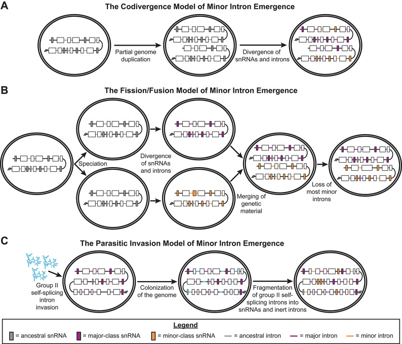
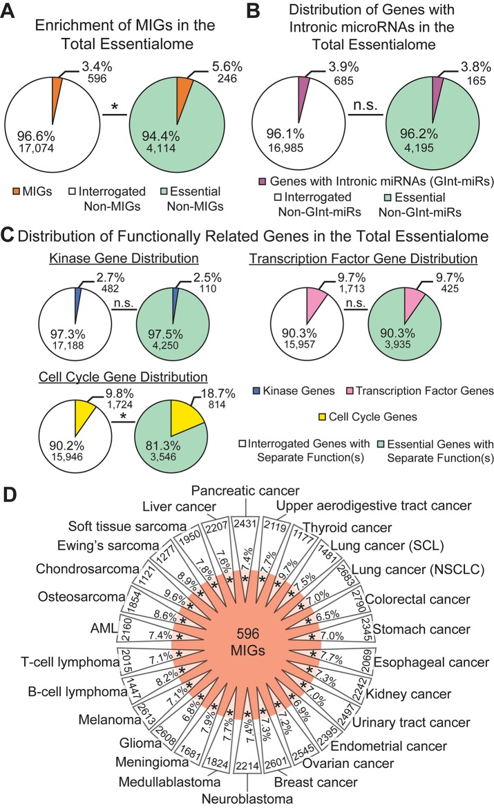
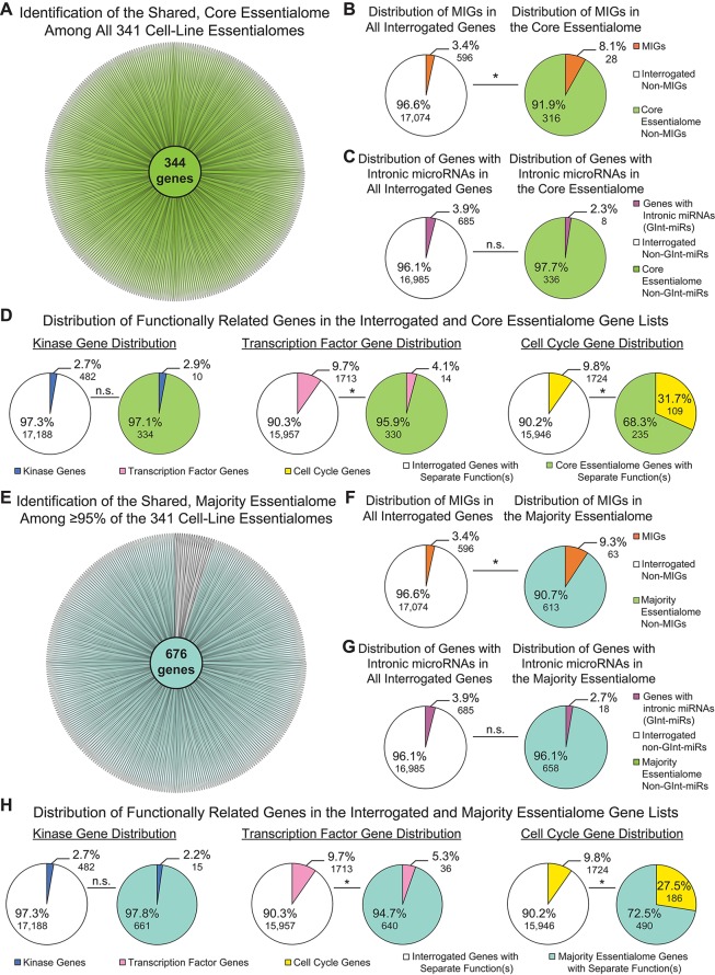
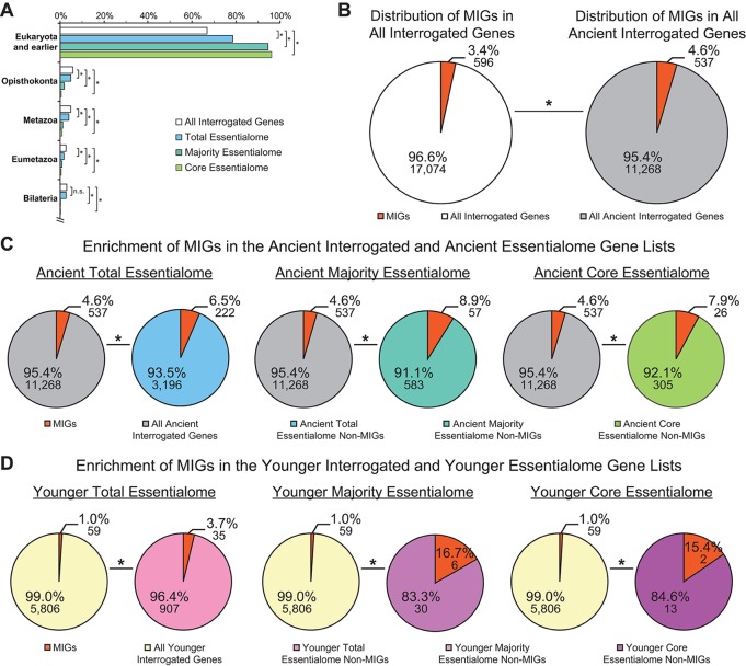
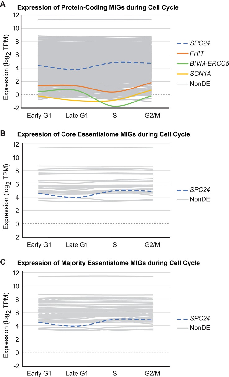
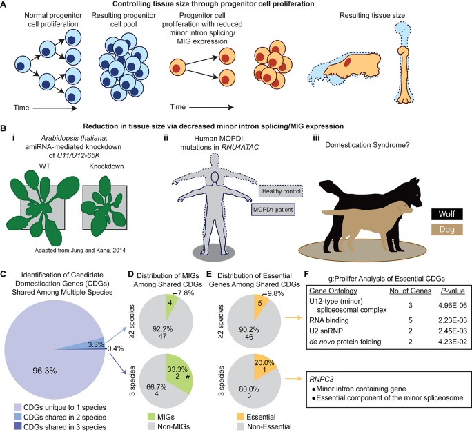
Minor introns constitute <0.5% of the introns in the human genome and have remained an enigma since their discovery. These introns are removed by a distinct splicing complex, the minor spliceosome. Both are ancient, tracing back to the last eukaryotic common ancestor (LECA), which is reflected by minor intron enrichment in specific gene families, such as the mitogen activated-protein kinase kinases, voltage-gated sodium and calcium ion channels, and E2F transcription factors. Most minor introns occur as single introns in genes with predominantly major introns. Due to this organization, minor intron-containing gene (MIG) expression requires the coordinated action of two spliceosomes, which increases the probability of missplicing. Thus, one would expect loss of minor introns via purifying selection. This has resulted in complete minor intron loss in at least nine eukaryotic lineages. However, minor introns are highly conserved in land plants and metazoans, where their importance is underscored by embryonic lethality when the minor spliceosome is inactivated. Conditional inactivation of the minor spliceosome has shown that rapidly dividing progenitor cells are highly sensitive to minor spliceosome loss. Indeed, we found that MIGs were significantly enriched in a screen for genes essential for survival in 341 cycling cell lines. Here, we propose that minor introns inserted randomly into genes in LECA or earlier and were subsequently conserved in genes crucial for cycling cell survival. We hypothesize that the essentiality of MIGs allowed minor introns to endure through the unicellularity of early eukaryotic evolution. Moreover, we identified 59 MIGs that emerged after LECA, and that many of these are essential for cycling cell survival, reinforcing our essentiality model for MIG conservation. This suggests that minor intron emergence is dynamic across eukaryotic evolution, and that minor introns should not be viewed as molecular fossils. We also posit that minor intron splicing was co-opted in multicellular evolution as a regulatory switch for en masse control of MIG expression and the biological processes they regulate. Specifically, this mode of regulation could control cell proliferation and thus body size, an idea supported by domestication syndrome, wherein MIGs are enriched in common candidate animal domestication genes.

Keywords: animal domestication; essential genes; eukaryotic evolution; human disease; minor introns; minor spliceosome; multicellularity; scaling.


Figures

Figure 1
Figure 1
Previously published models of minor intron emergence. Schematics showing the three models of minor intron emergence proposed by Burge et al. (1998), including (A) the codivergence model, (B) the fission/fusion model, and (C) the parasitic invasion model. Unicellular organisms are shown by the double-bordered ovals, while regions of the genome are displayed as lines. Black lines denote intergenic regions, gray lines for ancestral introns, blue lines for major introns, and purple lines for minor introns. Boxes represent coding regions of genes, with colored boxes denoting ancestral snRNA genes (gray), major spliceosomal snRNA genes (blue), or minor spliceosome-specific snRNA genes (purple). Arrows denote the passage of time; events occurring during this period of time are noted in text under the respective arrow. Reproduced with permission from Burge et al., 1998.
Figure 2
Figure 2
Minor intron-containing gene (MIGs) are enriched in the essentialome. (A) Pie charts showing the percentage of MIGs (orange) in all genes interrogated (left) and in all essential genes (right). (B) Pie charts showing the percentage of genes with intronic microRNA genes (GInt-miRs) in all interrogated genes (left) and in the total essentialome (right). (C) Pie charts showing the percentage of genes with kinase activity (top left), transcription factor activity (right), or a role in cell cycle regulation (bottom left) in all interrogated genes and in the total essentialome. (D) Daisy model representing the 26 cancer-type essentialomes (triangular petals) and the 596 MIGs (orange center). The overlap of the center with the petals indicates the enrichment of MIGs in the cell-line essentialomes, with the percentage of MIG enrichment in each petal. Gene number within each cancer-type essentialome is located on the outside of each petal. Statistical significance was determined by Fisher’s exact test. AML; acute myeloid leukemia, SCL; small cell lung cancer, NSCLC; non-small cell lung cancer. N.s., not significant; * P ≤ 5.45E−16.
Figure 3
Figure 3
Minor intron-containing genes (MIGs) are enriched in shared essential genes. (A) Daisy model showing the number of essential genes common to all 341 cell-line essentialomes (“core essentialome”; green circle). Each petal corresponds to one cell-line essentialome. (B) Pie charts showing the percentage of MIGs in all interrogated genes (left) and in the core essentialome (right). (C) Pie charts showing the percentage of genes with intronic microRNA genes (GInt-miRs) in all interrogated genes (left) and in the core essentialome (right). (D) Pie charts showing the percentage of genes with kinase activity (leftmost), transcription factor activity (middle), or a role in cell cycle regulation (rightmost) in all interrogated genes and in the core essentialome. (E) Daisy model showing the number of essential genes common to the majority (≥95%) of the 341 cell-line essentialomes (“majority essentialome”; teal circle). (F) Pie charts showing the percentage of MIGs in all interrogated genes (left) and in the majority essentialome (right). (G) Pie charts showing the percentage of genes with intronic microRNA genes (GInt-miRs) in all interrogated genes (left) and the majority essentialome (right). (H) Pie charts showing the percentage of genes with kinase activity (leftmost), transcription factor activity (middle), or a role in cell cycle regulation (rightmost) in all interrogated genes and in the majority essentialome. Statistical significance was determined by Fisher’s exact test. N.s., not significant, * P ≤ 1.40E−04.
Figure 4
Figure 4
Minor intron-containing genes (MIGs) are enriched in essential genes regardless of age. (A) Evolutionary age of all interrogated genes (white), the total essentialome (blue), the majority essentialome (teal), and the core essentialome (green). Clades are listed on the y-axis. The x-axis shows the percentage of genes that can be traced to the listed clade, but not to an older clade. (B) Pie charts showing the enrichment of MIGs (orange) in the all interrogated genes (left) and all ancient genes (tracing back to Eukaryota and earlier; right). (C) Pie charts showing the enrichment of MIGs (orange) in the ancient total essentialome (blue, left), the ancient majority essentialome (teal, middle), and the ancient core essentialome (green, right), compared to all ancient interrogated genes (gray). (D) Pie charts showing the enrichment of MIGs (orange) in the younger (tracing back to Opisthokonta or later) total essentialome (pink, left), the younger majority essentialome (light purple, middle), and the younger core essentialome (dark purple, right), compared to all younger interrogated genes (yellow). Statistical significance, relative to the percentage of MIGs present in the respective interrogated gene lists, was determined by Fisher’s exact test. N.s., not significant; * P ≤ 0.007.
Figure 5
Figure 5
Most minor intron-containing genes (MIGs) are expressed stably during cell cycle. Data plotted for (A) 624 protein-coding MIGs, (B) 28 core essentialome MIGs, and (C) 63 majority essentialome MIGs deemed to be expressed during cell cycle (as determined by TPM≥1 in at least one stage of cell cycle). Data obtained from analysis of RNAseq generated by Singh et al. (2013). Each line represents the expression of one MIG throughout successive stages of cell cycle (early G1, late G1, S, and G2/M). Colored lines indicate MIGs that showed differential expression in at least one stage of cell cycle relative to the previous stage, gray lines are MIGs that are non-differentially expressed (NonDE) in all stages, and dashed lines represent MIGs in the younger essentialome.
Figure 6
Figure 6
A potential role for the minor spliceosome in domestication. (A) A model demonstrating how tissue size can be controlled through progenitor cell proliferation. (B) Evidence for tissue size reduction upon impairment of minor intron splicing/minor intron-containing gene (MIG) expression. i) Plant size is smaller in A. thaliana upon knockdown of the minor spliceosome-specific U11/U12-65K. Reproduced with permission from Jung and Kang, 2014. ii) Mutations in RNU4ATAC cause three diseases in human, such as microcephalic osteodysplastic primordial dwarfism type 1 (MOPD1), wherein patients’ display reductions in multiple tissues causing a severely reduced body plan relative to healthy controls (theoretical output). iii) Previous studies suggest a link between reduced minor intron splicing/MIG expression and domestication syndrome, of which one prominent example is the domestication of dog (beige) from wolf (black). (C) Pie-chart representing the distribution of candidate domestication genes (CDGs) from dog, cat, cattle, horse, and anatomically modern human curated from Theofanopoulou et al. (2017) that are unique to one species (light purple), overlap in two species (blue), or are shared among three species (dark purple). (D) Pie-chart showing the number of MIGs (green) or non-MIGs (gray) found within the list of CDGs shared between two (top) or three (bottom) species. (E) Pie-chart showing the number of essential (yellow) or non-essential (gray) genes found within the list of CDGs shared between two (top) or three (bottom) species. (F) Description of gene enrichment (as determined through g:Profiler) for essential genes shared among two or three species (only one gene, RNPC3, is both essential and a shared CDG among three species). Significance determined by Fisher’s exact test; * P < 0.05.

References

    1. Alioto T. S. (2007). U12DB: a database of orthologous U12-type spliceosomal introns. Nucleic Acids Res. 35, D110–D115. 10.1093/nar/gkl796 - DOI - PMC - PubMed
    1. Argente J., Flores R., Gutierrez-Arumi A., Verma B., Martos-Moreno G. A., Cusco I., et al. (2014). Defective minor spliceosome mRNA processing results in isolated familial growth hormone deficiency. EMBO Mol. Med. 6, 299–306. 10.1002/emmm.201303573 - DOI - PMC - PubMed
    1. Bartschat S., Samuelsson T. (2010). U12 type introns were lost at multiple occasions during evolution. BMC Genomics 11, 106. 10.1186/1471-2164-11-106 - DOI - PMC - PubMed
    1. Basu M. K., Makalowski W., Rogozin I. B., Koonin E. V. (2008. a). U12 intron positions are more strongly conserved between animals and plants than U2 intron positions. Biol. Direct 3, 19. 10.1186/1745-6150-3-19 - DOI - PMC - PubMed
    1. Basu M. K., Rogozin I. B., Koonin E. V. (2008. b). Primordial spliceosomal introns were probably U2-type. Trends Genet. 24, 525–528. 10.1016/j.tig.2008.09.002 - DOI - PMC - PubMed