Novel frameshift mutation causes early termination of the thyroxine-binding globulin protein and complete thyroxine-binding globulin deficiency in a Chinese family: A case report
- PMID: 31799319
- PMCID: PMC6887617
- DOI: 10.12998/wjcc.v7.i22.3887
Novel frameshift mutation causes early termination of the thyroxine-binding globulin protein and complete thyroxine-binding globulin deficiency in a Chinese family: A case report
Abstract
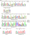
Background: Thyroxine-binding globulin (TBG; the gene product of SERPINA7) is the main transporter of thyroid hormones in humans. Mutations in the TBG gene may lead to inherited TBG deficiency. There have been 28 reported mutations that associate with complete TBG deficiency (TBG-CD). Here we identified a novel frameshift mutation causing early termination of the TBG protein and TBG-CD in a Chinese family.
Case summary: A 46-year-old Chinese man was referred to our hospital with normal free thyroxine, free triiodothyronine, thyrotropin, but lower total thyroxine and total triiodothyronine, and undetectable serum TBG, indicative of TBG-CD. Blood samples were obtained from the patient's family members and thyroid function and serum TBG were evaluated. Genomic DNA from peripheral blood was sequenced to detect possible TBG mutation(s). Quantitative PCR high-resolution melting curve analysis was used to screen TBG-Poly (L283F) among 117 Chinese men. A novel mutation of TBG (p.Phe135Alafs*21), a 19-nucleotide insertion in exon 1, was identified, which resulted in a truncated TBG protein product and caused TBG-CD. The other mutation, identified in the proband's father, is a known polymorphism, TBG-Poly (L283F). The frequency of the TBG-Poly allele among 117 unrelated Han Chinese men from northeast China was 21.37%.
Conclusion: A novel mutation in the TBG gene associated with the TBG-CD phenotype was identified in a Chinese family. Additionally, it was found that 21.37% of Chinese males had TBG-Poly (L283F).
Keywords: Case report; Complete thyroxine-binding globulin deficiency; Gene polymorphism; Partial thyroxine-binding globulin deficiency; Thyroxine-binding globulin.
©The Author(s) 2019. Published by Baishideng Publishing Group Inc. All rights reserved.
Conflict of interest statement
Conflict-of-interest statement: The authors declare that they have no competing interests.
Figures


Similar articles
-
Partial thyroxine-binding globulin deficiency in a family with coding region mutations in the TBG gene.J Endocrinol Invest. 2020 Dec;43(12):1703-1710. doi: 10.1007/s40618-020-01245-1. Epub 2020 Apr 7. J Endocrinol Invest. 2020. PMID: 32266677
-
Two novel mutations in the gene encoding thyroxine-binding globulin (TBG) as a cause of complete TBG deficiency in Taiwan.Clin Endocrinol (Oxf). 2003 Apr;58(4):409-14. doi: 10.1046/j.1365-2265.2003.01730.x. Clin Endocrinol (Oxf). 2003. PMID: 12641622
-
Identification of a novel mutation in thyroxine-binding globulin (TBG) gene associated with TBG-deficiency and its effect on the thyroid function.J Endocrinol Invest. 2022 Apr;45(4):731-739. doi: 10.1007/s40618-021-01697-z. Epub 2021 Nov 10. J Endocrinol Invest. 2022. PMID: 34761328
-
[Partial thyroxine binding globulin deficiency in test tube infants: report of cases and literature review].Zhonghua Er Ke Za Zhi. 2016 Jun 2;54(6):428-32. doi: 10.3760/cma.j.issn.0578-1310.2016.06.008. Zhonghua Er Ke Za Zhi. 2016. PMID: 27256229 Review. Chinese.
-
TBG deficiency: description of two novel mutations associated with complete TBG deficiency and review of the literature.J Mol Med (Berl). 2006 Oct;84(10):864-71. doi: 10.1007/s00109-006-0078-9. Epub 2006 Sep 1. J Mol Med (Berl). 2006. PMID: 16947003 Review.
Cited by
-
Partial Thyroid Hormone-Binding Globulin Deficiency: A Case Report and Literature Review.Diabetes Metab Syndr Obes. 2023 Jul 26;16:2225-2232. doi: 10.2147/DMSO.S413048. eCollection 2023. Diabetes Metab Syndr Obes. 2023. PMID: 37525823 Free PMC article.
-
Identification of Mutations in the Thyroxine-Binding Globulin (TBG) Gene in Patients with TBG Deficiency in Korea.Endocrinol Metab (Seoul). 2022 Dec;37(6):870-878. doi: 10.3803/EnM.2022.1591. Epub 2022 Dec 7. Endocrinol Metab (Seoul). 2022. PMID: 36475360 Free PMC article.
-
Compound hemizygous variants in SERPINA7 gene cause thyroxine-binding globulin deficiency.Mol Genet Genomic Med. 2021 Feb;9(2):e1571. doi: 10.1002/mgg3.1571. Epub 2021 Feb 7. Mol Genet Genomic Med. 2021. PMID: 33554479 Free PMC article.
References
-
- Refetoff S. Inherited thyroxine-binding globulin abnormalities in man. Endocr Rev. 1989;10:275–293. - PubMed
-
- Refetoff S. Thyroid Hormone Serum Transport Proteins. In: Feingold KR, Anawalt B, Boyce A, editors. Endotext. South Dartmouth: MDText.com, Inc; 2000.
-
- Robbins J, Rall JE. Zone electrophoresis in filter paper of serum I 131 after radioiodide administration. Proc Soc Exp Biol Med. 1952;81:530–536. - PubMed
-
- Mori Y, Miura Y, Oiso Y, Hisao S, Takazumi K. Precise localization of the human thyroxine-binding globulin gene to chromosome Xq22.2 by fluorescence in situ hybridization. Hum Genet. 1995;96:481–482. - PubMed
Publication types
LinkOut - more resources
Full Text Sources
Miscellaneous

