. 2020 Jan 24;48(2):817-829.
doi: 10.1093/nar/gkz1018.

Oxidative damage diminishes mitochondrial DNA polymerase replication fidelity

Affiliations

Oxidative damage diminishes mitochondrial DNA polymerase replication fidelity

Andrew P Anderson et al. Nucleic Acids Res. .

Abstract

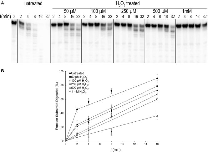
Mitochondrial DNA (mtDNA) resides in a high ROS environment and suffers more mutations than its nuclear counterpart. Increasing evidence suggests that mtDNA mutations are not the results of direct oxidative damage, rather are caused, at least in part, by DNA replication errors. To understand how the mtDNA replicase, Pol γ, can give rise to elevated mutations, we studied the effect of oxidation of Pol γ on replication errors. Pol γ is a high fidelity polymerase with polymerase (pol) and proofreading exonuclease (exo) activities. We show that Pol γ exo domain is far more sensitive to oxidation than pol; under oxidative conditions, exonuclease activity therefore declines more rapidly than polymerase. The oxidized Pol γ becomes editing-deficient, displaying a 20-fold elevated mutations than the unoxidized enzyme. Mass spectrometry analysis reveals that Pol γ exo domain is a hotspot for oxidation. The oxidized exo residues increase the net negative charge around the active site that should reduce the affinity to mismatched primer/template DNA. Our results suggest that the oxidative stress induced high mutation frequency on mtDNA can be indirectly caused by oxidation of the mitochondrial replicase.


Figures

Figure 1.
Figure 1.
Oxidation decreases Pol γ exo activity. (A) the time-dependent exonuclease activity on single-stranded DNA. (B) Quantification of digested substrate from each sample normalized by the total accounts within the lane. The means and standard errors are calculated from three independent experiments.
Figure 2.
Figure 2.
Oxidized Pol γ pol activity. (A) DNA synthesis activity of untreated and treated Pol γ measured on a singly primed M13mp18 ssDNA, (B) Quantification the fraction of full-length products that sums the product between 500 and 1000 nt then normalized by the total counts within the lane. Data were averaged from three independent reactions.
Figure 3.
Figure 3.
Effects of oxidation on mismatch extension and excision. (A) The schematic of the substrate with a 1-nt mismatch at the 3′-end primer used in primer extension and excision assays. The asterisk indicates 5′-32P labeling. (B) Time-dependent mismatched primer extension by untreated, treated and exo-deficient Pol γ. (C) Mismatched primer excision by untreated, H2O2 treated, and exo-deficient Pol γ. (D) Quantification of fraction of mismatch primer extension to full-length at 120 s from data presented in (B). (E) Quantification mismatched primer in panel (C). The mean and standard deviation were determined from three independent experiments.
Figure 4.
Figure 4.
Effect of oxidation on polymerase replication fidelity. (A) The scheme for distinguishing error-free and error-prone DNA synthesis on the designed mismatched primer/template DNA. The asterisk indicates 5′-32P labeling. (B) Products of mismatched DNA extension and digestion by HindIII by Pol γ treated with H2O2 and the exo-deficient variant. (C) Quantification of the undigested band divided by the sum of all density within the lane.
Figure 5.
Figure 5.
Structural illustration of oxidized Pol γ residues. (A) The overall fraction of oxidation (Fr, Equation 1) for 500 μM and 1 mM H2O2 treated Pol γ, relative to the untreated. (B) Consistency of oxidized residues from three different oxidation experiments (C) The average oxidation Fr of pol (red) and exo (green) active sites. (D) Oxidized residues of untreated (left) and 1 mM H2O2 treated (right) catalytic subunit displayed on Pol γ holoenzyme ternary complex structure (PDB ID: 4ztz). Oxidation of each residue is color-coded by Fr. Oxidation clusters are indicated. (E) Oxidation of the pol site. The asterisk marks the location of pol catalytic residues D890. (F) Oxidation of the exo site. The asterisk marks the exo catalytic residue D198.
Figure 6.
Figure 6.
Chemical structures of detected oxidized residues.
Figure 7.
Figure 7.
Cooperation for protein and DNA oxidative damage.

References

    1. Anderson S., Bankier A.T., Barrell B.G., de Bruijn M.H., Coulson A.R., Drouin J., Eperon I.C., Nierlich D.P., Roe B.A., Sanger F. et al. .. Sequence and organization of the human mitochondrial genome. Nature. 1981; 290:457–465. - PubMed
    1. Brown W.M., George M. Jr, Wilson A.C.. Rapid evolution of animal mitochondrial DNA. Proc. Natl. Acad. Sci. U.S.A. 1979; 76:1967–1971. - PMC - PubMed
    1. Brown W.M., Prager E.M., Wang A., Wilson A.C.. Mitochondrial DNA sequences of primates: tempo and mode of evolution. J. Mol. Evol. 1982; 18:225–239. - PubMed
    1. Gonzalez-Freire M., de Cabo R., Bernier M., Sollott S.J., Fabbri E., Navas P., Ferrucci L.. Reconsidering the role of mitochondria in aging. J. Gerontol. A Biol. Sci. Med. Sci. 2015; 70:1334–1342. - PMC - PubMed
    1. Wallace D.C. Mitochondrial DNA mutations in disease and aging. Environ. Mol. Mutagen. 2010; 51:440–450. - PubMed

Publication types

MeSH terms