. 2020 Jan 3;19(1):554-560.
doi: 10.1021/acs.jproteome.9b00759. Epub 2019 Dec 18.

Optimized Workflow for Multiplexed Phosphorylation Analysis of TMT-Labeled Peptides Using High-Field Asymmetric Waveform Ion Mobility Spectrometry

Affiliations

Optimized Workflow for Multiplexed Phosphorylation Analysis of TMT-Labeled Peptides Using High-Field Asymmetric Waveform Ion Mobility Spectrometry

Devin K Schweppe et al. J Proteome Res. .

Abstract

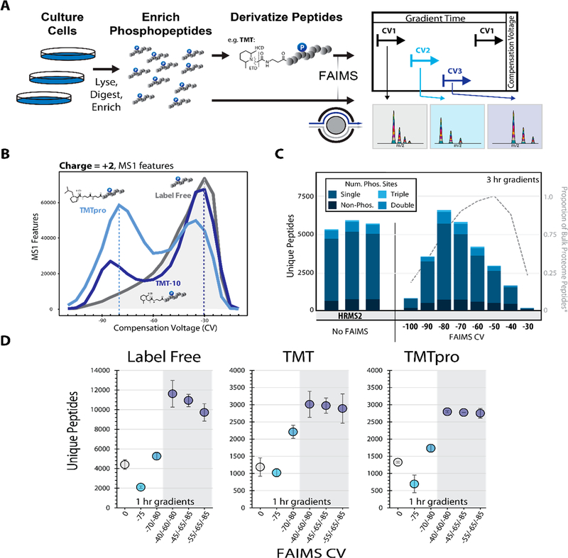
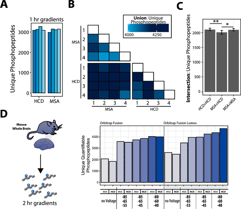
Phosphorylation is a post-translational modification with a vital role in cellular signaling. Isobaric labeling-based strategies, such as tandem mass tags (TMT), can measure the relative phosphorylation states of peptides in a multiplexed format. However, the low stoichiometry of protein phosphorylation constrains the depth of phosphopeptide analysis by mass spectrometry. As such, robust and sensitive workflows are required. Here we evaluate and optimize high-Field Asymmetric waveform Ion Mobility Spectrometry (FAIMS) coupled to Orbitrap Tribrid mass spectrometers for the analysis of TMT-labeled phosphopeptides. We determined that using FAIMS-MS3 with three compensation voltages (CV) in a single method (e.g., CV = -40/-60/-80 V) maximizes phosphopeptide coverage while minimizing inter-CV overlap. Furthermore, consecutive analyses using MSA-CID (multistage activation collision-induced dissociation) and HCD (higher-energy collisional dissociation) fragmentation at the MS2 stage increases the depth of phosphorylation analysis. The methodology and results outlined herein provide a template for tailoring optimized FAIMS-based methods.

Keywords: FAIMS; MSA; SL-TMT; SPS-MS3; TMTpro0; phosphorylation.


Figures

Figure 1.
Figure 1.
Optimizing the FAIMS compensation voltage (CV) for phosphopeptide analysis. A. General FAIMS workflow for phosphopeptide analysis. Samples were processed using a modified SL-TMT protocol in which phosphopeptides were enriched prior to TMT labeling. The resulting peptides were analyzed by a FAIMS-Pro-equipped Orbitrap Fusion or Fusion Lumos mass spectrometer. B. MS1 features (z = +2) detected at varying compensation voltages (−110 V to −10 V). C. Unique peptides (divided by degree of phosphorylation) identified by various data acquisition methods. All data were collected with high-resolution MS2 (HRMS2) without FAIMS (in triplicates) or with CVs ranging from −100 to −30 V (no peptides were identified at −110 V or −20 V). Dotted line represents the proportion of non-phosphorylated peptides. D. Comparison of unique peptides identified as label-free (left), and derivatized with TMT reagent, (i.e., TMT, middle or TMTpro, right). Analyses were performed in triplicate. Error bars are standard deviations.
Figure 2.
Figure 2.
Benchmarking the selection of optimal FAIMS compensation voltages (CV) for phosphoproteome analysis. A. Enriched-phosphopeptides from human cell lysate are TMT0-labeled, fragmented with different fragmentation schemes. B. Matrix of binary comparisons of the unions of phosphopeptides (total phosphopeptides identified from combining two runs) found in A. C. Intersection of unique phosphopeptides identified using either HCD or MSA fragmentation. Error bars: standard deviation. ANOVA, Tukey HSD: * p-value <0.05; ** p-value <0.01. D. Proteins extracted from mouse brain tissue, digested, enriched for phosphopeptides, and labeled with TMT. The data were collected with different methods (MSA or HCD, various CV combinations). Bar graphs illustrating the unique quantifiable phosphopeptides (unique, with summed TMT reporter ion signal-to-noise >200) with varying acquisition energy and compensation voltages (CV).

References

    1. Singh V; Ram M; Kumar R; Prasad R; Roy BK; Singh KK Phosphorylation: Implications in Cancer. Protein J. 2017, 36 (1), 1–6. - PubMed
    1. Manning G; Whyte DB; Martinez R; Hunter T; Sudarsanam S The protein kinase complement of the human genome. Science 2002, 298 (5600), 1912–34. - PubMed
    1. Sharma K; D’Souza RC; Tyanova S; Schaab C; Wisniewski JR; Cox J; Mann M Ultradeep human phosphoproteome reveals a distinct regulatory nature of Tyr and Ser/Thr-based signaling. Cell Rep. 2014, 8 (5), 1583–94. - PubMed
    1. Possemato AP; Paulo JA; Mulhern D; Guo A; Gygi SP; Beausoleil SA Multiplexed Phosphoproteomic Profiling Using Titanium Dioxide and Immunoaffinity Enrichments Reveals Complementary Phosphorylation Events. J. Proteome Res. 2017, 16 (4), 1506–1514. - PMC - PubMed
    1. Hogrebe A; von Stechow L; Bekker-Jensen DB; Weinert BT; Kelstrup CD; Olsen JV Benchmarking common quantification strategies for large-scale phosphoproteomics. Nat. Commun. 2018, 9 (1), 1045. - PMC - PubMed

Publication types

MeSH terms

Substances