. 2020 Jan 1;143(1):161-174.
doi: 10.1093/brain/awz365.

Adult loss of Cacna1a in mice recapitulates childhood absence epilepsy by distinct thalamic bursting mechanisms

Affiliations

Adult loss of Cacna1a in mice recapitulates childhood absence epilepsy by distinct thalamic bursting mechanisms

Qing-Long Miao et al. Brain. .

Erratum in

  • Erratum.
    [No authors listed] [No authors listed] Brain. 2020 Mar 1;143(3):e24. doi: 10.1093/brain/awaa007. Brain. 2020. PMID: 32333675 Free PMC article. No abstract available.

Abstract

Inborn errors of CACNA1A-encoded P/Q-type calcium channels impair synaptic transmission, producing early and lifelong neurological deficits, including childhood absence epilepsy, ataxia and dystonia. Whether these impairments owe their pathologies to defective channel function during the critical period for thalamic network stabilization in immature brain remains unclear. Here we show that mice with tamoxifen-induced adult-onset ablation of P/Q channel alpha subunit (iKOp/q) display identical patterns of dysfunction, replicating the inborn loss-of-function phenotypes and, therefore demonstrate that these neurological defects do not rely upon developmental abnormality. Unexpectedly, unlike the inborn model, the adult-onset pattern of excitability changes believed to be pathogenic within the thalamic network is non-canonical. Specifically, adult ablation of P/Q channels does not promote Cacna1g-mediated burst firing or T-type calcium current (IT) in the thalamocortical relay neurons; however, burst firing in thalamocortical relay neurons remains essential as iKOp/q mice generated on a Cacna1g deleted background show substantially diminished seizure generation. Moreover, in thalamic reticular nucleus neurons, burst firing is impaired accompanied by attenuated IT. Interestingly, inborn deletion of thalamic reticular nucleus-enriched, human childhood absence epilepsy-linked gene Cacna1h in iKOp/q mice reduces thalamic reticular nucleus burst firing and promotes rather than reduces seizure, indicating an epileptogenic role for loss-of-function Cacna1h gene variants reported in human childhood absence epilepsy cases. Together, our results demonstrate that P/Q channels remain critical for maintaining normal thalamocortical oscillations and motor control in the adult brain, and suggest that the developmental plasticity of membrane currents regulating pathological rhythmicity is both degenerate and age-dependent.

Keywords: CACNA1A; CACNA1H; ataxia; burst firing; childhood absence epilepsy.


Figures

Figure 1
Figure 1
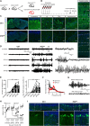
Adult Cacna1aflox/flox;CAG-ER-Cre mice lose CACNA1A after tamoxifen induction and develop spike-wave seizure and ataxia. (A) Breeding diagram of Cacna1aflox/flox;CAG-ER-Cre mice and control loxP (Cacna1aflox/flox) littermates. (B) Experimental timeline for (C). DP1i21, 28, 21 and 28. (C) Left: Confocal images showing loss of CACNA1A in an Cacna1aflox/flox;CAG-ER-Cre mouse (iKOp/q) compared with control littermate (Ctrl) after tamoxifen induction (DP1i25). Scale bars = 1 mm. Right: Magnified images show the loss in the cerebellum cortex, thalamic LD and TRN, hippocampus, and cerebral cortex. Nuclei were labelled with 4′,6-diamidino-2-phenylindole (DAPI). Scale bars = 20 μm. (D) Representative EEG traces from control and iKOp/q mice before (pre-tamo) and after tamoxifen induction. Right: Expanded traces show the three boxed areas. Note the development of spontaneous SWDs in iKOp/q mice. (E) Summary graphs for the total number (left) and total duration (right) of SWDs in 30 min for iKOp/q mice. Kolmogorov–Smirnov test, ***P < 0.001. (F) PSD (solid line, averaged; shadow, SEM) shows the dominant frequency (4–6 Hz) of SWDs in iKOp/q mice (n = 7) at DP1i21 compared with that before tamoxifen induction. PSD was normalized to total PSD under 50 Hz. (G) Representative EEG traces show that ethosuximide (200 mg/kg body weight, i.p.) abolished SWDs from iKOp/q mice (n = 6). (H) iKOp/q mice (n = 13), but not control (n = 17) show a progressively shorter latency to fall on a rotarod after tamoxifen induction. Two-way (genotype × trial) ANOVA with repeated measures, ***P < 0.001. (I) Confocal images show enhanced expression of TH in the vermis of cerebellum from iKOp/q mice compared with control. Scale bars = 20 μm. Mean ± SEM.
Figure 2
Figure 2
Adult Cacna1a loss reduces voltage sag and hyperpolarizes resting membrane potential in thalamic laterodorsal neurons. (A) Schematic of whole-cell recording experiments. Rec. = recording electrode. (B) Representative rebound spiking of thalamocortical neurons from tamoxifen-induced control loxP and iKOp/q mice in response to a hyperpolarizing current step. (C) Summary graph for the number of rebound spikes in the thalamic LD neurons of control and iKOp/q mice. On average, the number of rebound spikes is similar (control: n = 13; iKOp/q: n = 18). Kolmogorov–Smirnov test, P = 0.99. (D and E) current injected, membrane capacitance (Cm) and resistance (Rm) are similar. (F) Representative responses (solid line, averaged; shadow, repeats) to a hyperpolarizing current step into LD neurons (160 and 100 pA, respectively). (G and H) Summary graphs for sag ratio (G) and resting membrane potential (H). Depolarizing sag is smaller and the average resting membrane potential is more hyperpolarized in LD neurons from iKOp/q mice compared with control. Student’s t-test. (I) T-type calcium current (IT) recorded from LD neurons in response to test potentials ranging from −70 to −45 mV after being hyperpolarized (2–3 s) to −90 mV. Traces are aligned to the mean of the last 50 ms. (J) Summary graph of the peak amplitude of IT (control: n = 10; iKOp/q: n = 10). The average amplitude of IT in iKOp/q mice is similar to that in control mice. Student’s t-test. P = 0.99. *P < 0.05, **P < 0.01. n.s. = not significant. Mean ± SEM. TC = thalamocortical relay neuron.
Figure 3
Figure 3
α1G deficiency suppresses but does not prevent absence seizure in Cacna1a adult-ablated mice. (A) PCR results for genotyping Cacna1a flox/flox;CAG-ER-Cre;Cacna1G/ (iKOp/q+g) mice. The Cre primers generated a 100-bp product. The Cacna1a primers generated a 300-bp band for the wild-type or two bands (400 bp and 700 bp) for the loxP-flanked allele. The Cacna1g primers produced a 175-bp band for the wild-type, a 300-bp product for the floxed allele or a 700-bp product for knockout allele. (B) Representative responses to a hyperpolarizing current (200 pA) and a depolarizing current (100 pA) in LD neurons from iKOp/q and iKOp/q+g mice. No rebound spike was evoked in LD neurons (n = 9) from iKOp/q+g mice as shown in bar graph. Student’s t-test. ***P <0.001. (C) Representative T-type calcium current (IT) recorded from LD neurons in response to test potentials from −90 mV in tamoxifen induced iKOp/q (DP1i23) and iKOp/q+g (DP1i31) mice. Bottom right: Summary graph of the peak amplitude of IT (iKOp/q+g: n = 5). On average, the amplitude of IT in iKOp/q+g mice is 9.91 ± 7.51 pA. Student’s t-test. *P < 0.001. (D) Representative EEG traces (two channels, L and R) show spontaneous spike-wave-discharges (SWDs) from a tamoxifen induced iKOp/q+g mouse (DP1i21). Expanded traces show the boxed area. (E) PSD analysis shows that unlike iKOp/q mice, a similar PSD distribution in the iKOp/q+g mouse at DP1i21 to that before tamoxifen induction (Pre-tamo). (F) Summary graph for PSD before and after tamoxifen induction for iKOp/q+g mice. Mean ± SEM.
Figure 4
Figure 4
Adult Cacna1a ablation results in reduced rebound spiking and membrane excitability of neurons in TRN. (A) Schematic of whole-cell recording experiments. (B) Representative rebound spiking of TRN neurons from tamoxifen induced control loxP and iKOp/q mice in response to a hyperpolarizing current step. (C) Summary graph of the number of rebound spikes (control: n = 22; iKOp/q: n = 23). The average number of TRN rebound spikes in iKOp/q mice is smaller than that for control mice. Mann–Whitney U-test. **P < 0.01. (D and E) Current injected, membrane capacitance and resistance are similar. (F) As in B, but for tonic firing in response to a series of depolarizing current steps. (G) Summary graph shows firing frequency versus the amplitude of injected current (individual neuron: grey and light red lines; average: black filled square and red filled circle) for TRN neurons (control: n = 21; iKOp/q: n = 22). N-way ANOVA. ***P < 0.001. (H) T-type calcium current (IT) recorded from TRN neurons in response to test potentials ranging from −60 to −30 mV after being hyperpolarized (2–3 s) to −90 mV. Traces are aligned to the mean of the last 50 ms. (I) Summary graph of the peak amplitude of IT (control: n = 18; iKOp/q: n = 17). On average, the amplitude of IT in iKOp/q mice is smaller compared with control. Student’s t-test. *P < 0.05. Mean ± SEM. TC = thalamocortical relay neuron.
Figure 5
Figure 5
Cacna1h null mutation results in reduced TRN excitability and prolonged SWDs in Cacna1a adult-ablated mice. (A) PCR results for genotyping Cacna1aflox/flox;CAG-ER-Cre;Cacna1h/ (iKOp/q+h) mice. The Cre primers generated a 100-bp product. The Cacna1a primers generated a 300-bp band for the wild-type or two bands (400 bp and 700 bp) for the loxP-flanked allele. The Cacna1h primers produced a 480-bp band for the wild-type or a 330-bp product for the knockout allele. (B) Left: Representative responses to a hyperpolarizing step current injected into TRN neurons in iKOp/q+h mice without tamoxifen induction. Right: Summary graph shows fewer rebound spikes in TRN neurons from iKOp/q+h mice (n = 12) compared with Cacna1aflox/flox mice (Ctrl, dash bar). Mann–Whitney U-test. *P < 0.05. (C) As in B, but for tonic firing in response to a series of depolarizing current steps. (D) Summary graph shows firing frequency versus the amplitude of current injected (individual neuron: light magenta lines; averaged: magenta filled circle and grey line) for TRN neurons (iKOp/q+h: n = 7). TRN neuron from iKOp/q+h mice is less excitable compared with Cacna1aflox/flox mice (Ctrl, grey line). N-way ANOVA. ***P < 0.001. (E) Representative EEG traces show spontaneous SWDs from an iKOp/q+h mouse induced by tamoxifen injection. Expanded traces show the boxed areas. (F) PSD analysis shows the dominant frequency (4–6 Hz) of SWDs in iKOp/q+h mice (n = 5) at DP1i21 compared with that before tamoxifen induction (Pre-tamo). (G) Bar graph shows that the average duration of SWDs from iKOp/q+h mice is longer compared with iKOp/q mice. Student’s t-test. *P < 0.05, **P < 0.01. Mean ± SEM.

References

    1. Anderson MP, Mochizuki T, Xie J, Fischler W, Manger JP, Talley EM, et al.Thalamic Cav3.1 T-type Ca2+ channel plays a crucial role in stabilizing sleep. Proc Natl Acad Sci U S A 2005; 102: 1743–8. - PMC - PubMed
    1. Blumenfeld H. Cellular and network mechanisms of spike-wave seizures. Epilepsia 2005; 46: 21–33. - PubMed
    1. Bomben VC, Aiba I, Qian J, Mark MD, Herlitze S, Noebels JL. Isolated P/Q calcium channel deletion in layer VI corticothalamic neurons generates absence epilepsy. J Neurosci 2016; 36: 405–18. - PMC - PubMed
    1. Broicher T, Kanyshkova T, Meuth P, Pape HC, Budde T. Correlation of T-channel coding gene expression, IT, and the low threshold Ca2+ spike in the thalamus of a rat model of absence epilepsy. Mol Cell Neurosci 2008; 39: 384–99. - PubMed
    1. Cain SM, Tyson JR, Choi HB, Ko R, Lin PJC, LeDue JM, et al.CaV 3.2 drives sustained burst-firing, which is critical for absence seizure propagation in reticular thalamic neurons. Epilepsia 2018; 59: 778–91. - PMC - PubMed

Publication types

MeSH terms

Substances