

doi: 10.1093/cvr/cvz283.
Cardiac arrhythmogenesis: a tale of two clocks?
Affiliations
- PMID: 31800017
- PMCID: PMC7695354
- DOI: 10.1093/cvr/cvz283
Item in Clipboard
Cardiac arrhythmogenesis: a tale of two clocks?
Cardiovasc Res.
.
No abstract available
Keywords: Anti-arrhythmic drugs; Ca2+ homeostasis; Cardiac arrhythmias; Ion channels.
Figures
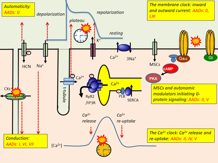
Molecular ion channel and transporter, and signalling components contributing to normal cardiac activity and arrhythmic events and their grouping (AADs 0-VII) into pharmacological targets in the updated Oxford classification scheme. This scheme maps onto a two-clock scheme for normal and abnormal cardiac electrophysiological function. A membrane (M) clock mediates surface electrophysiological changes and conduction of excitation but feeds forwards onto the calcium (C-) clock through producing alterations in cytosolic Ca2+. These trigger cycles of store Ca2+ release and re-uptake normally driven by activation and recovery processes in the M-clock. However, the C-clock also feeds back onto the M-clock, altering AP conduction, recovery, and post-recovery stability, besides exerting longer-term effects on channel expression. Autonomic sympathetic and parasympathetic control mechanisms act at different control points respectively situated in the C- and M-clocks. cAMP, cyclic 3′5-adenosine monophosphate; Cx, connexin; Gi, inhibitory G protein; Gs, stimulatory G-protein; HCN, hyperpolarization-activated cyclic nucleotide-gated channel; MSC, mechanically sensitive channel; PKA, protein kinase A; PLB, phospholamban; RyR2, cardiac ryanodine receptor, type 2; SERCA, sarcoplasmic reticulum Ca2+-ATPase; Na+, K+ and Ca2+ flux through Nav1.5, Kv and Ca1.2 channels also shown.


Similar articles
-
["Sicilian gambit": pathophysiological approach to drug therapy of arrhythmia (lecture]].Ter Arkh. 1999;71(8):67-74. Ter Arkh. 1999. PMID: 10515042 Russian. No abstract available.
-
Ranolazine in Cardiac Arrhythmia.Clin Cardiol. 2016 Mar;39(3):170-8. doi: 10.1002/clc.22476. Epub 2015 Oct 13. Clin Cardiol. 2016. PMID: 26459200 Free PMC article. Review.
-
Modernized Classification of Cardiac Antiarrhythmic Drugs.Circulation. 2018 Oct 23;138(17):1879-1896. doi: 10.1161/CIRCULATIONAHA.118.035455. Circulation. 2018. PMID: 30354657
-
The Relentless Pursuit of New Drugs to Treat Cardiac Arrhythmias.Circulation. 2020 May 12;141(19):1507-1509. doi: 10.1161/CIRCULATIONAHA.119.045149. Epub 2020 May 11. Circulation. 2020. PMID: 32392105 No abstract available.
-
Modulation of spiral wave reentry by K(+) channel blockade.Circ J. 2007;71 Suppl A:A26-31. doi: 10.1253/circj.71.a26. Circ J. 2007. PMID: 17587736 Review.
Cited by
-
Cardiac Connexin-43 Hemichannels and Pannexin1 Channels: Provocative Antiarrhythmic Targets.Int J Mol Sci. 2020 Dec 29;22(1):260. doi: 10.3390/ijms22010260. Int J Mol Sci. 2020. PMID: 33383853 Free PMC article. Review.
-
Feedback contributions to excitation-contraction coupling in native functioning striated muscle.Philos Trans R Soc Lond B Biol Sci. 2023 Jun 19;378(1879):20220162. doi: 10.1098/rstb.2022.0162. Epub 2023 May 1. Philos Trans R Soc Lond B Biol Sci. 2023. PMID: 37122213 Free PMC article. Review.
-
Dual calcium-voltage optical mapping of regional voltage and calcium signals in intact murine RyR2-R2474S hearts.J Mol Cell Cardiol Plus. 2024 Dec;10:100121. doi: 10.1016/j.jmccpl.2024.100121. J Mol Cell Cardiol Plus. 2024. PMID: 39697246 Free PMC article.
-
Ageing Increases Cardiac Electrical Remodelling in Rats and Mice via NOX4/ROS/CaMKII-Mediated Calcium Signalling.Oxid Med Cell Longev. 2022 Mar 28;2022:8538296. doi: 10.1155/2022/8538296. eCollection 2022. Oxid Med Cell Longev. 2022. PMID: 35387264 Free PMC article.
-
Electrophysiological and Proarrhythmic Effects of Hydroxychloroquine Challenge in Guinea-Pig Hearts.ACS Pharmacol Transl Sci. 2021 Aug 30;4(5):1639-1653. doi: 10.1021/acsptsci.1c00166. eCollection 2021 Oct 8. ACS Pharmacol Transl Sci. 2021. PMID: 34661080 Free PMC article.
References
-
- Kuriachan V, Sumner G, Mitchell L. Sudden cardiac death. Curr Probl Cardiol 2015;40:133–200. - PubMed
-
- Vaughan Williams E. Classification of antiarrhythmic drugs In Sandoe E, Flensted-Jensen E, Olsen K (eds). Symposium on Cardiac Arrhythmias. Elsinore, Denmark: Astra, 1970. pp. 449–472.
-
- Vaughan Williams E. Classification of antidysrhythmic drugs. Pharmacol Ther B 1975;1:115–138. - PubMed
-
- Carmeliet E, Vereecke J. Cardiac Cellular Electrophysiology. Dordrecht: Kluwer Academic Publishers; 2002.
-
- Gilmour RF, Zipes DP. Mechanisms of disease: new mechanisms of antiarrhythmic actions. Nat Rev Cardiol 2004;1:37–41. - PubMed
Publication types
MeSH terms
Substances
Grants and funding
- PG/12/21/29473/BHF_/British Heart Foundation/United Kingdom
- PG/16/67/32340/BHF_/British Heart Foundation/United Kingdom
- 105727/Z/14/Z/WT_/Wellcome Trust/United Kingdom
- MR/M001288/1/MRC_/Medical Research Council/United Kingdom
- PG/14/79/31102/BHF_/British Heart Foundation/United Kingdom
- G1002647/MRC_/Medical Research Council/United Kingdom
- PG/14/80/31106/BHF_/British Heart Foundation/United Kingdom
- WT_/Wellcome Trust/United Kingdom
- PG/11/59/29006/BHF_/British Heart Foundation/United Kingdom
- PG/15/12/31280/BHF_/British Heart Foundation/United Kingdom
- G1002082/MRC_/Medical Research Council/United Kingdom
LinkOut - more resources
Full Text Sources
Medical
Miscellaneous
