Review
. 2019 Dec 4;101(23):2141-2151.
doi: 10.2106/JBJS.19.00346.

Hip Joint Capsular Anatomy, Mechanics, and Surgical Management

Affiliations
Review

Hip Joint Capsular Anatomy, Mechanics, and Surgical Management

K C Geoffrey Ng et al. J Bone Joint Surg Am. .

Abstract

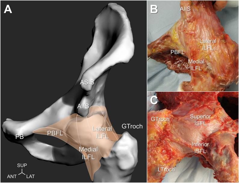
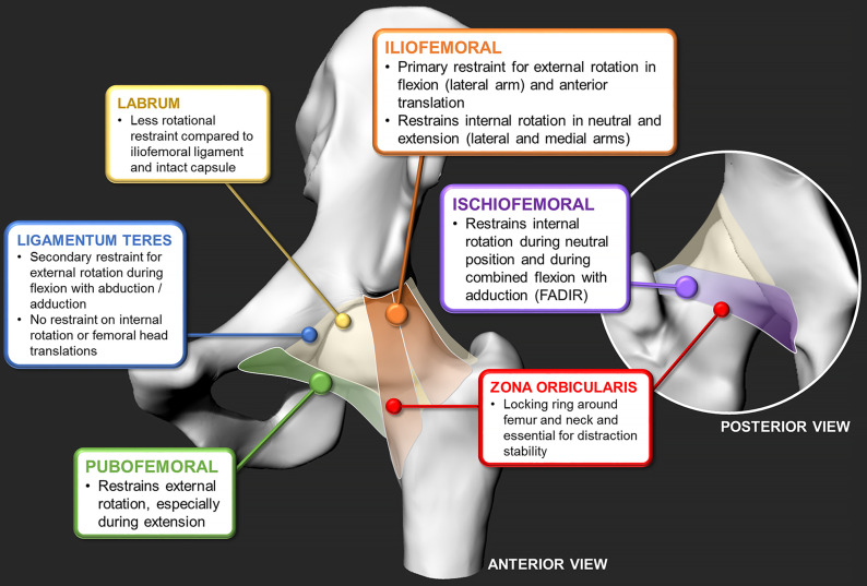
  1. Hip joint capsular ligaments (iliofemoral, ischiofemoral, and pubofemoral) play a predominant role in functional mobility and joint stability.

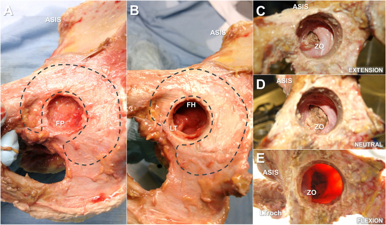
  2. The zona orbicularis resists joint distraction (during neutral positions), and its aperture mechanism stabilizes the hip from adverse edge-loading (during extreme hip flexion-extension).

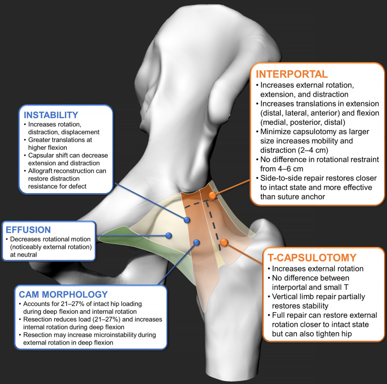
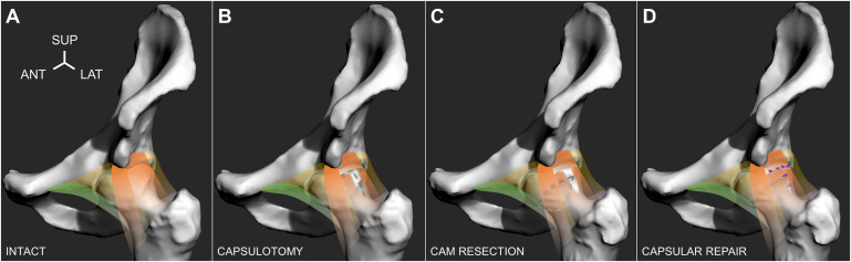
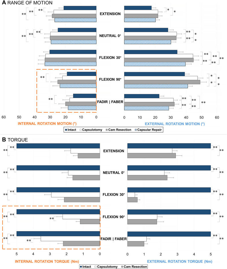
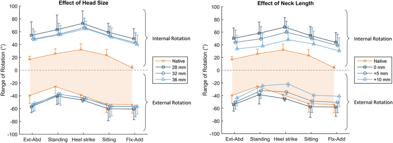
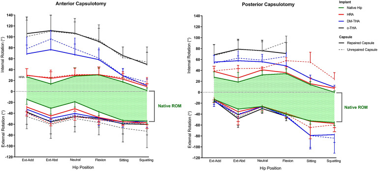
  3. To preserve joint function and stability, it is important to minimize capsulotomy size and avoid disrupting the zona orbicularis, preserve the femoral head size and neck length, and only repair when or as necessary without altering capsular tensions.

  4. It is not fully understood what the role of capsular tightness is in patients who have cam femoroacetabular impingement and if partial capsular release could be beneficial and/or therapeutic.

  5. During arthroplasty surgery, a femoral head implant that is nearly equivalent to the native head size with an optimal neck-length offset can optimize capsular tension and decrease dislocation risk where an intact posterior hip capsule plays a critical role in maintaining hip stability.


Figures

Fig. 1
Fig. 1
Anatomy of the capsular ligaments illustrated with a left-sided hip model in neutral position (Fig. 1-A) and showing the anterior view of a cadaveric hip specimen in external rotation (Fig. 1-B) and the posterior view of a cadaveric hip specimen in internal rotation (Fig. 1-C). The lateral and medial branches of the iliofemoral ligament (ILFL), pubofemoral ligament (PBFL), superior and inferior fibers of the ischiofemoral ligament (ISFL), anterior superior iliac spine (ASIS), anterior inferior iliac spine (AIIS), pubis (PB), and greater and lesser trochanters (GTroch and LTroch) are indicated. SUP = superior, ANT = anterior, and LAT = lateral.
Fig. 2
Fig. 2
Medial-to-lateral view of a right-sided cadaveric hip specimen through progressive portal sizes and depths of the cotyloid fossa and acetabulum, indicating the small portal view of the fat pad (FP), cushioning the innervations and proprioceptors (Fig. 2-A); ligamentum teres (LT) tethering the femoral head (FH) at the fovea pit (with the fat pad removed) (Fig. 2-B); large portal view of the tightened posterior collar of the zona orbicularis (ZO) during hip extension (with ligamentum teres, acetabulum, and femoral head removed) (Fig. 2-C); relaxed ZO during neutral position (Fig. 2-D); and tightened anterior collar of the ZO during deep hip flexion (Fig. 2-E). The acetabular cartilage (dashed lines), anterior superior iliac spine (ASIS), and lesser trochanter (LTroch) are indicated for reference.
Fig. 3
Fig. 3
The findings of in vitro cadaveric studies on individual capsular ligament contributions to joint stability, outlining iliofemoral, ischiofemoral, and pubofemoral ligaments,,; the zona orbicularis; ligamentum teres,; and labrum,. (For a detailed summary for each study’s scope, methods, and observations, see Appendix Table I.)
Fig. 4
Fig. 4
The findings of in vitro cadaveric studies on the effects of capsular conditions and surgical stages, outlining the contributions of the interportal capsulotomy,,,,,, and T-capsulotomy,,,, and the effects of instability,-,,, effusion, and cam morphology,. (For a detailed summary for each study’s scope, methods, and observations, see Appendix Tables II and III.)
Fig. 5
Fig. 5
Four surgical stages depicted on a left-sided hip, which included the intact hip with cam morphology, were tested (with orange indicating the iliofemoral ligament; green, the pubofemoral ligament; and yellow, the encapsulating tissue) (Fig. 5-A); capsulotomy, in which the iliofemoral ligament was incised to create a T-capsulotomy (dashed lines) (Fig. 5-B); cam resection, in which the cam morphology was resected through the incised capsule (dashed lines) (Fig. 5-C); and capsular repair, in which the incised portal was closed using simple, interrupted sutures (purple knots) (Fig. 5-D). ANT = anterior, LAT = lateral, and SUP = superior. (Adapted, under CC BY 4.0 [https://creativecommons.org/licenses/by/4.0/], from: Ng KCG, El Daou H, Bankes MJK, Rodriguez Y Baena F, Jeffers JRT. Hip joint torsional loading before and after cam femoroacetabular impingement surgery. Am J Sports Med. 2019 Feb;47[2]:420-30. Published by SAGE Publishing. https://doi.org/10.1177/0363546518815159.)
Fig. 6
Fig. 6
Range of motion (Fig. 6-A) and torque loading (Fig. 6-B) in internal and external rotation, at each stage of testing: intact, capsulotomy, cam resection, and capsular repair (range of motion only), reported as the mean and standard error. Cam resection further increased internal rotation in the deep flexion positions (highlighted dashed lines). *The difference is significant (p < 0.05). **The difference is significant (p < 0.01.) FADIR = flexion-adduction and internal rotation, and FABER = flexion-abduction and external rotation. (Adapted, under CC BY 4.0 [https://creativecommons.org/licenses/by/4.0/], from: Ng KCG, El Daou H, Bankes MJK, Rodriguez Y Baena F, Jeffers JRT. Hip joint torsional loading before and after cam femoroacetabular impingement surgery. Am J Sports Med. 2019 Feb;47[2]:420-30. Published by SAGE Publishing. https://doi.org/10.1177/0363546518815159.)
Fig. 7
Fig. 7
After total hip arthroplasty, the effects of head size (left), with anatomical neck length, and increasing neck lengths (right) on rotational range of motion are depicted. The x axis denotes the testing positions, and the y axis denotes the internal-external rotation (positive-negative). The shaded orange area dictates the measured range of native hip rotation. Increasing head size had little effect, whereas lengthening the neck tightened the hip capsule and reduced external rotation in some hip positions. The values are given as the mean, and the error bars indicate the standard deviation. Ext-Abd = extension and abduction, and Flx-Add = flexion and adduction. (Reproduced from: van Arkel RJ, Ng KCG, Muirhead-Allwood SK, Jeffers JRT. Capsular ligament function after total hip arthroplasty. J Bone Joint Surg Am. 2018 Jul 18;100[14]:e94.)
Fig. 8
Fig. 8
The effects of the anterior (left) and posterior approaches (right) are examined with various types of implants: hip resurfacing arthroplasty (HRA; red), dual mobility arthroplasty (DM-THA; blue), and conventional total hip arthroplasty (c-THA; black) after unrepaired (dashed) and repaired conditions (solid). The x axis denotes the testing positions, and the y axis denotes the internal-external rotation (positive-negative). The shaded green area dictates the measured range of native hip rotation. The larger head size of the hip resurfacing arthroplasty demonstrated a range of motion that was more similar to the intact condition. The posterior approach showed higher dislocation risks during deep flexion. The values are given as the mean, and the error bars indicate the standard deviation. Ext-Abd = extension and abduction, Flx-Add = flexion and adduction, and ROM = range of motion. (Reproduced, with permission, from: Logishetty K, van Arkel RJ, Ng KCG, Muirhead-Allwood SK, Cobb JP, Jeffers JRT. Hip capsule biomechanics after arthroplasty: the effect of implant, approach, and surgical repair. Bone Joint J. 2019 Apr;101-B(4):426-34. ©2019 The British Editorial Society of Bone & Joint Surgery).

References

    1. Hewitt J, Guilak F, Glisson R, Vail TP. Regional material properties of the human hip joint capsule ligaments. J Orthop Res. 2001. May;19(3):359-64. - PubMed
    1. Malagelada F, Tayar R, Barke S, Stafford G, Field RE. Anatomy of the zona orbicularis of the hip: a magnetic resonance study. Surg Radiol Anat. 2015. January;37(1):11-8. Epub 2014 Apr 29. - PubMed
    1. Wagner FV, Negrão JR, Campos J, Ward SR, Haghighi P, Trudell DJ, Resnick D. Capsular ligaments of the hip: anatomic, histologic, and positional study in cadaveric specimens with MR arthrography. Radiology. 2012. April;263(1):189-98. Epub 2012 Feb 27. - PubMed
    1. Walters BL, Cooper JH, Rodriguez JA. New findings in hip capsular anatomy: dimensions of capsular thickness and pericapsular contributions. Arthroscopy. 2014. October;30(10):1235-45. Epub 2014 Jul 23. - PubMed
    1. Bedi A, Galano G, Walsh C, Kelly BT. Capsular management during hip arthroscopy: from femoroacetabular impingement to instability. Arthroscopy. 2011. December;27(12):1720-31. Epub 2011 Nov 1. - PubMed