Review
. 2019 Nov 30;20(23):6047.
doi: 10.3390/ijms20236047.

Bone Pain in Cancer Patients: Mechanisms and Current Treatment

Affiliations
Review

Bone Pain in Cancer Patients: Mechanisms and Current Treatment

Renata Zajączkowska et al. Int J Mol Sci. .

Abstract

The skeletal system is the third most common site for cancer metastases, surpassed only by the lungs and liver. Many tumors, especially those of the breast, prostate, lungs, and kidneys, have a strong predilection to metastasize to bone, which causes pain, hypercalcemia, pathological skeletal fractures, compression of the spinal cord or other nervous structures, decreased mobility, and increased mortality. Metastatic cancer-induced bone pain (CIBP) is a type of chronic pain with unique and complex pathophysiology characterized by nociceptive and neuropathic components. Its treatment should be multimodal (pharmacological and non-pharmacological), including causal anticancer and symptomatic analgesic treatment to improve quality of life (QoL). The aim of this paper is to discuss the mechanisms involved in the occurrence and persistence of cancer-associated bone pain and to review the treatment methods recommended by experts in clinical practice. The final part of the paper reviews experimental therapeutic methods that are currently being studied and that may improve the efficacy of bone pain treatment in cancer patients in the future.

Keywords: cancer-induced bone pain; multimodal pain treatment; neuropathic pain; nociceptive pain.


Conflict of interest statement

The authors declare no conflicts of interest.

Figures

Figure 1
Figure 1
Receptors and ion channels on bone sensory nerves (According to [14]). Several types of receptors and ion channels are located on bone-innervating sensory nerves. They help to detect and transmit signals about noxious stimuli produced by tumor cells, stromal cells, and tumor-associated immune cells. These receptors (endothelin receptor ETAR, prostaglandin receptors, tropomyosin kinase A (TrkA) receptor, bradykinin receptor, cytokine receptors, chemokine receptors, TRPV1 (transient receptor potential channel, vanilloid subfamily member 1), acid-sensing ion channel 3 (ASIC3), purinergic receptor (P2X3)) are activated by mediators, e.g., endothelin (ET), prostaglandin E2 (PGE2), nerve growth factor (NGF), bradykinin, pro-inflammatory cytokines (tumor necrosis factor α (TNFα), interleukins (IL1β, IL6, IL8, IL15)), chemokines (CCL5, monocyte chemoattractant protein 1 (MCP-1), macrophage inflammatory protein-1a (MIP–1a)), H+, and extracellular adenosine triphosphate (ATP).
Figure 2
Figure 2
Interactions between tumor cells, osteoblasts, osteoclasts, stromal cells, and inflammatory cells at the site of bone metastasis (According to [12]). Tumor cells release endothelin (ET), which, via its appropriate receptors, interacts with osteoblasts to stimulate their proliferation. Activated osteoblasts release RANKL, which, in turn, provides a signal for the proliferation and maturation of osteoclasts and, hence, their destructive effect on bones. Osteoclasts produce ATP and acidosis-inducing H+, which activate appropriate receptors (P2X, TRPV1, ASICS3) located on bone-innervating neurons. Moreover, tumor cells, stromal cells, and activated immune cells secrete several mediators (endothelin, prostaglandin, NGF, bradykinin, pro-inflammatory cytokines, chemokines, H+, and ATP), which activate appropriate receptors (endothelin receptor, prostaglandin receptor, TrkA receptor, bradykinin receptor, cytokine receptors, chemokine receptors, TRPV1, ASIC3, P2X3) located on bone-innervating nerve fiber endings. These receptors help to detect and transmit signals about noxious stimuli to the spinal cord and then to the cerebral cortex, where perception takes place.
Figure 3
Figure 3
Role of the RANKL/RANK system and the mechanism of action of denosumab in bone pain due to cancer (According to [12]). RANKL is a protein secreted by tumor cells and the osteoblastic cell line (i.e., by mature osteoblasts and their precursors). RANKL binds to RANK receptors located on the osteoclast surface, which triggers the proliferation and maturation of osteoclasts, thereby initiating their damaging effect on bones. Denosumab is characterized by a high affinity and specificity for RANKL. It prevents the activation of RANK receptors on the surface of osteoclasts and their precursors, thereby inhibiting the formation, proliferation, and survival of osteoclasts, which reduces osteoclastic bone resorption. X = stop.
Figure 4
Figure 4
Pathological nerve sprouting in bone metastases and the mechanisms of action of tanezumab (according to [42]). After binding to the TrkA receptor, nerve growth factor (NGF) activates the pathological formation and growth of a network of new nerve fibers characterized by a unique morphology, organization, and density, which exceeds the density of nerve fibers in normal bone by up to 10–70 times. These new pathological nerve fibers are formed within the periosteum, mineralized bone tissue, and bone marrow (A). Tanezumab, a humanized anti-NGF monoclonal antibody, prevents NGF from binding to the TrkA receptor and consequently inhibits the pathological sprouting and formation of new pathological nerve fiber networks (B).
Figure 5
Figure 5
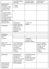
Treatment of pain due to bone metastases according to ESMO and WHO recommendations [44,45]. mSCC, metastatic spinal cord compression; SRE, skeletal-related events; BP, bisphosphonates; EBRT, external beam radiotherapy; SBRT, stereotactic body radiotherapy; HFRT, hypofractionated radiotherapy; RT, radiotherapy; (+) recommended; (+/−) experts have not made a recommendation for or against the given method of treatment.
Figure 6
Figure 6
Mechanisms of action of bisphosphonates (BPs) (According to [13]).

References

    1. Zhu X.C., Zhang J.L., Ge C.T., Yu Y.Y., Wang P., Yuan T.F., Fu C.Y. Advances in cancer pain from bone metastasis. Drug Des. Dev. Ther. 2015;9:4239–4245. - PMC - PubMed
    1. Mercadante S. Malignant bone pain: Pathophysiology and treatment. Pain. 1997;69:1–18. doi: 10.1016/S0304-3959(96)03267-8. - DOI - PubMed
    1. Roodman G.D. Mechanisms of bone metastasis. N. Engl. J. Med. 2004;350:1655–1664. doi: 10.1056/NEJMra030831. - DOI - PubMed
    1. Davila D., Antoniou A., Chaudhry M.A. Evaluation of Osseous Metastasis in Bone Scintigraphy. Semin. Nucl. Med. 2015;45:3–15. doi: 10.1053/j.semnuclmed.2014.07.004. - DOI - PubMed
    1. Li B.T., Wong M.H., Pavlakis N. Treatment and prevention of bone metastases from breast cancer: A comprehensive review of evidence for clinical practice. J. Clin. Med. 2014;3:1–24. doi: 10.3390/jcm3010001. - DOI - PMC - PubMed

MeSH terms