

Post-traumatic thoracolumbar spinal epidural haematoma in a child: a rare clinical entity
- PMID: 31801780
- PMCID: PMC7001729
- DOI: 10.1136/bcr-2019-232055
Post-traumatic thoracolumbar spinal epidural haematoma in a child: a rare clinical entity
Abstract
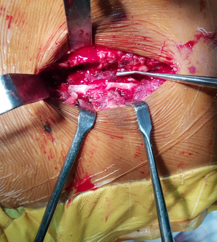
Post-traumatic spinal epidural haematoma (SEH) is a rare clinical entity in children. We are reporting the case of an 8-year-old child who presented with thoracolumbar SEH with neurological deficit. MRI confirmed SEH without bony disruption. Emergency evacuation of haematoma was done. There was an improvement in neurological status after removal of haematoma. Diagnosis of this rare condition is tricky in children owing to variable presenting symptoms, especially in an early stage with subtle neurological changes. There should be high clinical suspicion in children with atypical symptoms, and MRI should be done to confirm the diagnosis. Patients with acute neurological deficit should undergo urgent operative decompression. Conservative treatment has a limited role. Patients may be considered for non-operative management if they have medical contraindications, coagulation dysfunction or a small SEH without neurological deficit. These patients require serial MRI monitoring.
Keywords: back pain; neurological injury; orthopaedic and trauma surgery; paediatrics; spinal cord.
© BMJ Publishing Group Limited 2019. No commercial re-use. See rights and permissions. Published by BMJ.
Conflict of interest statement
Competing interests: None declared.
Figures


References
Publication types
MeSH terms
LinkOut - more resources
Full Text Sources
Medical