

Extremely Preterm Infants Have Significant Alterations in Their Conventional T Cell Compartment during the First Weeks of Life
- PMID: 31801814
- PMCID: PMC6926392
- DOI: 10.4049/jimmunol.1900941
Extremely Preterm Infants Have Significant Alterations in Their Conventional T Cell Compartment during the First Weeks of Life
Abstract
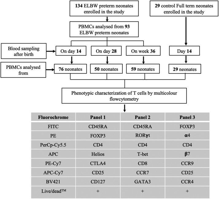
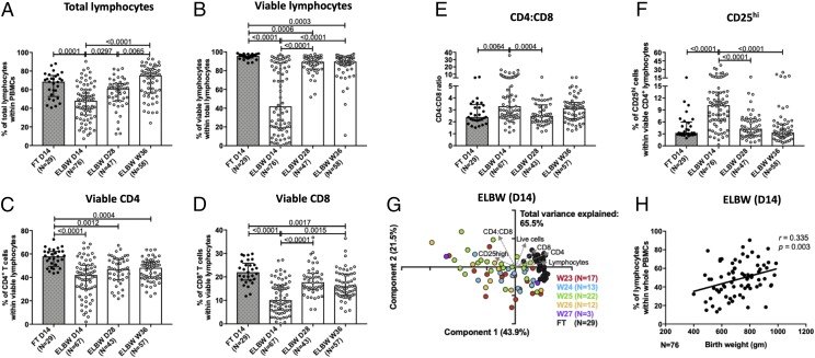
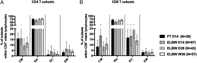
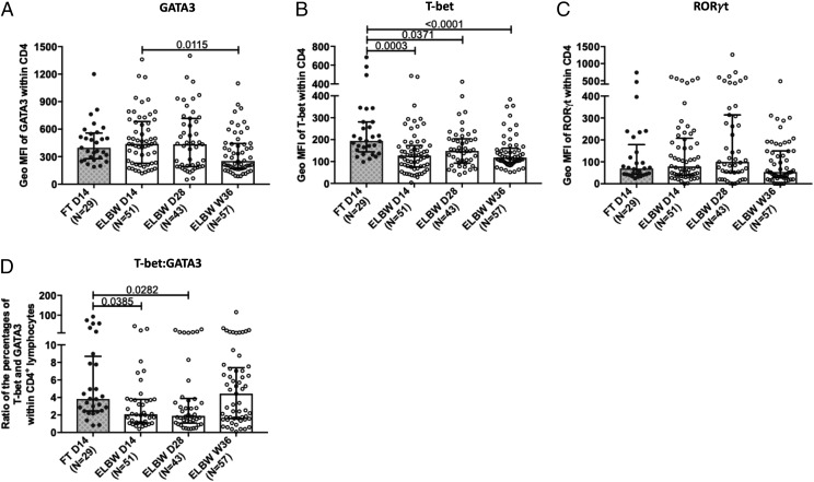
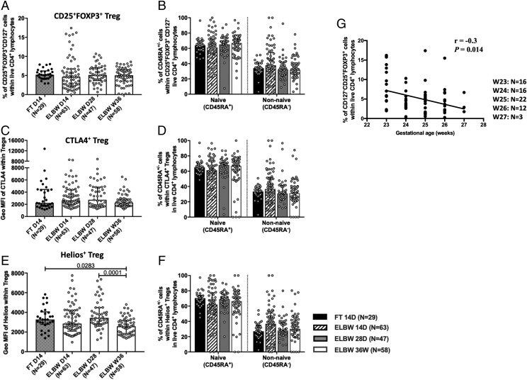
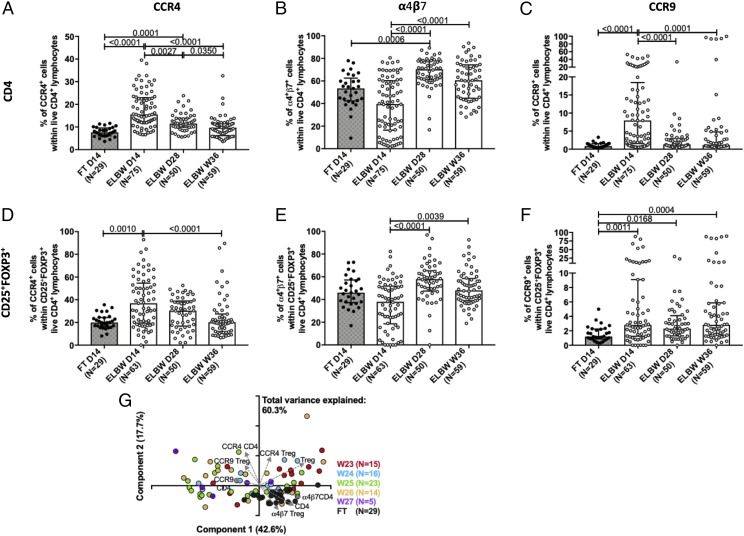
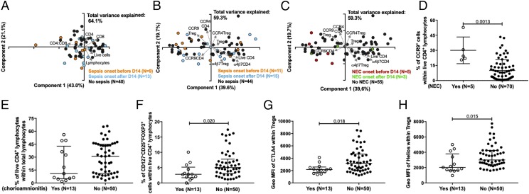
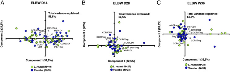
Extremely preterm neonates are particularly susceptible to infections, likely because of severely impaired immune function. However, little is known on the composition of the T cell compartment in early life in this vulnerable population. We conducted a comprehensive phenotypic flow cytometry-based longitudinal analysis of the peripheral conventional T cell compartment of human extremely low gestational age neonates (ELGAN) with extremely low birth weight (ELBW; <1000 g) participating in a randomized placebo-controlled study of probiotic supplementation. PBMCs from ELGAN/ELBW neonates were collected at day 14, day 28, and postmenstrual week 36. Comparisons were made with full-term 14-d-old neonates. Total CD4+ and CD8+ T cell frequencies were markedly lower in the preterm neonates. The reduction was more pronounced among the CD8+ population, resulting in an increased CD4/CD8 ratio. The preterm infants were also more Th2 skewed than the full-term infants. Although the frequency of regulatory T cells seemed normal in the ELGAN/ELBW preterm neonates, their expression of the homing receptors α4β7, CCR4, and CCR9 was altered. Notably, ELGAN/ELBW infants developing necrotizing enterocolitis before day 14 had higher expression of CCR9 in CD4+T cells at day 14. Chorioamnionitis clearly associated with reduced T regulatory cell frequencies and functional characteristics within the preterm group. Finally, probiotic supplementation with Lactobacillus reuteri did not impose any phenotypic changes of the conventional T cell compartment. In conclusion, notable immaturities of the T cell compartment in ELGAN/ELBW neonates may at least partially explain their increased susceptibility to severe immune-mediated morbidities.
Copyright © 2019 by The American Association of Immunologists, Inc.
Figures
Similar articles
-
Extreme prematurity and sepsis strongly influence frequencies and functional characteristics of circulating γδ T and natural killer cells.Clin Transl Immunology. 2021 Jun 10;10(6):e1294. doi: 10.1002/cti2.1294. eCollection 2021. Clin Transl Immunology. 2021. PMID: 34136218 Free PMC article.
-
High prevalence of abnormal motor repertoire at 3 months corrected age in extremely preterm infants.Eur J Paediatr Neurol. 2016 Mar;20(2):236-242. doi: 10.1016/j.ejpn.2015.12.009. Epub 2015 Dec 30. Eur J Paediatr Neurol. 2016. PMID: 26786751
-
Lactobacillus reuteri Colonisation of Extremely Preterm Infants in a Randomised Placebo-Controlled Trial.Microorganisms. 2021 Apr 24;9(5):915. doi: 10.3390/microorganisms9050915. Microorganisms. 2021. PMID: 33923278 Free PMC article.
-
Role of amino acid supplementation in the prevention of necrotizing enterocolitis in preterm neonates - a review of current evidences.J Matern Fetal Neonatal Med. 2018 Sep;31(17):2349-2366. doi: 10.1080/14767058.2017.1342797. Epub 2017 Jun 30. J Matern Fetal Neonatal Med. 2018. PMID: 28614987 Review.
-
Inhalation or instillation of steroids for the prevention of bronchopulmonary dysplasia.Neonatology. 2015;107(4):358-9. doi: 10.1159/000381132. Epub 2015 Jun 5. Neonatology. 2015. PMID: 26044104 Review.
Cited by
-
Premature Infants Have Normal Maturation of the T Cell Receptor Repertoire at Term.Front Immunol. 2022 May 30;13:854414. doi: 10.3389/fimmu.2022.854414. eCollection 2022. Front Immunol. 2022. PMID: 35707545 Free PMC article.
-
Extreme prematurity and sepsis strongly influence frequencies and functional characteristics of circulating γδ T and natural killer cells.Clin Transl Immunology. 2021 Jun 10;10(6):e1294. doi: 10.1002/cti2.1294. eCollection 2021. Clin Transl Immunology. 2021. PMID: 34136218 Free PMC article.
-
Impact of Extreme Prematurity, Chorioamnionitis, and Sepsis on Neonatal Monocyte Characteristics and Functions.J Innate Immun. 2024;16(1):470-488. doi: 10.1159/000541468. Epub 2024 Sep 14. J Innate Immun. 2024. PMID: 39278208 Free PMC article. Clinical Trial.
-
Depletion of Ly6G-Expressing Neutrophilic Cells Leads to Altered Peripheral T-Cell Homeostasis and Thymic Development in Neonatal Mice.Int J Mol Sci. 2023 Apr 24;24(9):7763. doi: 10.3390/ijms24097763. Int J Mol Sci. 2023. PMID: 37175470 Free PMC article.
-
Regulatory T Cells in Development and Prediction of Necrotizing Enterocolitis in Preterm Neonates: A Scoping Review.Int J Mol Sci. 2022 Sep 18;23(18):10903. doi: 10.3390/ijms231810903. Int J Mol Sci. 2022. PMID: 36142816 Free PMC article.
References
-
- Purisch S. E., Gyamfi-Bannerman C. 2017. Epidemiology of preterm birth. Semin. Perinatol. 41: 387–391. - PubMed
-
- Norman M., Hallberg B., Abrahamsson T., Björklund L. J., Domellöf M., Farooqi A., Foyn Bruun C., Gadsbøll C., Hellström-Westas L., Ingemansson F., et al. 2019. Association between year of birth and 1-year survival among extremely preterm infants in Sweden during 2004-2007 and 2014-2016. JAMA 321: 1188–1199. - PMC - PubMed
-
- Manuck T. A., Rice M. M., Bailit J. L., Grobman W. A., Reddy U. M., Wapner R. J., Thorp J. M., Caritis S. N., Prasad M., Tita A. T., et al. Eunice Kennedy Shriver National Institute of Child Health and Human Development Maternal-Fetal Medicine Units Network 2016. Preterm neonatal morbidity and mortality by gestational age: a contemporary cohort. Am. J. Obstet. Gynecol. 215: 103.e1–103.e14. - PMC - PubMed
-
- Zhang X., Zhivaki D., Lo-Man R. 2017. Unique aspects of the perinatal immune system. Nat. Rev. Immunol. 17: 495–507. - PubMed
Publication types
MeSH terms
LinkOut - more resources
Full Text Sources
Research Materials
