. 2020 Jan 10;295(2):529-538.
doi: 10.1074/jbc.RA119.010594. Epub 2019 Dec 4.

DNA polymerase β nucleotide-stabilized template misalignment fidelity depends on local sequence context

Affiliations

DNA polymerase β nucleotide-stabilized template misalignment fidelity depends on local sequence context

Michael J Howard et al. J Biol Chem. .

Abstract

DNA polymerase β has two DNA-binding domains that interact with the opposite sides of short DNA gaps. These domains contribute two activities that modify the 5' and 3' margins of gapped DNA during base excision repair. DNA gaps greater than 1 nucleotide (nt) pose an architectural and logistical problem for the two domains to interact with their respective DNA termini. Here, crystallographic and kinetic analyses of 2-nt gap-filling DNA synthesis revealed that the fidelity of DNA synthesis depends on local sequence context. This was due to template dynamics that altered which of the two template nucleotides in the gap served as the coding nucleotide. We observed that, when a purine nucleotide was in the first coding position, DNA synthesis fidelity was similar to that observed with a 1-nt gap. However, when the initial templating nucleotide was a pyrimidine, fidelity was decreased. If the first templating nucleotide was a cytidine, there was a significantly higher probability that the downstream template nucleotide coded for the incoming nucleotide. This dNTP-stabilized misalignment reduced base substitution and frameshift deletion fidelities. A crystal structure of a binary DNA product complex revealed that the cytidine in the first templating site was in an extrahelical position, permitting the downstream template nucleotide to occupy the coding position. These results indicate that DNA polymerase β can induce a strain in the DNA that modulates the position of the coding nucleotide and thereby impacts the identity of the incoming nucleotide. Our findings demonstrate that "correct" DNA synthesis can result in errors when template dynamics induce coding ambiguity.

Keywords: DNA polymerase; DNA repair; DNA structure; DNA synthesis; X-ray crystallography; base excision repair; kinetics; mutagenesis; mutagenesis mechanism; polymerase fidelity; structure–function.


Conflict of interest statement

The authors declare that they have no conflicts of interest with the contents of this article. The content is solely the responsibility of the authors and does not necessarily represent the official views of the National Institutes of Health

Figures
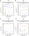
Figure 1.
Figure 1.
Discrimination plots for 2-nt gap-filling DNA synthesis by pol β. Steady-state catalytic efficiencies (kcat/Km,dNTP) are plotted on a log scale so that the distance between the efficiencies of correct and incorrect insertions is a measure of discrimination (29). The first column in each plot illustrates the catalytic efficiencies and discrimination for 1-nt gap filling; the templating base in the gap is indicated. The other columns show the catalytic efficiencies and discrimination for 2-nt gap filling; the templating sequence in the gap is indicated. A cartoon of the 2-nt gapped DNA substrate is shown above each plot, highlighting the identity of the first templating base and showing that the downstream template nucleotide (N) was varied. A, the identity of the first templating nucleotide is deoxyadenosine (A). B, the identity of the first templating nucleotide is deoxycytidine (C). C, the identity of the first templating nucleotide is deoxyguanosine (G). D, the identity of the first templating nucleotide is thymidine (T). The data are tabulated in Tables S1–S4, respectively. The identities of the incoming nucleotides are dATP (red), dCTP (green), dGTP (black), and dTTP (blue).
Figure 2.
Figure 2.
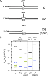
Effect of modifying pol β interactions with downstream DNA on DNA synthesis discrimination. A, steady-state catalytic efficiencies (kcat/Km,dNTP) were determined for 1-nt (templating C), 2-nt (CG in the gap), and open (no downstream oligonucleotide; i.e. no DNA gap) substrates; the relevant DNA sequence is given in the cartoon, and the full sequence is provided under “Experimental procedures.” In addition, 3KA, which perturbs 5′-phosphate gapped binding, was utilized. RB69exo (B family replicative pol) was used as a control. B, discrimination plot for the steady-state catalytic efficiencies tabulated in Table S5. The identities of the incoming nucleotides are dCTP (green), dGTP (black), and dTTP (blue).
Figure 3.
Figure 3.
Single-turnover analysis of mutagenic 2-nt gap-filling DNA synthesis. Single-turnover primer extension was assayed and quantified as described under “Experimental procedures.” A, illustration of the 5′-FAM–labeled 2-nt gapped DNA substrate. The sequence in the gap is 3′-CG-5′, and dCTP was added to initiate DNA synthesis. B, gel image illustrating the time course for 2-nt gap-filling (shown above). The substrate primer band (S) is extended by two dCMP insertions (+2 band). An intermediate band (+1) accumulates at short time intervals (<10 s). C, plot of the time course for the product bands (+1 and +2 as well as their sum, Total P). The progress curves were fit to either a single-exponential (+2 time course: A = −0.93, k = 0.07 s−1, C = 0.89; Total P time course: A = −0.88, k = 0.30 s−1, C = 0.91) or double-exponential equation (+1 time course: A1 = −0.78, k1 = 0.49 s−1, A2 = 0.75, k2 = 0.09 s−1, C = 0.05). D, illustration of the 5′-FAM–labeled 2-nt gapped DNA substrate. The sequence in the gap is 3′-GC-5′, and dGTP was added to initiate DNA synthesis. E, gel image illustrating the time course for 2-nt gap-filling (shown above). The substrate primer band (S) is extended by at least two dGMP insertions (P). An intermediate band can be observed that accumulates at later intervals. F, plot of the time course for all product bands (Total P). The progress curve was fitted to a single-exponential equation (A = −0.90, k = 0.0007 s−1, C = 0.90).
Figure 4.
Figure 4.
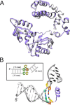
Structure of a stable binary pol β–DNA complex with an extrahelical templating cytosine. A, pol β ribbon representation of the binary pol β–DNA complex with an extrahelical templating cytosine (purple) superimposed with an open binary 1-nt gapped DNA substrate (gray; PDB code 3ISB) (18). The root mean square deviation over 320 Cα is 0.54 Å. B, DNA structure of the of resulting product after insertion of dCMP into a 2-nt gap (5′-CG-3′). The inserted cytosine (yellow) forms a stable base pair with the downstream template G (orange) but is distant from α-helix N (purple ribbon), which has repositioned itself to an open state following dCMP insertion. The substrate primer terminus base pair is colored green, and the extrahelical cytosine is also yellow. The inset illustrates the sequence alignment, color code, and general oligonucleotide arrangement.
Figure 5.
Figure 5.
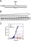
Single-turnover analysis of mutagenic 2-nt gap-filling DNA synthesis. Single-turnover primer extension was assayed and quantified as described under “Experimental procedures.” A, illustration of the 5′-FAM–labeled 2-nt gapped DNA substrate. The sequence in the gap is 3′-CT-5′, and dATP was added to initiate DNA synthesis. B, gel image illustrating the time course for 2-nt gap-filling (shown above). The substrate primer band (S) is extended by two dAMP insertions (+2 band). A weak intermediate band (+1) is also observed. C, plot of the time course for the product bands (+1 and +2 as well as their sum, Total P). The progress curves were fit to either a single-exponential (+2 time course: A = −0.84, k = 0.23 s−1, C = 0.82; Total P time course: A = −0.82, k = 0.28 s−1, C = 0.82) or double-exponential equation (+1 time course: A1 = −0.07, k1 = 2.1 s−1, A2 = 0.06, k2 = 0.10 s−1, C = 0.02). The inset expands the time course for the n+1 band.
Figure 6.
Figure 6.
Mutagenically correct nucleotide insertion. Shown is a diagram illustrating how correct nucleotide insertion can result in mutagenic DNA synthesis because of a dNTP-stabilized misalignment. In this general example, a 2-nt gapped DNA substrate is shown, with template nucleotides A (green) and B (red) in the gap. For pol β, when a purine occupies the first templating position (left, error-free path i), gap-filling DNA synthesis is faithful, inserting the complementary nucleotide (lowercase and in the same color; i.e. daTP opposite A and dbTP opposite B). In this situation, the downstream noncoding nucleotide (i.e. B) must relocate outside of the active site. Structures of pol λ with such an intermediate indicate that it occupies a position near α-helix N and has been termed scrunching (22). Translocation creates a 1-nt gap that is efficiently filled with high fidelity. In contrast to this error-free path, when a pyrimidine occupies the first templating position in a 2-nt gap (mutagenic path ii), template instability (looping out or dislocation) can lead to dNTP-stabilized misalignment (dotted box). In this case, the pyrimidine is moved out of the active site into an extrahelical position, permitting the downstream templating base to occupy the coding template position; in this case, B serves as the coding template base for the correct incoming nucleotide, dbTP. After insertion, the primer terminus could realign, generating a mispaired primer terminus and 1-nt gap. Correct insertion from this mispaired terminus completes gap filling but results in a base substitution error. Alternatively, if the first nascent base pair is stable and does not realign, then this pseudonicked structure provides a substrate for DNA ligation. Ligation of this pseudonicked substrate would generate a −1 deletion.

References

    1. Beard W. A., and Wilson S. H. (2006) Structure and mechanism of DNA polymerase β. Chem. Rev. 106, 361–382 10.1021/cr0404904 - DOI - PubMed
    1. Ray S., Breuer G., DeVeaux M., Zelterman D., Bindra R., and Sweasy J. B. (2018) DNA polymerase β participates in DNA end-joining. Nucleic Acids Res. 46, 242–255 10.1093/nar/gkx1147 - DOI - PMC - PubMed
    1. Prasad R., Beard W. A., and Wilson S. H. (1994) Studies of gapped DNA substrate binding by mammalian DNA polymerase β: dependence on 5′-phosphate group. J. Biol. Chem. 269, 18096–18101 - PubMed
    1. Beard W. A., Shock D. D., Yang X.-P., DeLauder S. F., and Wilson S. H. (2002) Loss of DNA polymerase β stacking interactions with templating purines, but not pyrimidines, alters catalytic efficiency and fidelity. J. Biol. Chem. 277, 8235–8242 10.1074/jbc.M107286200 - DOI - PubMed
    1. Beard W. A., Shock D. D., Batra V. K., Prasad R., and Wilson S. H. (2014) Substrate-induced DNA polymerase β activation. J. Biol. Chem. 289, 31411–31422 10.1074/jbc.M114.607432 - DOI - PMC - PubMed

Publication types