. 2020 Feb 14;94(5):e01531-19.
doi: 10.1128/JVI.01531-19. Print 2020 Feb 14.

PACSIN2 Interacts with Nonstructural Protein 5A and Regulates Hepatitis C Virus Assembly

Affiliations

PACSIN2 Interacts with Nonstructural Protein 5A and Regulates Hepatitis C Virus Assembly

Lap P Nguyen et al. J Virol. .

Abstract

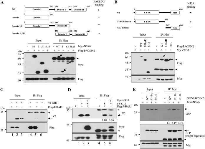
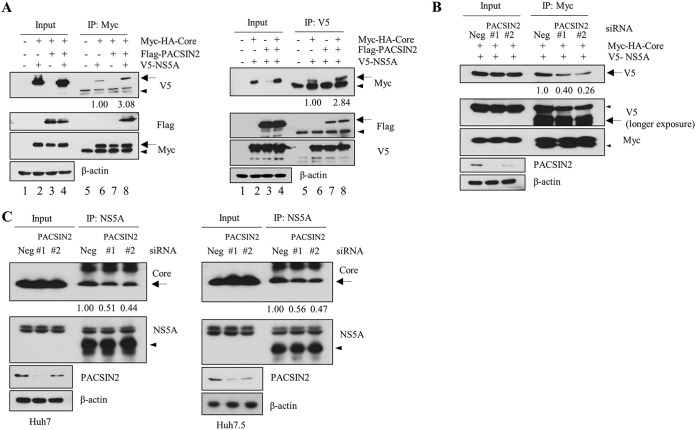
Hepatitis C virus (HCV) is a major etiologic agent of chronic liver diseases. HCV is highly dependent on cellular machinery for viral propagation. Using protein microarray analysis, we previously identified 90 cellular proteins as nonstructural 5A (NS5A) interacting partners. Of these, protein kinase C and casein kinase substrate in neurons protein 2 (PACSIN2) was selected for further study. PACSIN2 belongs to the PACSIN family, which is involved in the formation of caveolae. Protein interaction between NS5A and PACSIN2 was confirmed by pulldown assay and further verified by both coimmunoprecipitation and immunofluorescence assays. We showed that PACSIN2 interacted with domain I of NS5A and the Fer-CIP4 homology (FCH)-Bin/amphiphysin/Rvs (F-BAR) region of PACSIN2. Interestingly, NS5A specifically attenuated protein kinase C alpha (PKCα)-mediated phosphorylation of PACSIN2 at serine 313 by interrupting PACSIN2 and PKCα interaction. In fact, mutation of the serine 313 to alanine (S313A) of PACSIN2 increased protein interaction with NS5A. Silencing of PACSIN2 decreased both viral RNA and protein expression levels of HCV. Ectopic expression of the small interfering RNA (siRNA)-resistant PACSIN2 recovered the viral infectivity, suggesting that PACSIN2 was specifically required for HCV propagation. PACSIN2 was involved in viral assembly without affecting other steps of the HCV life cycle. Indeed, overexpression of PACSIN2 promoted NS5A and core protein (core) interaction. We further showed that inhibition of PKCα increased NS5A and core interaction, suggesting that phosphorylation of PACSIN2 might influence HCV assembly. Moreover, PACSIN2 was required for lipid droplet formation via modulating extracellular signal-regulated kinase 1/2 (ERK1/2) phosphorylation. Taken together, these data indicate that HCV modulates PACSIN2 via NS5A to promote virion assembly.IMPORTANCE PACSIN2 is a lipid-binding protein that triggers the tubulation of the phosphatidic acid-containing membranes. The functional involvement of PACSIN2 in the virus life cycle has not yet been demonstrated. We showed that phosphorylation of PACSIN2 displayed a negative effect on NS5A and core interaction. The most significant finding is that NS5A prevents PKCα from binding to PACSIN2. Therefore, the phosphorylation level of PACSIN2 is decreased in HCV-infected cells. We showed that HCV NS5A interrupted PKCα-mediated PACSIN2 phosphorylation at serine 313, thereby promoting NS5A-PACSIN2 interaction. We further demonstrated that PACSIN2 modulated lipid droplet formation through ERK1/2 phosphorylation. These data provide evidence that PACSIN2 is a proviral cellular factor required for viral propagation.

Keywords: NS5A; PACSIN2; hepatitis C virus; protein microarray; viral propagation.


Figures

FIG 1
FIG 1
(A) Identification of PACSIN2 in a protein microarray. (B) NS5A interacts with PACSIN2. HEK293T cells were transiently transfected with Flag-tagged PACSIN2 expression plasmid, and total cell lysates harvested at 24 h after transfection were incubated with either glutathione S-transferase (GST) or GST-tagged NS5A. After pulldown by GST beads, bound protein was detected by immunoblot analysis with an anti-Flag antibody. (C) HEK293T cells were cotransfected with Myc-tagged NS5A and Flag-tagged PACSIN2 expression plasmids. At 48 h after transfection, total cell lysates were immunoprecipitated with an anti-Myc antibody (upper) or an anti-Flag antibody (lower), and then bound protein was detected by immunoblot analysis with an anti-Flag antibody or an anti-Myc antibody. (D) Huh7 cells were either electroporated with Jc1 RNA or infected with Jc1. Cell lysates were then immunoprecipitated with either an anti-PACSIN2 antibody (left) or an anti-NS5A antibody (right). Bound protein was analyzed by immunoblotting with an anti-NS5A antibody and an anti-PACSIN2 antibody. All immunoblots were performed in triplicate. (E) Huh7 cells seeded onto coverslips were either mock infected or infected with Jc1. At 4 days postinfection, cells were fixed in cold methanol at −20°C for 10 min, and immunofluorescence staining was performed using an anti-PACSIN2 monoclonal antibody and tetramethyl rhodamine isocyanate (TRITC)-conjugated goat anti-mouse IgG to detect PACSIN2 (red), and a rabbit anti-NS5A antibody and fluorescein isothiocyanate (FITC)-conjugated goat anti-rabbit IgG to detect NS5A (green). Dual staining showed endogenous colocalization of PACSIN2 and NS5A as yellow fluorescence in the merged image. Cells were counterstained with 4′,6-diamidino-2-phenylindole (DAPI) to label nuclei (blue). Colocalization of PACSIN2 and HCV NS5A in the cytoplasm was quantified by both Pearson’s and Manders’ overlap coefficients using ImageJ analysis. (F) Huh7 cells harboring the HCV subgenomic replicon derived from genotype 1b were seeded onto coverslips and fixed in cold methanol at −20°C for 10 min. Immunofluorescence staining was performed as described in the panel E legend. Higher magnifications of the boxed areas are shown in the cropped image. Arrows denote the interaction bands, and arrowheads indicate the position of the IgG.
FIG 2
FIG 2
HCV NS5A interacts with PACSIN2 through domain I of NS5A and amino acid region 1 to 362 of PACSIN2, and protein interaction is dependent on phosphorylation status of PACSIN2. (A) PACSIN2 interacts with domain I of NS5A. (Upper) Schematic illustration of both wild-type and mutant forms of the NS5A. (Lower) HEK293T cells were cotransfected with Flag-tagged PACSIN2 and Myc-tagged NS5A expression plasmids. At 48 h after transfection, cell lysates were immunoprecipitated with an anti-Flag antibody, and bound protein was immunoblotted with an anti-Myc antibody. Protein expressions of Myc-tagged NS5A and Flag-tagged PACSIN2 were verified by immunoblotting with an anti-Myc or an anti-Flag antibody using the same cell lysates. (B) NS5A interacts with the amino acid region from 1 to 362 of PACSIN2. (Upper) Schematic illustration of both wild-type and mutant PACSIN2. (Lower) HEK293T cells were cotransfected with Myc-tagged NS5A and Flag-tagged PACSIN2 expression plasmids. At 48 h after transfection, cell lysates were immunoprecipitated with an anti-Myc antibody, and bound protein was detected by an anti-Flag antibody. (C) HEK293T cells were cotransfected with the V5-tagged SH3 and Flag-tagged F-BAR domains of PACSIN2 expression plasmids. At 48 h after transfection, cell lysates were immunoprecipitated with an anti-Flag antibody, and bound protein was detected by an anti-V5 antibody. (D) HEK293T cells were cotransfected with the V5-tagged SH3 and Flag-tagged F-BAR domains of PACSIN2 expression plasmids in the absence or presence of Myc-tagged NS5A. At 48 h after transfection, cell lysates were immunoprecipitated with an anti-Flag antibody, and bound protein was detected by an anti-V5 antibody. (E) HEK293T cells were cotransfected with Myc-tagged NS5A and green fluorescent protein (GFP)-tagged wild-type or phosphoinactive mutant (S313A) or phosphomimetic mutant (S313E) of PACSIN2 expression plasmids. At 48 h after transfection, cell lysates were immunoprecipitated with an anti-Myc antibody, and bound protein was detected by an anti-GFP antibody. Protein band intensity of GFP/IgG was determined by using ImageJ. Arrows indicate the interaction bands, and arrowheads denote the position of the IgG. All immunoblot and immunoprecipitation assays were performed in triplicate.
FIG 3
FIG 3
HCV NS5A protein interrupts PKCα-mediated PACSIN2 phosphorylation at serine 313 to promote binding with PACSIN2. (A) HEK293T cells were cotransfected with Myc-tagged NS5A and Flag-tagged PACSIN2 plasmids. At 48 h after transfection, total cell lysates were immunoprecipitated with an anti-Flag antibody, and bound protein was detected by immunoblotting with an anti-Myc antibody. Phosphorylation of PACSIN2 was analyzed using an anti-p-serine antibody. Protein band intensity of p-serine/IgG was determined using ImageJ. (B) HEK293T cells were cotransfected with Flag-tagged PACSIN2 and each of the Myc-tagged HCV protein expression plasmids. At 48 h after transfection, cell lysates were immunoprecipitated with an anti-Flag antibody, and bound proteins were detected by immunoblot assay. Protein band intensity of p-serine/IgG was determined using ImageJ. (C) Huh7.5 cells were either mock-infected or infected with Jc1. At 72 h postinfection, cell lysates were immunoprecipitated with an anti-PACSIN2 antibody. Phosphorylation levels of total serine and serine 313 were detected by an anti-phosphoserine antibody and an antiphospho-serine 313 antibody, respectively. Protein band intensities of p-serine/PACSIN2 and p-S313/PACSIN2 were determined using ImageJ. (D) HEK293T cells were cotransfected with GFP-tagged PACSIN2 and Flag-tagged PKCα. At 48 h after transfection, cell lysates were immunoprecipitated with an anti-GFP antibody, and bound protein was detected by an anti-Flag antibody. PACSIN2 phosphorylation was analyzed by an anti-phosphoserine 313 antibody. (E) Huh7.5 cells were either mock infected or infected with Jc1. Total cell lysates harvested at various time points were immunoblotted with the indicated antibodies (dpi, days postinfection). (F) HEK293T cells were cotransfected with the indicated combinations of plasmids. At 48 h after transfection, cell lysates were immunoprecipitated with an anti-GFP antibody, and bound proteins were detected by immunoblotting with either an anti-Flag antibody or an anti-Myc antibody. PACSIN2 phosphorylation was determined by an anti-phosphoserine 313 antibody. Protein band intensity of Flag/IgG was determined by using ImageJ. (G) Huh7.5 cells were either mock infected or infected with Jc1. At 48 h postinfection, cell lysates were immunoprecipitated with either mouse IgG or an anti-PACSIN2 antibody. Bound proteins were detected by immunoblotting with either an anti-PKCα antibody or an anti-NS5A antibody. Immunoprecipitation efficiency was verified by immunoblot analysis using an anti-PACSIN2 antibody. Protein band intensities of PKCα/IgG and p-S313/β-actin were determined using ImageJ. (H) HEK293T cells were cotransfected with Myc-tagged NS5A and GFP-tagged wild-type or mutant forms of PACSIN2 expression plasmid, as indicated. At 48 h after transfection, cell lysates were immunoprecipitated with an anti-GFP antibody and bound protein was detected by an anti-Myc antibody. Protein band intensity of Myc/IgG was determined by using ImageJ. Interaction bands are indicated by arrows. Arrowheads denote the position of the IgG. All immunoblot and immunoprecipitation assays were performed in triplicate.
FIG 4
FIG 4
PACSIN2 is required for HCV propagation. (A) Huh7.5 cells were transfected with 20 nM the indicated siRNAs. At 2 days after transfection, cells were either mock infected or infected with Jc1 for 4 h. At 48 h postinfection, both RNA (left) and protein (right) levels were determined by reverse transcription-quantitative PCR (qRT-PCR) and immunoblot analysis, respectively. Negative, universal negative-control siRNA; positive, HCV-specific siRNA targeting the 5′ nontranslated region (NTR) of the Jc1 (5′-CCU CAA AGA AAA ACC AAA CUU-3′). (B) Naive Huh7.5 cells were infected with Jc1-containing supernatant harvested from the experiment described in the panel A legend. At 48 h postinfection, viral RNA (left) and protein (right) levels were determined. (C) Huh7.5 cells were transfected with 20 nM the indicated siRNAs. At 2 days after transfection, cell viability was assessed by water-soluble tetrazolium salt (WST) assay. (D) Huh7.5 cells were transfected with the indicated siRNAs. At 24 h after transfection, cells were further transfected with either vector or Flag-tagged PACSIN2 siRNA-resistant mutant plasmid for 24 h and then infected with Jc1. At 48 h postinfection, HCV RNA (left) and protein (right) levels were determined by qRT-PCR and immunoblot analysis, respectively. (E) Huh7.5 cells were transfected with either an empty vector or Flag-tagged PACSIN2 plasmid. At 24 h after transfection, cells were either mock infected or infected with Jc1. At 48 h postinfection, protein levels were analyzed by immunoblot analysis using the indicated antibodies. (F) Huh7.5 cells were transfected with increasing amounts of Flag-tagged PACSIN2 expression plasmid for 24 h. Cells were then electroporated with 10 μg of Jc1 RNA. At 48 h after electroporation, both RNA and protein levels were analyzed. Data represent averages from triplicate experiments. The asterisks indicate significant differences (ns, nonsignificant; *, P < 0.05; **, P < 0.01; ***, P < 0.001). qRT-PCR and immunoblot data were normalized to β-actin. All immunoblots were performed in triplicate.
FIG 5
FIG 5
PACSIN2 is not involved in entry, translation, and replication steps of the HCV life cycle. (A) Huh7.5 cells were transfected with increasing concentrations of PACSIN2-specific siRNA and then infected with either VSVpp or HCVpp derived from genotype 1a (H77D) and 2a (JFH1) for 6 h. At 48 h postinfection, cells were harvested, and viral entry was determined by luciferase activity. (B, top) A schematic diagram of the pRL-HL plasmid. (Bottom) Huh7.5 cells were transfected with either negative-control siRNA or PACSIN2-specific siRNAs, and then further cotransfected with pRL-HL dual luciferase and a pCH110-galactosidase plasmid. At 48 h after transfection, luciferase activity was determined. (C and D) Huh7 cells harboring HCV replicon derived from genotype 1b (C) or Huh6 cells harboring HCV replicon derived from genotype 2a (D) were transfected with 20 nM indicated siRNAs. Both RNA (left) and protein (right) levels were determined by qRT-PCR and immunoblot assay, respectively. (E) Huh7 cells were electroporated with HCV replicon harboring an assembly-defective (JFH1ΔE1E2) HCV RNA. At 48 h after electroporation, cells were transfected with 20 nM the indicated siRNAs. At 48 h after transfection, protein expressions were detected by immunoblot analysis. Data represent averages from triplicate experiments. P values are indicated by asterisks (ns, nonsignificant; *, P < 0.05; **, P < 0.01; ***, P < 0.001).
FIG 6
FIG 6
PACSIN2 is required for the assembly step of the HCV life cycle. (A) Huh7.5 cells were infected with Jc1. At 2 days postinfection, cells were transfected with 20 nM the indicated siRNAs. At 48 h after transfection, intracellular RNA levels (left) and protein levels (right) were analyzed. (B) Naive Huh7.5 cells were infected with Jc1-containing culture supernatant harvested from the cells for the experiment shown in panel A. At 2 days postinfection, viral infectivity was determined by measuring intracellular HCV RNA (left) and protein (right) levels. (C) Extracellular infectivity of culture supernatant harvested from the cells for panel A was determined by 50% tissue culture infective dose (TCID50) assay. (D) Huh7.5 cells infected with Jc1 were transfected with the indicated siRNAs. At 48 h after transfection, cells were lysed by four cycles of freezing and thawing. Intracellular infectivity was determined by TCID50 assay. (E) Percentages of intracellular and extracellular infectivity relative to the total were determined. The asterisks indicate significant differences (*, P < 0.05; **, P < 0.01; ***, P < 0.001). Experiments were carried out in triplicate. Error bars indicate standard deviations.
FIG 7
FIG 7
PACSIN2 mediates protein interplay between NS5A and core. (A) HEK293T cells were cotransfected with the indicated combinations of plasmids. At 48 h after transfection, cell lysates were immunoprecipitated with either an anti-Myc antibody (left) or an anti-V5 antibody (right), and bound proteins were detected by immunoblotting with the indicated antibodies. Protein band intensities of V5/IgG and Myc/IgG were analyzed using ImageJ. (B) Huh7.5 cells treated with 20 nM the indicated siRNAs were transfected with Myc-HA-tagged core and V5-tagged NS5A. At 48 h after transfection, cell lysates were immunoprecipitated with an anti-Myc antibody, and bound proteins were analyzed by immunoblotting with either an anti-V5 antibody or an anti-Myc antibody. Protein band intensity of V5/IgG was determined by using ImageJ. (C) Huh7 cells (left) or Huh7.5 cells (right) were infected with Jc1 for 48 h. Cells were further transfected with 20 nM the indicated siRNAs. At 48 h after transfection, cell lysates were immunoprecipitated with an anti-NS5A antibody, and bound protein was detected by immunoblotting with an anti-core antibody. Pulldown efficiency was verified by immunoblotting with an anti-NS5A antibody. Protein band intensity of core/IgG was analyzed using ImageJ. Data represent averages from three independent experiments. The asterisks indicate significant difference (*, P < 0.05; **, P < 0.01; ***, P < 0.001). qRT-PCR and immunoblot data were normalized to β-actin. All immunoblot and immunoprecipitation assays were performed in triplicate. Interaction band is indicated by an arrow. Arrowheads denote the position of the IgG.
FIG 8
FIG 8
PACSIN2 phosphorylation at serine 313 plays a negative role in HCV assembly. (A) Huh7 cells were either mock infected or infected with Jc1. At 48 h postinfection, cells were treated with dimethyl sulfoxide (DMSO), 5 nM PMA, 1 nM BIM I. At 24 h after chemical treatment, intracellular HCV RNA levels (left) and protein levels (right) were determined by either qRT-PCR or immunoblot assay using the indicated antibodies. Protein band intensity of PACSIN2 p-S313/PACSIN2 was determined using ImageJ. (B) Naive Huh7 cells were infected with Jc1-containing culture supernatant harvested from the cells used in the experiment shown in panel A. At 48 h postinfection, intracellular HCV RNA levels (upper) and protein levels (bottom) were determined. (C) Extracellular infectivity of de novo viral particles from the experiment shown in panel A was determined by TCID50 assay. (D) Huh7.5 cells were infected with Jc1. At 48 h postinfection, cells were either left untreated or treated with DMSO, 5 nM PMA, and 1 nM BIM I. At 24 h after chemical treatment, protein levels were analyzed by immunoblot assay using the indicated antibodies. Protein band intensity of PACSIN2 p-S313/PACSIN2 was determined using ImageJ. (E) Cell lysates harvested from the experiment shown in panel D were immunoprecipitated with an anti-NS5A antibody and bound protein was detected by immunoblotting with an anti-core antibody. Immunoprecipitation efficiency was verified by immunoblotting with an ani-NS5A antibody. Protein band intensity of core/IgG was determined using ImageJ. (F) Cell lysates harvested from the experiment shown in panel D were disrupted by repetitive cycles of freezing and thawing and centrifuged at 15,000 × g for 15 min at 4°C. Supernatants were collected to infect naive Huh7.5 cells. At 48 h postinfection, intracellular HCV RNA levels were quantified by qRT-PCR. (G) Naive Huh7.5 cells were infected with Jc1-containing culture supernatant harvested from the cells for the experiment shown in panel D. At 2 days postinfection, protein levels were analyzed by immunoblotting with the indicated antibodies. Data represent averages from three independent experiments. The asterisks indicate significant difference (*, P < 0.05; **, P < 0.01). Arrows denote interaction bands, and arrowheads indicate the position of the IgG. Experiments were performed in triplicate. Error bars indicate standard deviations.
FIG 9
FIG 9
PACSIN2 is required for LD formation via modulating ERK phosphorylation. (A) Huh7 cells seeded onto coverslips were transfected with either negative-control siRNA or PACSIN2-specific siRNAs. At 2 days after transfection, cells were fixed in cold methanol at −20°C for 10 min, and immunofluorescence staining was performed to detect PACSIN2 (red) and LDs (green) using boron-dipyrromethene (BODIPY). Dual staining showed endogenous colocalization of PACSIN2 and LDs as yellow fluorescence in the merged image. Higher magnifications of the boxed areas are shown in the cropped images. BODIPY (green) intensity was determined using ImageJ. (B) Huh7.5 cells were treated as described in the panel A legend. Cells were then counterstained with DAPI to label nuclei (blue). BODIPY (green) intensity was determined using ImageJ. (C) Huh7.5 cells were transfected with PACSIN2-specific siRNAs. At 48 h after transfection, mRNA levels of genes involved in LD biogenesis were determined by qRT-PCR. (D) Huh7.5 cells were transfected with either negative-control siRNA or PACSIN2-specific siRNAs. At 48 h after transfection, cells were either mock infected or infected with Jc1. At 48 h postinfection, protein expression levels were immunoblotted with the indicated antibodies. Protein band intensity of p-ERK/ERK was determined using ImageJ. Data represent averages from three independent experiments. qRT-PCR and immunoblot data were normalized to β-actin levels. The asterisks indicate significant difference (**, P < 0.01; ***, P < 0.001).

References

    1. Pietschmann T, Brown R. 2019. Hepatitis C virus. Trends Microbiol 27:379–380. doi:10.1016/j.tim.2019.01.001. - DOI - PubMed
    1. Zeisel MB, Crouchet E, Baumert TF, Schuster C. 2015. Host-targeting agents to prevent and cure hepatitis C virus infection. Viruses 7:5659–5685. doi:10.3390/v7112898. - DOI - PMC - PubMed
    1. Iyengar S, Tay-Teo K, Vogler S, Beyer P, Wiktor S, de Joncheere K, Hill S. 2016. Prices, costs, and affordability of new medicines for hepatitis C in 30 countries: an economic analysis. PLoS Med 13:e1002032. doi:10.1371/journal.pmed.1002032. - DOI - PMC - PubMed
    1. Bartenschlager R, Baumert TF, Bukh J, Houghton M, Lemon SM, Lindenbach BD, Lohmann V, Moradpour D, Pietschmann T, Rice CM, Thimme R, Wakita T. 2018. Critical challenges and emerging opportunities in hepatitis C virus research in an era of potent antiviral therapy: considerations for scientists and funding agencies. Virus Res 248:53–62. doi:10.1016/j.virusres.2018.02.016. - DOI - PubMed
    1. Nguyen LP, Park C, Luong TTD, Park E-M, Choi D-H, Han KM, Mai HN, Nguyen HC, Lim Y-S, Hwang SB. 2019. 5-Oxo-1-[(2,3,6,7-tetramethoxy-9-phenanthrenyl)methyl]-l-proline inhibits hepatitis C virus entry. Sci Rep 9:7288. doi:10.1038/s41598-019-43783-6. - DOI - PMC - PubMed

Publication types

MeSH terms

Substances