. 2019 Dec 17;116(51):25634-25640.
doi: 10.1073/pnas.1901864116. Epub 2019 Dec 4.

Mix-and-inject XFEL crystallography reveals gated conformational dynamics during enzyme catalysis

Affiliations

Mix-and-inject XFEL crystallography reveals gated conformational dynamics during enzyme catalysis

Medhanjali Dasgupta et al. Proc Natl Acad Sci U S A. .

Abstract

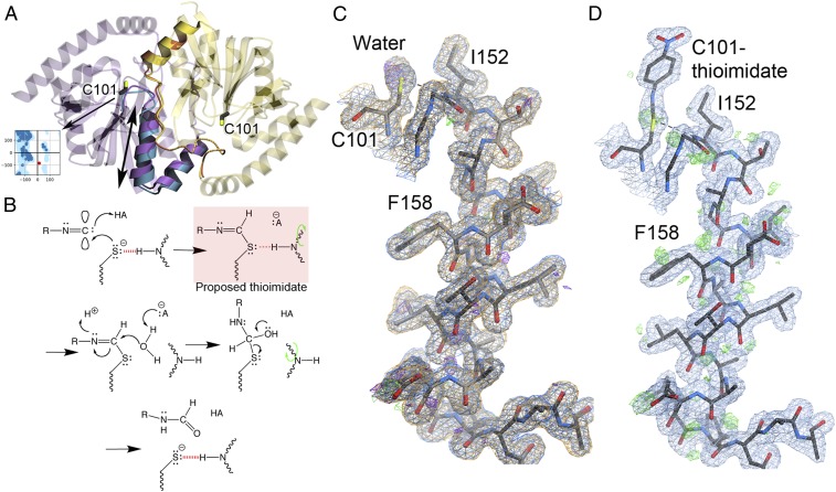
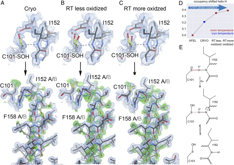
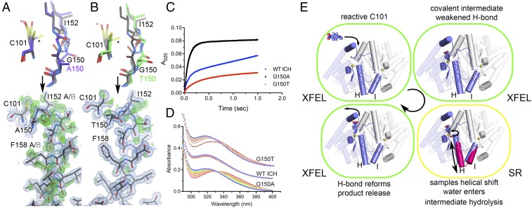
How changes in enzyme structure and dynamics facilitate passage along the reaction coordinate is a fundamental unanswered question. Here, we use time-resolved mix-and-inject serial crystallography (MISC) at an X-ray free electron laser (XFEL), ambient-temperature X-ray crystallography, computer simulations, and enzyme kinetics to characterize how covalent catalysis modulates isocyanide hydratase (ICH) conformational dynamics throughout its catalytic cycle. We visualize this previously hypothetical reaction mechanism, directly observing formation of a thioimidate covalent intermediate in ICH microcrystals during catalysis. ICH exhibits a concerted helical displacement upon active-site cysteine modification that is gated by changes in hydrogen bond strength between the cysteine thiolate and the backbone amide of the highly strained Ile152 residue. These catalysis-activated motions permit water entry into the ICH active site for intermediate hydrolysis. Mutations at a Gly residue (Gly150) that modulate helical mobility reduce ICH catalytic turnover and alter its pre-steady-state kinetic behavior, establishing that helical mobility is important for ICH catalytic efficiency. These results demonstrate that MISC can capture otherwise elusive aspects of enzyme mechanism and dynamics in microcrystalline samples, resolving long-standing questions about the connection between nonequilibrium protein motions and enzyme catalysis.

Keywords: X-ray crystallography; XFEL; cysteine modification; enzyme conformational dynamics; radiation damage.


Conflict of interest statement

The authors declare no competing interest.

Figures

Fig. 1.
Fig. 1.
Catalytic intermediate captured with MISC. (A) The ICH dimer is shown as an overlay of WT ICH (purple) and C101A ICH (blue). The “B” protomer is shown in yellow. The mobile helix H in C101A and areas exhibiting correlated backbone–side-chain disorder are rendered opaque. Ile152 is a Ramachandran outlier (Inset) whose backbone torsion angles move with helical displacement. (B) Postulated reaction mechanism for ICH, beginning with Cys101 thiolate attack at the electrophilic carbenic carbon atom of isocyanide substrates and proceeding in the direction of the arrows. A previously postulated thioimidate intermediate (red box) eliminates the charge on Cys101 and weakens the H-bond to Ile152 (dashed red line). This relieves backbone torsional strain at Ile152 and permits sampling of shifted helix H conformations (green curved arrow), allowing water access to the intermediate for hydrolysis. (C) ICH completes a full catalytic cycle in the crystal during MISC. Helix H is shown with 2mFo-DFc electron density (0.8 rmsd) prior to the introduction of substrate (blue) and after substrate has been exhausted (orange). These maps overlap almost perfectly, indicating that ICH is fully restored to its resting conformation after catalysis. The hydrogen bond between the peptide backbone of Ile152 and Cys101 is shown in a dotted line. The helix is not mobile in these resting structures, indicated by the absence of features in the mFo-DFc difference electron density (2.5 rmsd prior to substrate [green] and after catalysis is complete [purple]) (D) MISC confirms that ICH forms a covalent Cys101-thioimidate intermediate 15 s after substrate mixing. Difference mFo-DFc electron density contoured at 2.5 rmsd (green) supports sampling of shifted helix H conformations upon intermediate formation.
Fig. 2.
Fig. 2.
X-ray–induced cysteine oxidation drives helical motion in ICH. (AC, Upper) The environment of Cys101 with varying degrees of oxidation to Cys101-sulfenic acid. 2mFo-DFc electron density is contoured at 0.7 rmsd (blue), and the hydrogen bond between the peptide backbone of Ile152 and Cys101 is shown in a dotted line. “Cryo” is synchrotron data collected at 100 K (PDB 3NON); “RT less oxidized” is synchrotron data collected at 274 K with an absorbed dose of 2.4 × 10⁴ Gy; and “RT more oxidized” is synchrotron data collected at 277 K with an absorbed dose of 3.7 × 10⁵ Gy. (Lower) helix H in its strained (black) and relaxed, shifted conformations (gray). 2mFo-DFc electron density is contoured at 0.8 rmsd (blue) and omit mFo-DFc electron density for the shifted helical conformation is contoured at 3.0 rmsd (green). At 274–277 K, increased Cys101 oxidation disrupts the hydrogen bond to Ile152 and results in stronger difference electron density for the shifted helix conformation. (D) The refined occupancy of helix H in each X-ray dataset indicates that increases in temperature and Cys101 oxidation result in higher occupancy for the shifted (relaxed) helix conformation. (E) Mechanism of X-ray–induced covalent modification of C101 and weakening of the S-HN hydrogen bond.
Fig. 3.
Fig. 3.
Cysteine modification results in a protein-wide conformational response. (A) Fluctuations of the C101SG-I152H distance in simulations of ICH crystals in the Cys101-S (red) or Cys101-SOH (blue) state. Each ICH dimer is represented by a semitransparent line. Opaque red and blue lines denote the average C101SG-I152H distance across the dimers. (B) Conformational shift of helix H in the Cys101-S (red) or Cys101-SOH (blue) state. The dark gray lines in A and B represent the trajectory selected for Movie S1. (C) rmsfs calculated from MD simulations indicate highest fluctuations in linker I′-J′ of the B protomer. Helix H of the A protomer just underneath the linker also shows elevated rmsfs. (D) An isomorphous Fo(SR) – Fo(XFELFREE) difference map reveals features (green, positive; red, negative) distributed throughout the dimer, suggesting broadly altered structure and dynamics upon formation of the Cys101-SOH in the 274-K synchrotron radiation (FoRT) dataset. The “A” conformer is shown in slate blue, and the “B” conformer is shown as a gray semitransparent cartoon. Helices H and I are opaque in both conformers. The catalytic nucleophile is shown in spheres. Difference electron density features are nonuniformly distributed, with stronger features near helix H in the A protomer, and along region B169–B189, which contacts the N-terminal end of helix H. Maps are contoured at ±3.0 rmsd. (E) CONTACT analysis identifies allosteric coupling across the dimer interface, in striking agreement with isomorphous difference maps and rmsfs from D. The A protomer is color-coded in blue; the B protomer, in red. Residues identified in the CONTACT analysis are projected onto the cartoon.
Fig. 4.
Fig. 4.
Mutations at Glu150 alter helical mobility and reduce ICH catalytic turnover. (A and B) The environment of Cys101 in Gly150A and G150T ICH (Top). 2mFo-DFc electron density is contoured at 0.7 rmsd (blue) and the hydrogen bond appears as a dotted line. Both G150 mutations permit unmodified Cys101 to sample conformations (asterisk) that sterically conflict with Ile152 in the strained helical conformation (black), requiring the helix to sample shifted conformations (gray) in the absence of Cys101 modification. (A and B, Lower) The helix in its strained (black) and relaxed, shifted conformations (gray). 2mFo-DFc electron density is contoured at 0.8 rmsd (blue) and omit mFo-DFc electron density is contoured at +3.0 rmsd (green). (C) Pre-steady-state enzyme kinetics of WT (blue circles), G105A (black squares), and G150T (red triangles) ICH at 160 μM p-NPIC shows a pronounced burst phase for each protein with differing burst and steady-state rate constants. The divergent pre-steady-state profiles indicate that G150A impacts steps after the first chemical step, while G150T affects both early and later steps. (D) Single-turnover spectra of ICH enzymes with p-NPIC substrate shown from early (red) to later (blue) timepoints in 5-s increments. At early times, G105A and G150T accumulate a species with λmax = 335 nm, likely the thioimidate intermediate that resolves to product in the blue spectra with λmax = 320 nm. (E) The catalytic cycle of ICH. Substrate binds with Cys101-S poised for nucleophilic attack and with the Cys101-Ile152 H-bond intact. Formation of the thioimidate intermediate weakens the Cys101-Ile152 H-bond and causes helical motion (magenta helices H and I) that promotes intermediate hydrolysis (red sphere; curved arrow). Hydrolysis of the thioimidate intermediate restores the reactive C101-S and strengthens the Cys101-Ile152 H-bond, favoring the strained helical conformation.

References

    1. Danyal K., Mayweather D., Dean D. R., Seefeldt L. C., Hoffman B. M., Conformational gating of electron transfer from the nitrogenase Fe protein to MoFe protein. J. Am. Chem. Soc. 132, 6894–6895 (2010). - PMC - PubMed
    1. Henzler-Wildman K. A., et al. , Intrinsic motions along an enzymatic reaction trajectory. Nature 450, 838–844 (2007). - PubMed
    1. Aviram H. Y., et al. , Direct observation of ultrafast large-scale dynamics of an enzyme under turnover conditions. Proc. Natl. Acad. Sci. U.S.A. 115, 3243–3248 (2018). - PMC - PubMed
    1. Kim T. H., et al. , The role of dimer asymmetry and protomer dynamics in enzyme catalysis. Science 355, eaag2355 (2017). - PubMed
    1. Hammes G. G., Multiple conformational changes in enzyme catalysis. Biochemistry 41, 8221–8228 (2002). - PubMed

Publication types