. 2019 Dec 5;4(23):e133103.
doi: 10.1172/jci.insight.133103.

Discovery of specialized NK cell populations infiltrating human melanoma metastases

Affiliations

Discovery of specialized NK cell populations infiltrating human melanoma metastases

Lucas Ferrari de Andrade et al. JCI Insight. .

Abstract

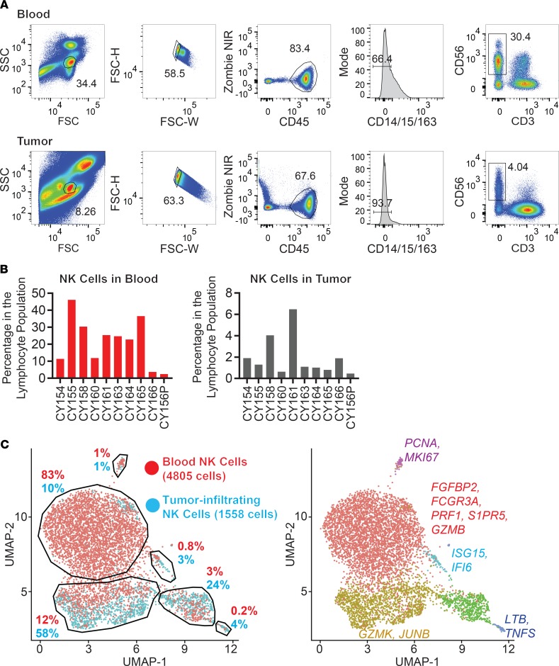
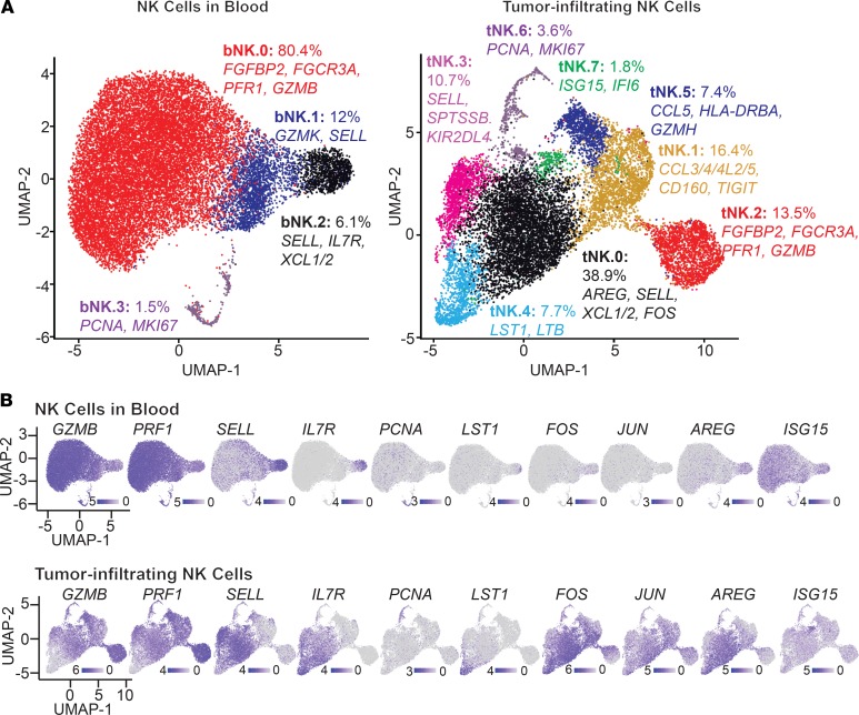
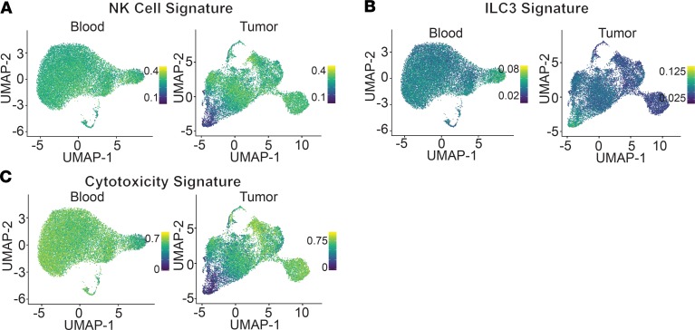
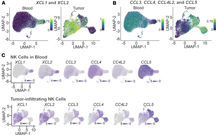
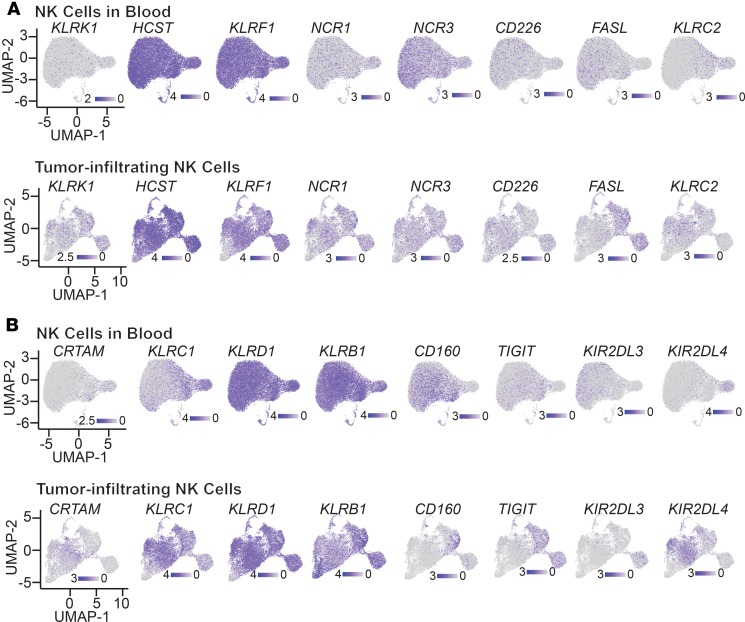
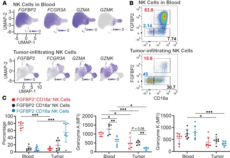
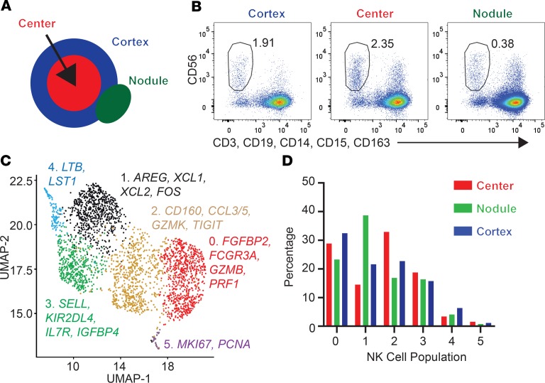
NK cells contribute to protective antitumor immunity, but little is known about the functional states of NK cells in human solid tumors. To address this issue, we performed single-cell RNA-seq analysis of NK cells isolated from human melanoma metastases, including lesions from patients who had progressed following checkpoint blockade. This analysis identified major differences in the transcriptional programs of tumor-infiltrating compared with circulating NK cells. Tumor-infiltrating NK cells represented 7 clusters with distinct gene expression programs indicative of significant functional specialization, including cytotoxicity and chemokine synthesis programs. In particular, NK cells from 3 clusters expressed high levels of XCL1 and XCL2, which encode 2 chemokines known to recruit XCR1+ cross-presenting DCs into tumors. In contrast, NK cells from 2 other clusters showed a higher level of expression of cytotoxicity genes. These data reveal key features of NK cells in human tumors and identify NK cell populations with specialized gene expression programs.

Keywords: Immunology; Innate immunity; Melanoma; NK cells; Oncology.


Conflict of interest statement

Conflict of interest: KWW serves on the scientific advisory boards of TCR2 Therapeutics, TScan Therapeutics, and Nextech Invest and receives sponsored research funding from Bristol-Myers Squibb and Novartis. He is a scientific cofounder of Immunitas Therapeutics.

Figures

Figure 1
Figure 1. Isolation and scRNA-seq characterization of NK cells from human melanoma metastases and matching blood samples.
(A) Gating strategy for the isolation of NK cells from blood and melanoma metastasis from patient CY158 by flow cytometry. (B) Percentage of blood (left) and tumor-infiltrating lymphocytes (right) from multiple patients that were NK cells (after exclusion of myeloid cells). (C) Comparison of NK cell clusters in blood versus melanoma metastasis (patient CY158) by scRNA-seq. UMAP plots were used to visualize blood and tumor-infiltrating NK cell clusters, and the percentage of NK cells in each cluster is indicated for blood and tumor NK cells (left). NK cell clusters are color coded, and key differentially expressed genes are shown for each cluster (right).
Figure 2
Figure 2. Single-cell RNA-seq analysis of NK cells using integrated data from 5 patients.
(A) NK cell populations in blood (left) and metastases (right) analyzed by scRNA-seq using merged data from 5 patients (CD129.2, CY155, CY158, CY160, and CY164). NK cell clusters in blood and metastases were visualized using UMAP plots. The percentage of NK cells in each cluster and key differentially expressed genes for each cluster are indicated. (B) mRNA transcripts for selected genes in blood (top) and tumor-infiltrating (bottom) NK cell populations were visualized using UMAP plots. The intensity of the blue color indicates the level of expression for indicated genes in individual cells and is scaled separately between blood and tumor-infiltrating NK cells within the integrated data set from 5 patients.
Figure 3
Figure 3. Identification of NK cells and ILC3-like cells in melanoma metastases.
(A and B) Identification of NK cell and ILC3 cell populations. Published single-cell data from innate lymphocytes isolated from human tonsil tissue were used to define gene expression signatures for NK cells and ILC3 (16). UMAP plots show the degree of similarity between NK cell (A) and ILC3 (B) gene expression signatures for blood and tumor NK cells. (C) Cytotoxicity gene expression signature (GZMA, GZMB, GZMH, GZMK, GZMM, PRF1, GNLY, and NKG7) for NK cells isolated from blood (left) and melanoma metastases (right). UMAP plots indicate the scores for this signature across NK cell clusters and are scaled separately between blood and tumor-infiltrating NK cells for the integrated data set from 5 patients.
Figure 4
Figure 4. Expression of chemokine genes by blood and tumor-infiltrating NK cells.
(A and B) UMAP plots showing expression of chemokine genes, XCL1 and XCL2 (A) as well as CCL3, CCL4, CCL4L2, and CCL5 (B), in blood and tumor-infiltrating NK cells. (C) Expression of each one of the chemokine genes by NK cells isolated from blood (top) and melanoma metastases (bottom). The intensity of the blue color indicates the level of expression for indicated genes in individual cells and is scaled separately between blood and tumor-infiltrating NK cells for the integrated data set from 5 patients.
Figure 5
Figure 5. Expression of genes encoding activating and inhibitory surface receptors on NK cells.
(A and B) Expression of activating (A) and inhibitory (B) receptors in blood (top) and tumor (bottom) specimens. The intensity of the blue color indicates the level of expression of selected genes in individual cells and is scaled separately between blood and tumor-infiltrating NK cells within the integrated data set from 5 patients.
Figure 6
Figure 6. Validation of NK cell subpopulations by flow cytometry.
(A) Expression of genes used for identification of NK cell populations (FGFBP2 and FCGR3A) and effector molecules (GZMA and GZMK). The intensity of the blue color indicates the level of expression for selected genes in individual cells and is scaled separately between blood and tumor-infiltrating NK cells within the integrated data set from 5 patients. (B) Validation of 3 NK cell populations identified by scRNA-seq in blood samples by flow cytometry using FGFBP2 and CD16a as markers. NK cells were identified by gating on CD45+ and CD56+ cells that were negative for CD3ε, CD19, CD14, CD15, CD163, and a dead cell marker. A representative analysis is shown for patient CY165. (C) Quantification of 3 NK cell populations in blood and tumor samples based on FGFBP2 and CD16a markers. Labeling for granzymes A and K is also shown for each of the 3 populations. The genes named FCGR3A, FGFBP2, GZMA, and GZMK encode the proteins CD16a, FGFBP2, granzyme A, and granzyme K, respectively. Each dot represents an individual patient. MFI, mean fluorescence intensity. Statistical analysis was performed by 2-way ANOVA, Bonferroni’s post hoc tests; *P < 0.05, **P < 0.01, ***P < 0.001.
Figure 7
Figure 7. Regional analysis of NK cells in a melanoma metastasis.
(A) Illustration of metastasis regions from which NK cells were isolated (patient CY129.2). (B) Flow cytometry analysis of NK cells isolated from each of the tumor regions. (C) UMAP representation of single-cell data for identification of NK cell clusters. NK cells from all 3 regions of the metastasis were combined for this UMAP plot. (D) Percentage of NK cell populations assigned to clusters (defined in C) in the 3 regions of this metastasis.

References

    1. Chiossone L, Dumas PY, Vienne M, Vivier E. Natural killer cells and other innate lymphoid cells in cancer. Nat Rev Immunol. 2018;18(11):671–688. doi: 10.1038/s41577-018-0061-z. - DOI - PubMed
    1. André P, et al. Anti-NKG2A mAb Is a checkpoint inhibitor that promotes anti-tumor immunity by unleashing both T and NK cells. Cell. 2018;175(7):1731–1743.e13. doi: 10.1016/j.cell.2018.10.014. - DOI - PMC - PubMed
    1. Zhang Q, et al. Blockade of the checkpoint receptor TIGIT prevents NK cell exhaustion and elicits potent anti-tumor immunity. Nat Immunol. 2018;19(7):723–732. doi: 10.1038/s41590-018-0132-0. - DOI - PubMed
    1. Blake SJ, et al. Suppression of metastases using a new lymphocyte checkpoint target for cancer immunotherapy. Cancer Discov. 2016;6(4):446–459. doi: 10.1158/2159-8290.CD-15-0944. - DOI - PubMed
    1. Ferrari de Andrade L, et al. Antibody-mediated inhibition of MICA and MICB shedding promotes NK cell-driven tumor immunity. Science. 2018;359(6383):1537–1542. doi: 10.1126/science.aao0505. - DOI - PMC - PubMed

Publication types