. 2019 Dec;576(7787):446-451.
doi: 10.1038/s41586-019-1816-9. Epub 2019 Dec 4.

Frontal cortex neuron types categorically encode single decision variables

Affiliations

Frontal cortex neuron types categorically encode single decision variables

Junya Hirokawa et al. Nature. 2019 Dec.

Abstract

Individual neurons in many cortical regions have been found to encode specific, identifiable features of the environment or body that pertain to the function of the region1-3. However, in frontal cortex, which is involved in cognition, neural responses display baffling complexity, carrying seemingly disordered mixtures of sensory, motor and other task-related variables4-13. This complexity has led to the suggestion that representations in individual frontal neurons are randomly mixed and can only be understood at the neural population level14,15. Here we show that neural activity in rat orbitofrontal cortex (OFC) is instead highly structured: single neuron activity co-varies with individual variables in computational models that explain choice behaviour. To characterize neural responses across a large behavioural space, we trained rats on a behavioural task that combines perceptual and value-guided decisions. An unbiased, model-free clustering analysis identified distinct groups of OFC neurons, each with a particular response profile in task-variable space. Applying a simple model of choice behaviour to these categorical response profiles revealed that each profile quantitatively corresponds to a specific decision variable, such as decision confidence. Additionally, we demonstrate that a connectivity-defined cell type, orbitofrontal neurons projecting to the striatum, carries a selective and temporally sustained representation of a single decision variable: integrated value. We propose that neurons in frontal cortex, as in other cortical regions, form a sparse and overcomplete representation of features relevant to the region's function, and that they distribute this information selectively to downstream regions to support behaviour.


Conflict of interest statement

The authors declare no competing financial interests.

Figures

Extended Data Figure 1|
Extended Data Figure 1|. Rat behavior reflects an integration of evidence and reward-size.
a-e, Average psychometric functions in unbiased, left and right bias blocks as a function of the odor percept (n=67 sessions). The decision-variable model (a) and actual data from the same data (b) (replication of Fig. 1c). c, Odor sampling time was larger for small rewards than for large rewards. Errors are shown as mean +/− SEM (n=3 rats). d, The model provides an excellent fit of choice patterns for each session. e, Choice driven by the relative value of left choice (replication of Fig.1e). f, Histogram across sessions, comparing the ratio of actual reward obtained to a model relying on odor stimuli but ignoring reward size. g, Histogram across sessions, comparing the ratio of actual reward obtained to a model relying on reward size but ignoring odor stimuli. h-l, Same convention with a-e but reporting the bias arising from the outcome of the previous trial during the control block without reward bias (n=67 sessions). h-I, The probability for left choice as a function of odor percept for all trials or separated by which choice (left/right) was rewarded in the previous trial. “Left reinforced” indicates that animals are rewarded (correct) on the left side in the previous trial or not rewarded (error) on the right side, regardless of the stimulus conditions used in previous trials. The decision-variable model accurately predicts changes in choice probability (h-i) arising due to previous outcome. j, Odor sampling time was larger after a previously unrewarded choice than for a previously rewarded choice. Errors are shown as mean +/− SEM (n=3 rats). The model provides an accurate fit across sessions (k) driven by the relative value of the left choice (l).
Extended Data Figure 2|
Extended Data Figure 2|. Recording sites in lateral OFC and functional clustering across rats.
a, Recording sites in lateral OFC for all seven rats from Cohort 1 and Cohort 2. Histological section shown in left (rat C068), in which red arrows indicate the tip of the tetrode bundle.
Extended Data Figure 3|
Extended Data Figure 3|. Profiles of individual neuron responses and population profiles in OFC.
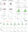
a, Example response profiles are shown for several individual neurons. In each case, a schematic tuning curve representing a plausible decision-variable is shown in left panels, while a matching neuronal response profile is shown in right panels. b, PCA decomposition of the OFC dataset reveals high dimensionality, with 21 principal components required to retain 90% of response profile variability. c, Diversity of tuning vectors for several example neurons in the space of the first three principal components. d, The 3 dominant principal components arising from a probabilistic PCA decomposition of 485 OFC response profiles, which account for ~40% of population variance. e, Neuronal response profile with 42 behavioral conditions was vectored for further analysis (same neuron shown as Fig.2b) f, Spectral clustering identifies robust clusters of response profiles within high-dimensional response space. g, Distribution of firing rates for all neurons across all conditions. Firing rates for observed data (red dots) show a right-tailed distribution, with strong activation of most neurons for only a small subset of conditions. This pattern of activation is significantly sparser than expected from a normal distribution (black line) or trial-shuffled data (gray dots). h, Coefficients arising from PCA analysis show a similar long-tailed distribution compared to a normal distribution (blue line) or trail-shuffled data (gray dots).
Extended Data Figure 4|
Extended Data Figure 4|. Testing for random mixed selectivity.
a-d, Exemplar distributions of toy model neurons showing mixed selectivity, presented using the coefficients for two dominant eigenvectors (Dimension 1 and Dimension 2). Two neuronal subpopulations are shown in red and blue. Populations in the top panels (a,b) have equal variance in both dimensions, while populations in the bottom panels (c,d) do not. Populations in the left panels (a,c) can be said to show random mixed selectivity, while distributions in the right panels (b,d) do not. Only the distribution shown in (a) can be said to show spherical symmetry, necessitating the development of modified tests. e-h Comparison of the PAIRS to the modified ePAIRS test, which accounts for elliptical distributions. e-f Sensitivity analysis of PAIRS and ePAIRS, tested across several datasets whose variance structure matched the OFC data (green), including a spherically uniform Gaussian (blue) and collections of 5 randomly oriented von Mises distributions with varying κ; blue, orange, yellow). Datasets were truncated at a given dimensionality, and mean p-values are reported across 30 replicates. Results show that spherical PAIRS generates false-positive results when tested on non-spherical but otherwise i.i.d Gaussian data. The modified ePAIRS test successfully identifies the non-uniformity of strongly clustered data (κ = 10) but not weaker clustering. The dimensionality required to reconstruct 70% and 90% of the variance in the full dataset is shown (gray lines). g-h The ePAIRS measure, nearest-neighbor angles, is shown for OFC data (green) and bootstrap distributions (black). OFC data showed smaller angles than expected for both 1 and 10 nearest neighbors, suggesting strong clustering. i-l Comparison of the random projection (RP) test to the elliptical random projection (eRP) test (eRP). i-j We compared the RP and eRP tests on several datasets including an elliptical Gaussian distribution (dark blue) and collections of 5 randomly oriented von Mises distributions with κ=3 (light blue), and observed OFC data (green). Other parameters matched panels e-f. Results show that spherical RP generates false-positive results when tested on non-spherical but otherwise i.i.d Gaussian data (dark blue). The modified eRP test successfully identifies the non-uniformity of von Mises clusters, as well as OFC data, while rejecting spherically uniform Gaussian data. k-l Cumulative distribution function (CDF) of Kolmogorov–Smirnov statistics arising in the eRP test. Results are shown for test distributions that are spherically-uniform Gaussians (k), as well as for OFC data (l). m, Analysis of individual animals from Cohort 1 and Cohort 2. All animals showed significant deviation from uniformity for both ePAIRS and eRP tests. Top: Histogram of nearest-neighbor angles for observed data (green) and bootstrap samples with similar elliptical distribution (black). All animals showed significant differences, assessed using a rank sum test. Middle: Cumulative distribution of Kolmogorov-Smirnov test statistics from eRP for bootstrap samples and observed data. Here each observation is the K-S statistic derived from comparing the distribution of projected angles onto a single random vector between observed data and a matching elliptical Gaussian distribution. This procedure is repeated for a set of k random vectors to generate the plotted distribution. This calculation is performed both for observed data (green) and for a simulated spherically-uniform bootstrap distribution with matching samples size and ellipticity (black). Bottom: Comparison of the observed median K-S statistic from observed data (green), to the distribution of medians observed across several realizations of a bootstrap distribution (gray). For all eRP estimates, higher K-S statistics denote greater deviation from uniform distribution, and all animals showed significant differences using a bootstrap test.
Extended Data Figure 5|
Extended Data Figure 5|. Stability of cluster identification.
a, Clusters are well distributed across animals from Cohort 1. Cells in each cluster were generally drawn from all three animals, and rarely showed significant animal-specific bias. b, Table of the distribution of the neurons across rats from Cohort 1 and across clusters identified through spectral clustering (see Fig.2). c, Neurons in each cluster were plotted against the relative recording depths (normalized to 0–1 from recording starting point to end point shown in Extended Data Figure 2) for each animal. d-h Null relationships between spatial proximity and response correlation. d For each pair of cells between all pairs of rats, we compared the pairwise mediolateral distances (x-axis) to the pairwise correlations in response profiles (y-axis). There is no significant relationship between mediolateral distance and magnitude of response correlation e This plot is identical to that in panel (d), except that correlations are normalized for sign. f Average magnitude of correlation between pairs of cells, analyzed for all pairs of rats. Rats are ordered by electrode position, from medial to lateral. There is no obvious relationship between ML position order and average pair-wise correlation of neuronal response profiles. f Same as panel (c) but normalized for each rat (i.e. relative to within-rat response variability). h Average magnitude of correlation between a given rat, and all other rats, with error bars representing SEM. This is equivalent to the row average of panel (f). g, Missing values are shown for all 485 neurons in Cohort 1. Top: missing values are common for some conditions, because animals rarely make errors against high-reliability cues, and are further biased against certain errors during bias blocks. Six such conditions are dropped due to excessive missing data (black dots). Bottom: after removing these conditions, 42 conditions remain for analysis, with sporadic missing values. h, Missing values are imputed during preprocessing using probabilistic PCA. If these missing values influence clustering, we expect to see a consistent pattern of missing values in certain clusters. Although there is variation in missing values across clusters, there is no obvious pattern of missing data.
Extended Data Figure 6|
Extended Data Figure 6|. Response profiles of OFC neurons from Cohort 2 replicate the results of Cohort 1.
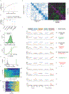
a, Distribution of variance across the first 29 eigenvectors for Cohort 2, see Extended Data Fig. 7e for corresponding panel for Cohort 1. b, Average correlations of individual cell response profiles with a set of canonical response profiles corresponding to decision variables side choice, confidence, reward size (trial-by-trial), previous outcome, and reward size (block average). The sign of the correlation was discarded and normalized across cells by the strongest correlation (i.e., side choice). Two representations (Previous Outcome and Block-wise Reward Size) showed reduced representation in animals from Cohort 2. c, ePAIRS test, showing the distribution of nearest-neighbor distances between observed data in Cohort 2 (green) and a bootstrap distribution derived from simulated data with a matching elliptical Gaussian (black). P < 0.001, Rank Sum Test. d, cumulative distribution function (CDF) for observed data (green) and a bootstrap distribution (black). Median values are shown with vertical lines. e, Distribution of median values for the K-S statistic across a set of bootstrap distributions (gray), compared to the median value for observed data (green). P < 0.001, bootstrap test. f, ARI for spectral clustering across k (k-nearest neighbors used to generate the adjacency matrix) and c (number of clusters), showing marked clustering around c=11 clusters. g, Proportion of cells from each cnimal associated with each cluster. h, Left: Dendrogram of inter-cluster distances. Middle: Adjacency matrix, derived from k = 21 using a correlation distance. Right: Between- and within-cluster correlations. i, Average response profiles for each cluster in Cohort 2. The format of this figure matches Fig. 3 in the main text. Overall, we identified 11 clusters, of which the top 7 (each containing > 5% of the cells in the dataset) correspond to separable representations of choice, confidence, and value.
Extended Data Figure 7|
Extended Data Figure 7|. Analysis of OFC response structure across behavioral epochs (Cohort 1).
a, Clustering results and hyperparameter selection for responses in the stimulus epoch (most stable configuration: c = 5 clusters and k = 16 nearest neighbors). Each combination of parameters is evaluated for stability using the adjusted rand index (ARI) (see Methods and Fig. 2). b, Clustering results for the stimulus epoch. The relationship between the 5 clusters can be examined visually by observation of the nearest-neighbor graph (top) and the within-cluster and between-cluster correlation coefficient (bottom). c, Clustering results and hyperparameter selection for responses in the feedback epoch (most stable configuration: c=5 clusters and k = 16 nearest neighbors). Each combination of parameters is evaluated for stability using the adjusted rand index (ARI) (see Methods and Fig. 2). d, Clustering results for the feedback epoch. The relationship between the 5 clusters can be examined visually by observation of the nearest-neighbor graph (top) and the within-cluster and between-cluster correlation coefficient (bottom). e-g, Full cluster response profiles for all three epochs. e, Average response profiles of each of the 9 identified response clusters in the Anticipation epoch (compare to Fig. 2). For each cluster, the normalized firing rate is shown for all 42 behavioral conditions used to generate the clustering results (responses conditioned on stimulus and choice, unbiased, left bias, and right bias blocks; conditioned on outcome of the previous choice and the evidence supporting the current choice, previous outcome). In addition, normalized firing rates are shown conditioned on the size of the reward associated with the choice port (reward size). For each cluster, we also note the corresponding putative decision variable. f, Average response profiles of each of the 5 identified response clusters in the stimulus epoch. Conventions same as in e. Two of the clusters did not obviously map on a putative decision variable. g, Average response profiles of each of the 5 identified response clusters in the feedback epoch. Conventions same as in panel e. Two of the clusters did not map on a putative decision variable. h-i, Transition probabilities for neurons in a given cluster across subsequent epochs (compare to Fig. 5a). h, Transition probability for neurons belonging to a given cluster in the stimulus epoch to belong to a given cluster in the Anticipation epoch (normalized per row). i, Transition probability between anticipation and feedback epochs (normalized per row). j, Neuron-based similarity measures across epochs. Neuronal response profiles are more similar across epochs for paired responses from the same neuron (red) compared to responses of two different neurons (black). Left: comparison of Stimulus and Anticipation epochs; right: comparison of anticipation and feedback epochs. Two-sample Kolmogorov-Smirnov test. k, Cluster-based similarity measures across epochs. Clusters derived from different epochs are more likely to share members if the average response profiles of each cluster are similar. Left: comparison of Stimulus and Anticipation epochs; right: comparison of anticipation and feedback epochs.
Extended Data Figure 8|
Extended Data Figure 8|. Negative confidence is quantitatively represented in a cluster of OFC neurons.
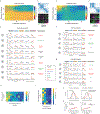
a, Response profile of neurons in Cluster 2 correspond to the decision-variable confidence (+), i) Peri-stimulus time histogram of normalized firing rate, grouped based on the degree of evidence supporting choice. ii) Trial-by-trial fit of each neuron in Cluster 2 to choice evidence reveals significant representation of statistical decision confidence variable (R2 with P < 0.05 based on bootstrap). iii-vi) Normalized average tuning curves for neurons in Cluster 2. iii) Firing rate as a function of stimulus difficulty and choice (vevaiometric curve). iv) Choice accuracy as a function of stimulus difficulty and firing rate (conditioned psychometric curve), v) Choice accuracy as a function of firing rate (calibration curve), and vi) Firing rate as a function of evidence supporting choice. Note that panel (iii) and (v) are replications of Fig.4a bottom panels. b, Response profile of neurons in Cluster 1, corresponding to a decision variable representation of Confidence (−): Same convention as a. c, Response profile of neurons in Cluster 3, corresponding to a decision-variable of integrated value (−). i-v) The representation of integrated value is analyzed similarly to confidence in panel (a), with the following changes: ii) Trial-by-trial fit of integrated value reveals significant representation of negative integrated value (R2 with P < 0.05 based on bootstrap). vi) Firing rate as a function of negative integrated value. v) Choice accuracy as a function of firing rate (* P<0.01, t-test), and vi) Firing rate as a function of integrated value. Note that panel (iii) and (v) are replications of Fig.4b bottom panels. d, Single-neurons encode coherent combinations of confidence and reward size. Each neuron’s response profile was fit to a two-parameter model representing confidence and reward size. For most neurons, regression coefficients (ß) for each component share the same sign. Data are shown for all neurons (gray) and neurons with significant beta coefficients for both components are shown in blue (P < 0.01 threshold). Polar histogram is significantly different from uniform (P < 0.01). e, Elementary task variables defined for the regression model. Each task variable was z-scored according to the weight of its non-masked conditions, with masked conditions were subsequently set to zero. f, Detailed results of the LASSO model from Fig. 4g for neuronal clusters, using both the canonical design matrix (corresponding to decision variables), and null models (corresponding to random rotations of the design matrix). Errors are shown as median +/− SEM. P-value calculated as paired sign-rank test. g, Detailed results of the LASSO model shown in Fig. 4h for single neurons for both the canonical design matrix (corresponding to decision variables), and null models (corresponding to random rotations of the design matrix). Errors are shown as median +/− SEM. P-value calculated as a two-sided t-test.
Extended Data Figure 9|
Extended Data Figure 9|. Time course clustering and positively outcome selective OFC-striatum projection neurons.
a,b The ePAIRS (a) and eRP (b) tests reveals significant non-random clustering in the OFC population based on response profiles with temporal but not task-related information (see Methods). For ePAIRS, nearest neighbor angles were smaller than expected, suggestive of clustering (rather than dispersion). c, Clustering results and hyperparameter selection for temporal clustering (most stable configuration: c = 8 clusters and k = 13 nearest neighbors; compare to Fig. 3f; see Methods). d, Analysis of temporal response profiles (left); spectral clustering of temporal response profiles without tuning information reveals 8 clusters with high within-cluster similarities (n = 7 rats combined, Methods). Dynamics of the trial averaged time course for single neurons in the 8 clusters for error trials (mid) and rewarded trials (right). We separated rewarded and error trials for this analysis as the actions performed during the outcome period are very different (drinking water vs return to center port). e, Average dynamics of the trial averaged time course for the 8 clusters (green: rewarded trials, red: error trials). f, Average activity in correct trials (top) and average PSTH grouped by outcome (error, small reward, large reward) of identified OFC-striatum projecting neurons that positively encoded outcome. Lower panels show that neurons are positively tuned to integrated value in the anticipation period and positive tuning to outcome in the feedback epoch and ITI. Conventions same as in Figure 5h. g, Average PSTH of neurons in Cluster A whose dynamics match those of optogenetically identified neurons encoding positive outcome (excluding optogenetically identified OFC-striatum projection neurons). Note that the coding of integrated value is weaker than for the negative population but still significant (r(90) = 0.1, p = 0.02). Conventions same as in panel f.
Extended Data Figure 10 |
Extended Data Figure 10 |. Optogenetic identification of OFC-striatum projection neurons.
a-e: An example neuron showing reliable light-evoked responses. a, For an example neuron, average waveforms of spontaneous (yellow) and light-evoked spikes (blue) across four tetrodes are very similar. b, Spike raster (top) and peri-stimulus time histogram (PSTH) (bottom) for the light-activated cell in panel a aligned to light onset (1ms duration, first stimulus in a train). c, Reliability of the evoked responses to the first stimulus as a function of pulse duration. d, Probability of light-evoked spikes as a function of stimulation frequency (1ms duration, 20 repetitions). e, Spike raster (top) and (PSTH) (bottom) aligned to light onset for stimulation trials at 10Hz and 40Hz. f, Histogram of Pearson’s correlation coefficients between the waveforms of spontaneous and light-evoked for identified OFC-striatum projecting neurons. g, Quantification of light-evoked responses, showing latency and jitter of light-evoked spikes for neurons tagged neurons as a function of the reliability of evoking a response to light. Putative OFC-striatum projection neurons are shown (blue points). h, Z-scored PSTH of all identified OFC-striatum projection neurons in response to 1–3ms blue light stimulation.
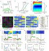
Figure 1 |
Figure 1 |. Reward-biased psychometric odor discrimination task to probe decision-variable integration.
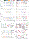
a, Task design, single trial. We primarily analyzed the 1-s Reward Anticipation window. b, Behavioral performance is modified by block-wise changes in reward size. Choice biases emerged rapidly across blocks (5.45 ± 0.48 trials to bias-shift, mean +/− SEM, n = 67 sessions from 3 rats). c, Average psychometric functions in unbiased, left-, and right-bias blocks reveal systematic bias (30 sessions from one rat (C068)). d, Decision-variable model of choice behavior in this task integrates variables from reward context (choice, size, presence of reward) and sensory evidence to generate internal variables representing expected reward value and decision confidence. These combine to estimate integrated value, which determines choice probability. e, The relative value derived from the model explains choice behavior (n=67 sessions from 3 rats).
Figure 2 |
Figure 2 |. OFC neurons form 9 discrete clusters.
a-b, Response during reward anticipation for one neuron (Unit 521). Activity in unbiased block reflected evidence supporting animal’s choice (a), and matched expected response profile of representation of decision confidence (varying with choice evidence, but not reward size (b). c-d, Distribution of nearest-neighbor angles (c) and projection angles (d) reveal non-random distribution in OFC population with angle magnitudes suggesting clustering (p<0.001 for both tests; Methods, Extended Fig. 4). e, Spectral clustering reveals nine robust groups of neurons, with high cluster stability across a range of hyperparameters (Adjusted Rand Index, ARI; see Methods for details of cross-validation). f, Sorted adjacency and correlation matrices reveal strong within-cluster similarity and between-cluster antagonism.
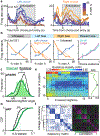
Figure 3 |
Figure 3 |. OFC neuron clusters represent putative decision variables.
Average response profiles for 4 of 9 clusters (see Extended Data Fig. 7e for others). Each row represents a cluster, with average normalized firing rate plotted across behavioral conditions. First three conditions represent combinations of stimulus and choice in different reward blocks (unbiased, left, right bias). Reward size column represents tuning as a function of choice evidence and reward size blocks (left and right bias). Previous outcome column represents tuning as a function of choice evidence and previous trial outcome (correct rewarded, error not rewarded). Tuning profiles suggest specific decision variables represented. For each cluster, we highlight expected shape of response profile that indicates which putative decision variable is represented.
Figure 4 |
Figure 4 |. OFC clusters quantitatively encode full decision-variable model.
a, Response profile of neurons in Cluster 2 correspond to decision variable confidence (+). Peri-stimulus time histogram of normalized firing rate, grouped by evidence supporting choice (top). Firing rate as a function of stimulus difficulty and choice (left bottom). Choice accuracy as a function of firing rate (right bottom). b, Same convention as (a) for Cluster 3, corresponding to decision variable integrated value (−). c, Cartoon of decision-variable model regression fit. d, Mean response profiles of single neurons and averaged clusters were fit: error was lower for actual neurons than for trial-shuffled control (P < 0.001), and dramatically lower for fits of cluster averages than for single neurons, clusters of trial-shuffled neurons, or random clusters. Error distributions were compared under cross-validation by bootstrap test. e, Least absolute shrinkage and selection operator (LASSO) regression analysis of individual clusters. We used two sets of decision-variable models to fit average cluster response profile: canonical model using elementary task variable such as stimulus and choice side, and alternate models in which these elementary task variables are randomly mixed (e.g. choice-side + reward block type – stimulus evidence). f,g, Fits of canonical decision-variable model to clusters required smaller mixture penalty (L1, P < 0.001, Sign rank test) than rotated variables (i.e. mixtures of decision variables), with similar fitting error. h, Individual neurons are also better fit to canonical decision variables than their rotated versions: distribution of regression coefficients derived from fitting individual neuronal responses has higher kurtosis when fit with canonical decision-variable model than with rotated models (P < 0.001; bootstrap test).
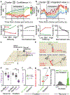
Figure 5 |
Figure 5 |. OFC-to-striatum projection neurons encode integrated value across task epochs.
a, Cluster transitions across epochs; each box represents neuron cluster in one epoch; lines between them represent neurons that belong to same cluster in two epochs. Transitions in cluster identities across trial epochs are highly structured, with two neurons in a given cluster likely to remain in same clusters over time. b, Dynamics of response time course for 8 temporal clusters (see Extended Figure 9d). c, Retrograde targeting of ChR2 to striatum-projecting neurons in lateral OFC for optogenetic identification. d, Laser pulse aligned responses for optogenetically identified unit. e, Latency and timing jitter of spikes from laser pulse onset for identified OFC-STR neurons was short and low. f, Spike raster and average activity in single striatum-projecting neuron aligned to outcome (reward or error sound, left) or to stimulus delivery in the next trial (right). Lower panels show analysis of firing rates in three different epochs: left Anticipation, for which firing rate varies with integrated value; middle, Feedback, for which firing rates reflects trial outcome with negative valence; right, Inter-trial interval (ITI), for which firing rates reflect graded outcomes with negative valence (NR, no reward; SR, small reward; LR, large reward). g, Average activity in error trials for optogenetically identified OFC-STR projection neurons (negative outcome selective, 18/24, AUC < 0 with P <0.01 permutation test) and their average PSTH grouped by error, correct small and large reward trials. h, Average dynamics and tuning of neurons in temporal Cluster B (panel b) match those of optogenetically identified striatum projecting neurons in (f-g). To avoid double counting, identified OFC-STR projection neurons were excluded from Cluster B.

References

    1. Hubel DH & Wiesel TN Receptive fields of single neurones in the cat’s striate cortex. J. Physiol 148, 574–91 (1959). - PMC - PubMed
    1. Bruce C, Desimone R & Gross CG Visual properties of neurons in a polysensory area in superior temporal sulcus of the macaque. J. Neurophysiol 46, 369–84 (1981). - PubMed
    1. O’Keefe J & Dostrovsky J The hippocampus as a spatial map. Preliminary evidence from unit activity in the freely-moving rat. Brain Res. 34, 171–5 (1971). - PubMed
    1. Abe H & Lee D Distributed coding of actual and hypothetical outcomes in the orbital and dorsolateral prefrontal cortex. Neuron 70, 731–41 (2011). - PMC - PubMed
    1. Feierstein CE, Quirk MC, Uchida N, Sosulski DL & Mainen ZF Representation of spatial goals in rat orbitofrontal cortex. Neuron 51, 495–507 (2006). - PubMed

References (Methods)

    1. Kepecs A, Uchida N, Zariwala HA & Mainen ZF Neural correlates, computation and behavioural impact of decision confidence. Nature 455, 227–31 (2008). - PubMed
    1. Sanders JI & Kepecs A A low-cost programmable pulse generator for physiology and behavior. Front. Neuroeng 7, 1–8 (2014). - PMC - PubMed
    1. Gradinaru V et al. Molecular and cellular approaches for diversifying and extending optogenetics. Cell 141, 154–165 (2010). - PMC - PubMed
    1. Li SJ, Vaughan A, Sturgill JF & Kepecs A A Viral Receptor Complementation Strategy to Overcome CAV-2 Tropism for Efficient Retrograde Targeting of Neurons. Neuron 98, 905–917.e5 (2018). - PubMed
    1. Soudais C, Laplace-Builhe C, K K & Kremer E. Preferential transduction of neurons by canine adenovirus vectors and their efficient retrograde transport in vivo. FASEB J. 15:, 2283–5 (2001). - PubMed

Publication types

MeSH terms