Review
. 2020 Jan;40(1):110-124.
doi: 10.1002/pd.5591. Epub 2020 Jan 6.

The MRI spectrum of congenital cytomegalovirus infection

Affiliations
Review

The MRI spectrum of congenital cytomegalovirus infection

Mariana C Diogo et al. Prenat Diagn. 2020 Jan.

Abstract

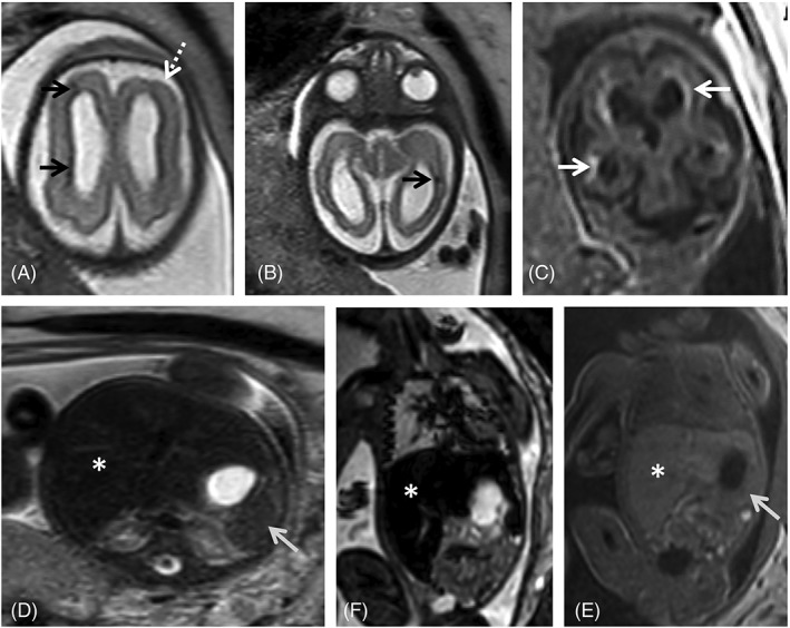
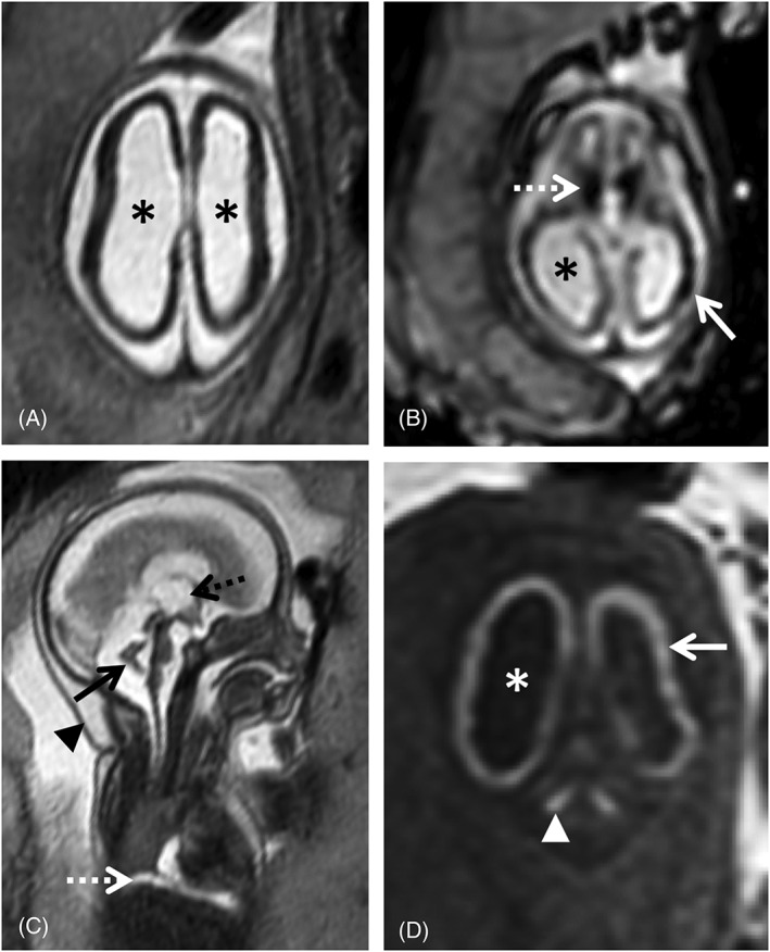
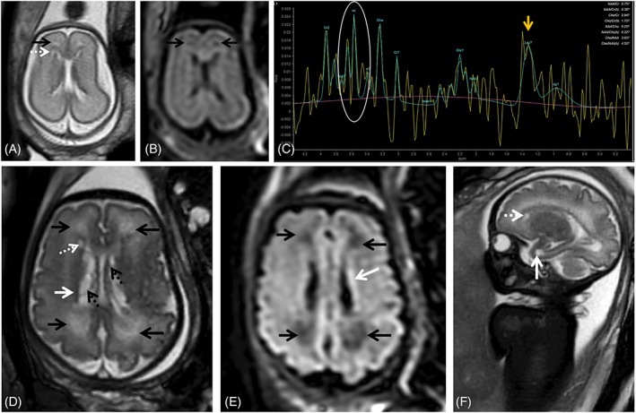
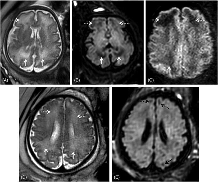
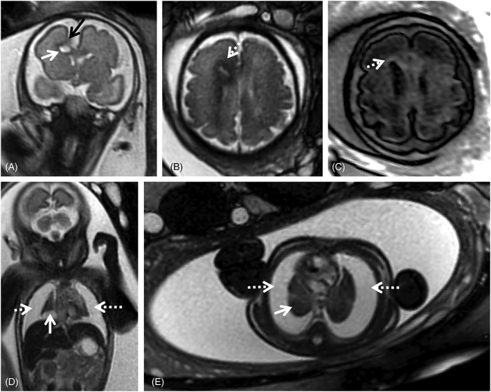
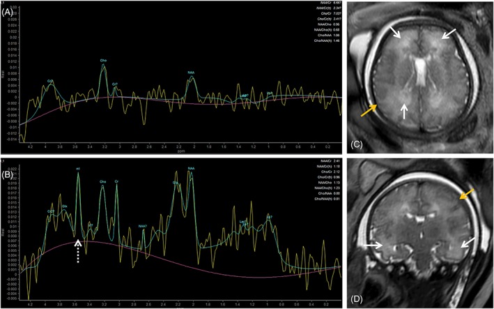
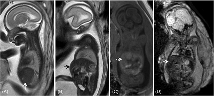
Human cytomegalovirus (CMV) is an ubiquitous pathogen, with a high worldwide seroprevalence. When acquired in the prenatal period, congenital CMV (cCMV) is a major cause of neurodevelopmental sequelae and hearing loss. cCMV remains an underdiagnosed condition, with no systematic screening implemented in pregnancy or in the postnatal period. Therefore, imaging takes a prominent role in prenatal diagnosis of cCMV. With the prospect of new viable therapies, accurate and timely diagnosis becomes paramount, as well as identification of fetuses at risk for neurodevelopmental sequelae. Fetal magnetic resonance imaging (MRI) provides a complementary method to ultrasound (US) in fetal brain and body imaging. Anterior temporal lobe lesions are the most specific finding, and MRI is superior to US in their detection. Other findings such as ventriculomegaly, cortical malformations and calcifications, as well as hepatosplenomegaly, liver signal changes and abnormal effusions are unspecific. However, when seen in combination these should raise the suspicion of fetal infection, highlighting the need for a full fetal assessment. Still, some fetuses deemed normal on prenatal imaging are symptomatic at birth or develop delayed cCMV-associated symptoms, leaving room for improvement of diagnostic tools. Advanced MR sequences may help in this field and in determining prognosis, but further studies are needed.


Conflict of interest statement

The authors declare no conflicts of interest.

Figures

Figure 1
Figure 1
Fetal magnetic resonance imaging (MRI) at 29 gestational weeks referred for microcephaly and splenomegaly. There is parenchymal loss, with global thinning of the cerebral mantle and consequent bilateral ventriculomegaly. Small areas of focal signal anomaly can be detected on T2WI (A, B, black arrows), as well as frontal polymicrogyria (A, white dashed arrow). Calcifications can be identified on T1WI (c, white arrows). Enlarged spleen (gray arrow) and liver (asterisk) can be identified on T2W steady state free precession (D), echo planar imaging (E) and T1W images (F), with slight signal intensity anomaly on the latter
Figure 2
Figure 2
24 gestational weeks fetus referred for fetal magnetic resonance imaging (MRI) for suspected lissencephaly and cerebellar hypoplasia. T2w single shot fast spin echo axial (A) and sagittal (C), axial echo planar imaging (EPI) (B) and coronal T1WI (D) show a marked reduction of the cerebral parenchyma thickness, with diffuse low SI on T2WI (A) and severe ventriculomegaly (asterisk), including dilatation of the third ventricle (C, black dashed arrow). There is diffuse high SI on T1WI of the supratentorial parenchyma (D, white arrow) compatible with presence of calcifications, confirmed on EP images (B) and particularly evident in the basal ganglia (B, white dashed arrow). There is cerebellar hypoplasia (C, black arrow) associated with deep gray nuclei calcifications (D, white arrowhead). Furthermore, small pleural effusion can be seen on sagittal T2WI (C, white dashed arrow). Findings were confirmed on postmortem MRI (not shown). Additionally, there is skin edema/thickening of the skin (C, black arrow head)
Figure 3
Figure 3
Fetal magnetic resonance imaging at 24 gestational weeks (GW) referred for maternal cytomegalovirus seroconversion and normal brain ultrasound. On T2 weighted images (WI) slight periventricular caps can be identified frontally (A, black arrow) as well as a small periventricular cyst. PRESS spectroscopy performed with a short TE (35 ms) depicts a myoinositol peak (myo‐inositol, white circle, C) and a lactate peak (Lac, yellow arrow, C), raising the suspicion of more extensive brain involvement. Follow up at 34 gestational weeks shows progression of white matter signal changes in the frontal and parieto‐occipital regions bilaterally (black arrows, D) that also have translation on the FLAIR image (black arrows, E). The presence of the small periventricular cyst can be confirmed (D, F, white dashed arrow) and there is the additional finding of a temporal pole cyst (F, white arrow) and irregularity and signal alteration of the ventricular lining (hypointense on T2WI, D, white arrow; and hyperintense on t2W FLAIR images, E, white arrow) with intraventricular septations/pseudocysts (D, black dashed arrow)
Figure 4
Figure 4
Two fetuses referred for fetal magnetic resonance imaging at 34 gestational weeks for congenital cytomegalovirus (cCMV) infection (A‐C) and abdominal cyst (D‐E). White matter hyperintensities can be identified on T2WI in the frontal (A, D, white dashed arrows) and parietal‐occipital parieto‐occipital (A, D, white arrows) regions. On T2w echo planar imaging‐FLAIR images (B, E) there is corresponding hypointensity in the cCMV patient (B, white/dashed arrows), as well as low SI on the zoom diffusion weighted image (C, white/dashed arrows), but not the control (E) except in the expected gyral crests corresponding to remnants of the subplate (E, black dashed and full arrows)
Figure 5
Figure 5
Magnetic resonance imaging of fetus referred at 31 gestational weeks for hydrothorax and suspected frontal brain lesion. On T2WI over the fetal brain (A,B) it is possible to identify a destructive lesion of the right frontal lobe, with focal parenchymal loss (A, black arrow), with associated intraparenchymal white matter cystic lesion (A, white arrow) and altered signal of the surrounding parenchyma, with T2 hypointensity (B, white dashed arrow) and high signal on T1‐weighted FLAIR imaging (C, white arrow), suggesting gliotic changes. There are extensive bilateral pleural effusions (D, E, dashed white arrows), with severe reduction of the lung volume (D, E, white arrows)
Figure 6
Figure 6
Comparison of intermediate (A, TE 144) and short (B, TE 31) TE spectroscopy of fetus referred for magnetic resonance imaging due to confirmed fetal cytomegalovirus infection and fetal growth restriction at 36 gestational weeks. T2 single shot fast spin echo images (C, axial; D, coronal) showed moderate brain swelling with extracerebral CSF spaces effacement (yellow arrows). There is white matter T2‐hyperintensity in the frontal, parietal‐occipital (C, white arrows) and to a lesser extent in the temporal lobe (D, white arrows). Magnetic resonance spectroscopy at TE 144 shows no significant changes; at TE 31 there is a myo‐inositol peak (B, white dashed arrow)
Figure 7
Figure 7
Fetal magnetic resonance imaging of a cytomegalovirus (CMV) positive fetus at 24 gestational weeks. T2WI depict splenomegaly (A, white arrow) and hepatomegaly (B, black arrow), protruding in the anterior abdominal wall. There is associated abnormal liver signal: isointense on T1WI (C, dashed white arrow) and on echo planar images (D, dashed white arrow), which cannot be identified on T2WI. These findings are often found in fetal infections and may help guide the diagnosis when brain anomalies are found, in the absence of a definite diagnosis of CMV infection

References

    1. Sert Y, Ozgu‐Erdinc AS, Saygan S, Engin UY. Antenatal cytomegalovirus infection screening results of 32,188 patients in a tertiary referral center: A retrospective cohort study. Fetal Pediatr Pathol. 2019;38(2):112‐120. - PubMed
    1. Griffiths P, Baraniak I, Reeves M. The pathogenesis of human cytomegalovirus. J Pathol. 2015;235(2):288‐297. - PubMed
    1. Cannon MJ, Schmid DS, Hyde TB. Review of cytomegalovirus seroprevalence and demographic characteristics associated with infection. Rev Med Virol. 2010;20(4):202‐213. - PubMed
    1. Buxmann H, Hamprecht K, Meyer‐Wittkopf M, Friese K. Primary human cytomegalovirus (HCMV) infection in pregnancy. Deutsches Aerzteblatt Online. 2017;144:45‐52. 10.3238/arztebl.2017.0045 - DOI - PMC - PubMed
    1. Cannon MJ, Griffiths PD, Aston V, Rawlinson WD. Universal newborn screening for congenital CMV infection: what is the evidence of potential benefit? Rev Med Virol. 2014;24(5):291‐307. - PMC - PubMed

MeSH terms