. 2020 Sep;23(5):e12928.
doi: 10.1111/desc.12928. Epub 2019 Dec 30.

Cerebral blood flow in 5- to 8-month-olds: Regional tissue maturity is associated with infant affect

Affiliations

Cerebral blood flow in 5- to 8-month-olds: Regional tissue maturity is associated with infant affect

M Catalina Camacho et al. Dev Sci. 2020 Sep.

Abstract

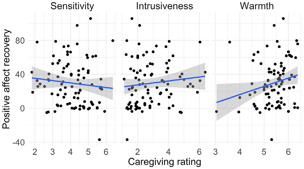
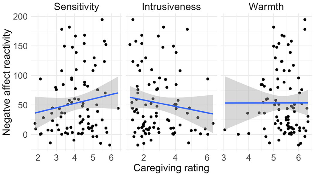
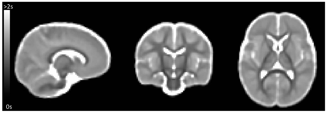
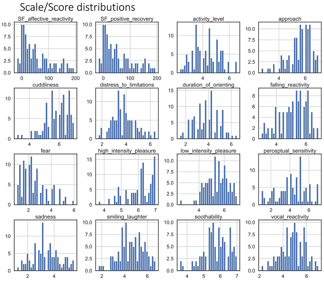
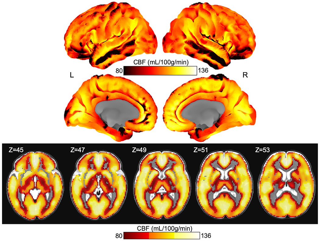
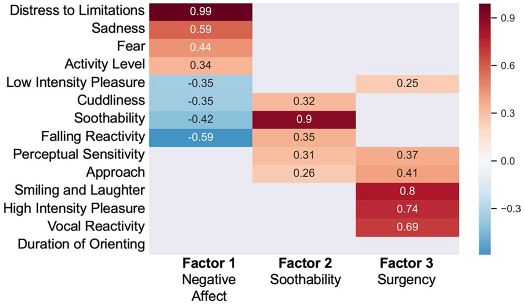
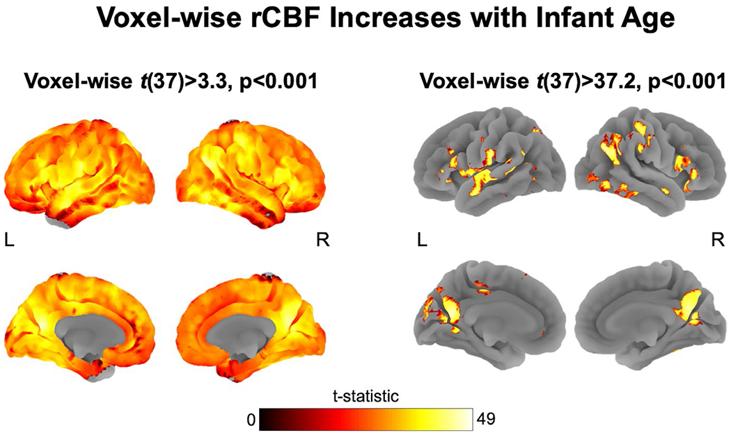
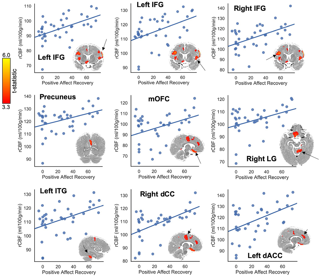
Infancy is marked by rapid neural and emotional development. The relation between brain function and emotion in infancy, however, is not well understood. Methods for measuring brain function predominantly rely on the BOLD signal; however, interpretation of the BOLD signal in infancy is challenging because the neuronal-hemodynamic relation is immature. Regional cerebral blood flow (rCBF) provides a context for the infant BOLD signal and can yield insight into the developmental maturity of brain regions that may support affective behaviors. This study aims to elucidate the relations among rCBF, age, and emotion in infancy. One hundred and seven mothers reported their infants' (infant age M ± SD = 6.14 ± 0.51 months) temperament. A subsample of infants completed MRI scans, 38 of whom produced usable perfusion MRI during natural sleep to quantify rCBF. Mother-infant dyads completed the repeated Still-Face Paradigm, from which infant affect reactivity and recovery to stress were quantified. We tested associations of infant age at scan, temperament factor scores, and observed affect reactivity and recovery with voxel-wise rCBF. Infant age was positively associated with CBF in nearly all voxels, with peaks located in sensory cortices and the ventral prefrontal cortex, supporting the formulation that rCBF is an indicator of tissue maturity. Temperamental Negative Affect and recovery of positive affect following a stressor were positively associated with rCBF in several cortical and subcortical limbic regions, including the orbitofrontal cortex and inferior frontal gyrus. This finding yields insight into the nature of affective neurodevelopment during infancy. Specifically, infants with relatively increased prefrontal cortex maturity may evidence a disposition toward greater negative affect and negative reactivity in their daily lives yet show better recovery of positive affect following a social stressor.

Keywords: cerebral blood flow; emotion; infant brain development; still-face; temperament.


Conflict of interest statement

CONFLICT OF INTEREST

The authors have no conflicts of interest to declare.

Figures

FIGURE A1
FIGURE A1
Associations between maternal caregiving ratings and Positive Affect Recovery
FIGURE A2
FIGURE A2
Associations between maternal caregiving ratings and Negative Affect Reactivity
FIGURE A3
FIGURE A3
Average T1 created from a quantitative T1 sequence obtained from a subset of infants
FIGURE A4
FIGURE A4
Histogram distributions for the affective codes from the Still-Face Paradigm (SF Negative Reactivity and SF Positive Recovery) and for each scale of the Infant Behavior Questionnaire-Revised, Short Form. Bin widths are plotted to fit 30 bins
FIGURE A5
FIGURE A5
Scree plots of actual and simulated factor analyses generated using the psych library in R. From this graphic, a 2-factor model was recommended for these data
FIGURE A6
FIGURE A6
Infant positive and negative affect across the SFP-R in the full sample (N = 93). Notes: P = play, SF1 = first still-face, R1 = first reunion, SF2 = second still-face, R2 = second reunion. Replicating previous research, infants exhibited increased negative affect in still-face relative to non-still-face episodes and increased positive affect in reunion relative to still-face episodes
FIGURE A7
FIGURE A7
Whole brain rCBF projected onto the cortical surface and in slices. Cortical projections are colorbar centered at 80–136, thus regions that are black indicate an rCBF of 80 ml 100 g−1 min−1 or below. Slices of the insula and subcortical structures are included below the cortical surface projections with the Z-direction slice number indicated in the upper left corner of each image. The slice images are thresholded at 80 ml 100 g−1 min−1 for ease of reading, thus clear voxels on the overlay indicate either signal below 80 ml 100 g−1 min−1 or excluded voxels (such as in the case of white matter)
FIGURE A8
FIGURE A8
Final CFA for the 3-factor model. Standardized loading values are shown. Duration of Orienting was excluded from this model for not meeting the 0.3 threshold in the EFA. ✦p < .05; *p < .01; **p < .005; ***p < .001
FIGURE 1
FIGURE 1
Factor structure for the Infant Behavior Questionnaire-Revised Short Form (IBQ-R-SF) derived from an exploratory three-factor analysis. Low loadings between −0.25 and 0.25 are omitted for clarity. Based on guidelines provided in de Winter, Dodou, and Wieringa (2009), this analysis is well-powered to detect loadings that are approximately 0.5 or higher
FIGURE 2
FIGURE 2
Almost every investigated voxel of gray matter cerebral blood flow (CBF) was positively associated with age after accounting for other predictors in the general linear model (GLM) (peak t(37) = 41.2, p < .001) and in a stand-alone re-analysis with only age and sex terms in the model (peak t(37) = 50.4, p < .001). 3a: The full statistical map thresholded at voxel-wise p < .001. 3b: the statistical map thresholded at the 95th percentile (t(37) = 37.2), highlighting cortical regions most significantly predicted by age at scan
FIGURE 3
FIGURE 3
Cerebral blood flow (CBF) clusters associated with each affect measure that met the conservative 98.6 minimum cluster size. Clusters are FWE corrected p < .05, voxel-wise threshold p < .001. The scatterplot illustrates the positive linear association between the mean regional (rCBF) in these clusters and average increases in positive affect (Positive Affect Recovery) from the stressor to the reunion periods of the Still-Face Paradigm (SFP-R). The plotted cluster is indicated for each plot with a black arrow. dACC, dorsal anterior cingulate; dCC, medial dorsal cingulate; IFG, inferior frontal gyrus; ITG, inferior temporal gyrus; LG, lingual gyrus; mOFC, medial orbitofrontal cortex

References

    1. Ahmed SP, Bittencourt-Hewitt A, & Sebastian CL (2015). Neurocognitive bases of emotion regulation development in adolescence. Developmental Cognitive Neuroscience, 15, 11–25. 10.1016/j.dcn.2015.07.006 - DOI - PMC - PubMed
    1. Alsop DC, Detre JA, Golay X, Günther M, Hendrikse J, Hernandez-Garcia L, … Zaharchuk G (2015). Recommended implementation of arterial spin-labeled Perfusion MRI for clinical applications: A consensus of the ISMRM Perfusion Study group and the European consortium for ASL in dementia. Magnetic Resonance in Medicine, 73(1), 102–116. 10.1002/mrm.25197 - DOI - PMC - PubMed
    1. Andersson JLR, Skare S, & Ashburner J (2003). How to correct susceptibility distortions in spin-echo echo-planar images: Application to diffusion tensor imaging. NeuroImage, 20(2), 870–888. 10.1016/S1053-8119(03)00336-7 - DOI - PubMed
    1. Arichi T, Fagiolo G, Varela M, Melendez-Calderon A, Allievi A, Merchant N, … Edwards AD (2012). Development of BOLD signal hemodynamic responses in the human brain. NeuroImage, 63(2), 663–673. 10.1016/j.neuroimage.2012.06.054 - DOI - PMC - PubMed
    1. Arichi T, Moraux A, Melendez A, Doria V, Groppo M, Merchant N, … Edwards AD (2010). Somatosensory cortical activation identified by functional MRI in preterm and term infants. NeuroImage, 49(3), 2063–2071. 10.1016/j.neuroimage.2009.10.038 - DOI - PubMed

Publication types