. 2019 Oct 31:14:8573-8588.
doi: 10.2147/IJN.S224047. eCollection 2019.

CTGF Loaded Electrospun Dual Porous Core-Shell Membrane For Diabetic Wound Healing

Affiliations

CTGF Loaded Electrospun Dual Porous Core-Shell Membrane For Diabetic Wound Healing

Robin Augustine et al. Int J Nanomedicine. .

Abstract

Purpose: Impairment of wound healing is a major issue in type-2 diabetes that often causes chronic infections, eventually leading to limb and/or organ amputation. Connective tissue growth factor (CTGF) is a signaling molecule with several roles in tissue repair and regeneration including promoting cell adhesion, cell migration, cell proliferation and angiogenesis. Incorporation of CTGF in a biodegradable core-shell fiber to facilitate its sustained release is a novel approach to promote angiogenesis, cell migration and facilitate wound healing. In this paper, we report the development of CTGF encapsulated electrospun dual porous PLA-PVA core-shell fiber based membranes for diabetic wound healing applications.

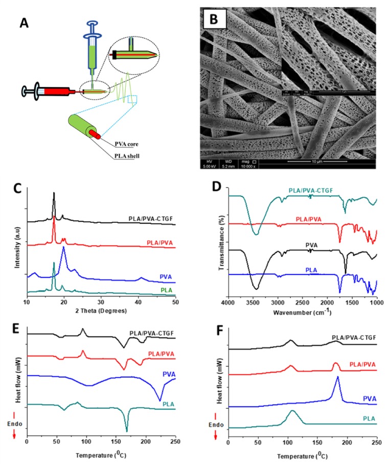
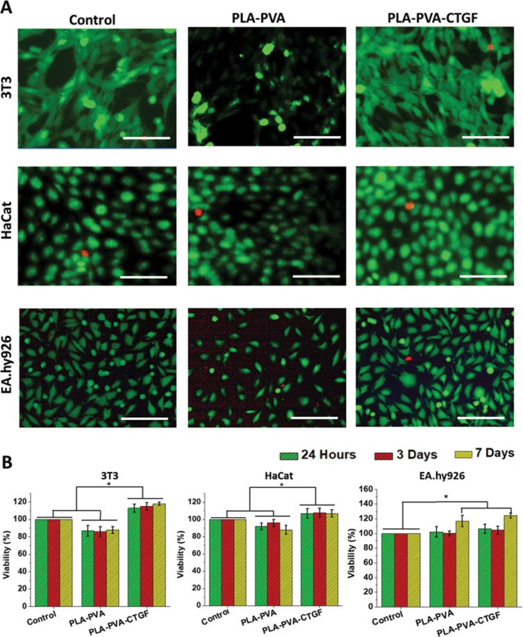
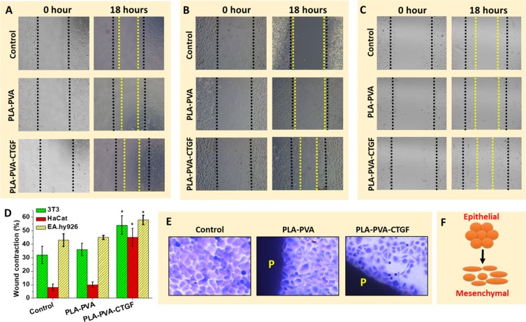
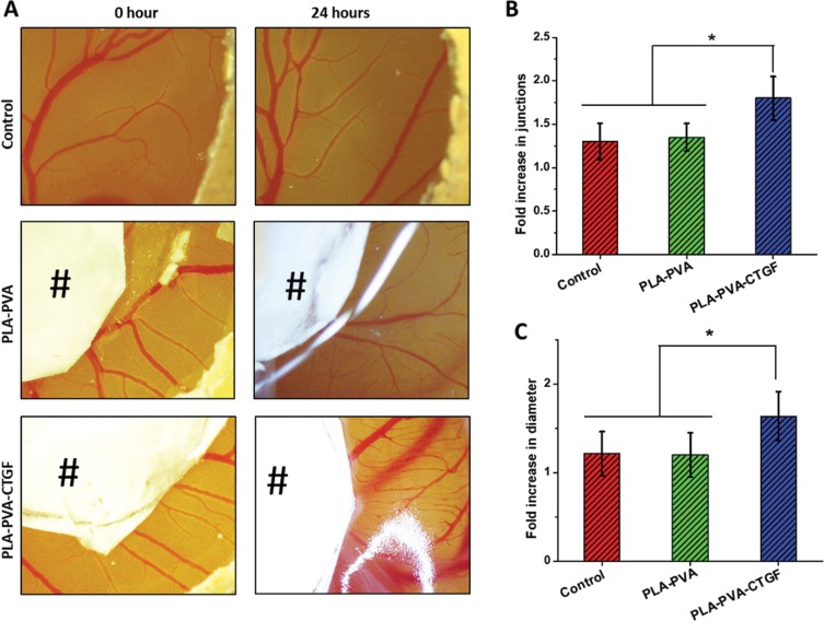
Methods: The membranes were fabricated by a core-shell electrospinning technique. CTGF was entrapped within the PVA core which was coated by a thin layer of PLA. The developed membranes were characterized by techniques such as Scanning Electron Microscopy (SEM), Fourier Transform Infrared Spectroscopy (FTIR) and X-Ray Diffraction (XRD) analysis. In vitro cell culture studies using fibroblasts, keratinocytes and endothelial cells were performed to understand the effect of CTGF loaded membranes on cell proliferation, cell viability and cell migration. A chicken chorioallantoic membrane (CAM) assay was performed to determine the angiogenic potential of the membranes.

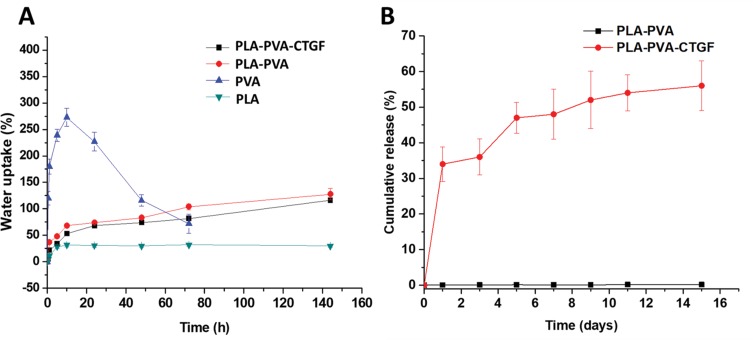
Results: Results showed that the developed membranes were highly porous in morphology with secondary pore formation on the surface of individual fibers. In vitro cell culture studies demonstrated that CTGF loaded core-shell membranes improved cell viability, cell proliferation and cell migration. A sustained release of CTGF from the core-shell fibers was observed for an extended time period. Moreover, the CAM assay showed that core-shell membranes incorporated with CTGF can enhance angiogenesis.

Conclusion: Owing to the excellent cell proliferation, migration and angiogenic potential of CTGF loaded core-shell PLA-PVA fibrous membranes, they can be used as an excellent wound dressing membrane for treating diabetic wounds and other chronic ulcers.

Keywords: CTGF; PLA; PVA; diabetic wound; electrospinning.


Conflict of interest statement

The authors declare no competing interests in this work.

Figures

Figure 1
Figure 1
Results of morphological and physical characterization of the developed core-shell fiber-based membranes. Schematic representation of the fabrication process of the core-shell membranes (A). SEM image of coaxial PLA-PVA-CTGF membranes showing the morphology (B). XRD patterns (C), FTIR spectra (D), DSC heating thermogram (E) and DSC cooling thermogram (F) of fabricated membranes.
Figure 2
Figure 2
Mechanical testing results of the developed membranes. Stress-strain curve (A), Elongation at break (B), Ultimate tensile stress (C) and Young’s modulus (D) of PLA, PVA, PLA-PVA and PLA-PVA-CTGF membranes. P-values were calculated using Student’s t-test where (*) indicates a significant difference from other group of comparison (p ≤ 0.05).
Figure 3
Figure 3
Water uptake capacity (A) and cumulative release (B) of PLA-PVA-CTGF membranes.
Figure 4
Figure 4
Results of in vitro cell culture studies on the developed membranes. Live/Dead assay results showing the viability of 3T3 fibroblasts, HaCat keratinocytes and EA.hy926 endothelial cells which were cultured with the PLA-PVA and PLA-PVA-CTGF membranes (A). Scale bars = 200 µm. Viability of 3T3, HaCat and EA.hy926 cells upon culturing with the scaffolds (B). P-values were calculated using Student’s t-test where (*) indicates a significant difference from the other group of comparison (p ≤ 0.05).
Figure 5
Figure 5
Results showing the effect of PLA-PVA and PLA-PVA-CTGF scaffolds on in vitro wound healing using 3T3 fibroblasts (A), HaCat keratinocytes (B) and EA.hy926 endothelial cells (C). Wound contraction (%) after treatment with the developed membranes (D). Morphological changes in HaCat cells which were incubated with PLA-PVA and PLA-PVA-CTGF membranes (E). Schematic representation of phenotypic changes of keratinocyte cells upon incubation with PLA-PVA-CTGF scaffolds (F). P-values were calculated using a Student’s t-test where (*) indicates a significant difference from the control (p ≤ 0.05). The images were taken at 100X magnification.
Figure 6
Figure 6
Effect of PLA-PVA-CTGF on angiogenesis in vivo was evaluated by in ovo model of chicken chorioallantoic membrane (CAM) assay (A). Fold increase in blood vessel junctions (B) and fold increase in blood vessel diameter (C) after 24 hrs of treatment with samples. # indicates the core-shell membranes. P-values were calculated using one-way ANOVA where (*) indicates a significant difference from the other group of comparison (p ≤ 0.05). The images were taken at 10X magnification.

References

    1. Borys S, Hohendorff J, Frankfurter C, Kiec-Wilk B, Malecki MT. Negative pressure wound therapy use in diabetic foot syndrome—from mechanisms of action to clinical practice. Eur J Clin Invest. 2019;e13067. doi:10.1111/eci.13067 - DOI - PubMed
    1. Cunha M, Queirós P, Cardoso D, Santos E, Rodrigues M, Apóstolo J. The effectiveness of cleansing solutions for wound treatment: a systematic review. JBI Database System Rev Implement Rep. 2014;12(10):121–151. doi:10.11124/jbisrir-2014-1746 - DOI
    1. Xu F, Zhang C, Graves DT. Abnormal cell responses and role of TNF- α in impaired diabetic wound healing. Biomed Res Int. 2013;2013:1–9. doi:10.1155/2013/754802 - DOI - PMC - PubMed
    1. Bao P, Kodra A, Tomic-Canic M, Golinko MS, Ehrlich HP, Brem H. The role of vascular endothelial growth factor in wound healing. J Surg Res. 2009;153(2):347–358. doi:10.1016/j.jss.2008.04.023 - DOI - PMC - PubMed
    1. Hardwicke J, Schmaljohann D, Boyce D, Thomas D. Epidermal growth factor therapy and wound healing - past, present and future perspectives. Surgeon. 2008;6(3):172–177. doi:10.1016/S1479-666X(08)80114-X - DOI - PubMed