Case Reports
. 2019 Nov 1:15:1283-1289.
doi: 10.2147/TCRM.S225832. eCollection 2019.

Eosinophilic Pneumonia Associated With Natalizumab In A Patient With Multiple Sclerosis: A Case Report And Literature Review

Affiliations
Case Reports

Eosinophilic Pneumonia Associated With Natalizumab In A Patient With Multiple Sclerosis: A Case Report And Literature Review

Yuichiro Yasuda et al. Ther Clin Risk Manag. .

Abstract

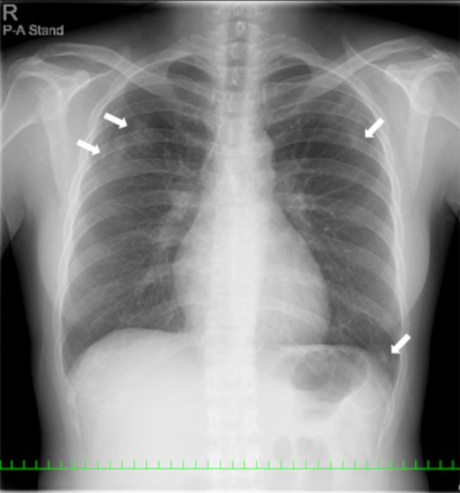
We herein report the case of a 39-year-old Japanese female with eosinophilic pneumonia associated with natalizumab. The patient with bronchial asthma had multiple sclerosis and was treated using natalizumab. The patient was referred to our department because of a persistent cough. A chest computed tomography (CT) scan revealed bilateral patchy consolidation surrounded by ground-glass opacity. A bronchoalveolar lavage (BAL) was performed. Eosinophil levels in the BAL fluid were increased and the patient was consequently diagnosed as eosinophilic pneumonia associated with natalizumab. Therefore, natalizumab treatment was discontinued. Subsequent chest CT findings showed a remarkable improvement without any treatment.

Keywords: eosinophilic pneumonia; multiple sclerosis; natalizumab.


Conflict of interest statement

All of the authors report that they have no conflicts of interest to disclose in this work.

Figures

Figure 1
Figure 1
A chest X-ray revealed patchy consolidation in the bilateral upper and left lower lung field (white arrow).
Figure 2
Figure 2
CT scans showed bilateral patchy consolidation surrounded by ground-glass opacity (white arrow).
Figure 3
Figure 3
BAL fluid showed increased numbers of eosinophils with Papanicolaou staining (A, ×400). Under high magnification, the eosinophils were stained light green with nuclear localization (B, ×800).
Figure 4
Figure 4
A chest X-ray and chest CT images showed an improvement in lung infiltrates after the discontinuation of natalizumab.
Figure 5
Figure 5
The clinical course of the patient.

References

    1. Sawcer S, Franklin RJ, Ban M. Multiple sclerosis genetics. Lancet Neurol. 2014;13:700–709. doi:10.1016/S1474-4422(14)70041-9 - DOI - PubMed
    1. Osoegawa M, Kira J, Fukazawa T, et al. Temporal changes and geographical differences in multiple sclerosis phenotypes in Japanese: nationwide survey results over 30 years. Mult Scler. 2009;15:159–173. doi:10.1177/1352458508098372 - DOI - PubMed
    1. Compston A, Coles A. Multiple sclerosis. Lancet. 2008;372:1502–1517. doi:10.1016/S0140-6736(08)61620-7 - DOI - PubMed
    1. Polman CH, O’Connor PW, Havrdova E, et al. A randomized, placebo-controlled trial of natalizumab for relapsing multiple sclerosis. N Engl J Med. 2006;354:899–910. doi:10.1056/NEJMoa044397 - DOI - PubMed
    1. Ransohoff RM. Natalizumab for multiple sclerosis. N Engl J Med. 2007;356:2622–2629. doi:10.1056/NEJMct071462 - DOI - PubMed

Publication types