Review
. 2019 Nov 12:10:1188.
doi: 10.3389/fneur.2019.01188. eCollection 2019.

Corticoreticular Tract in the Human Brain: A Mini Review

Affiliations
Review

Corticoreticular Tract in the Human Brain: A Mini Review

Sung Ho Jang et al. Front Neurol. .

Abstract

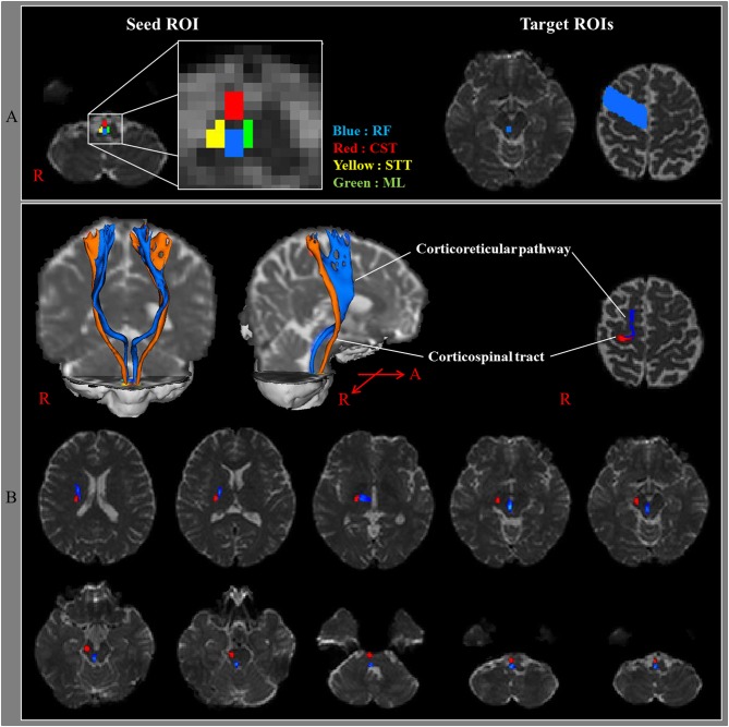
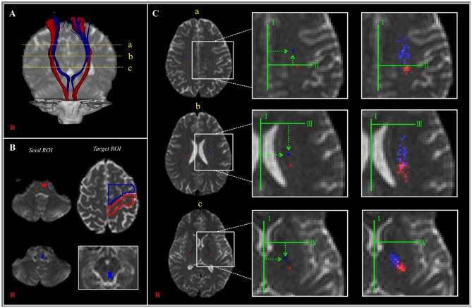
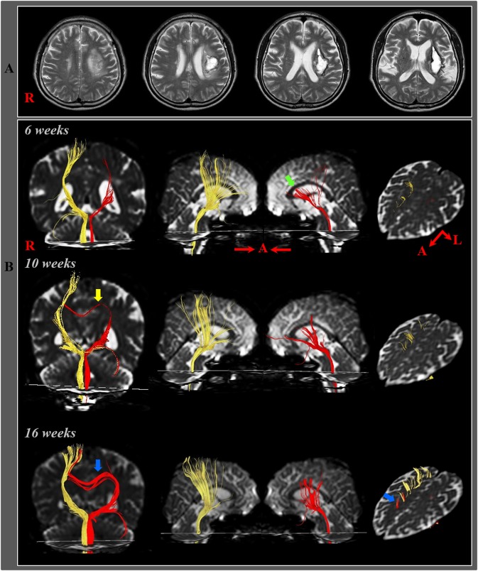
Previous studies have suggested that the corticoreticular tract (CRT) has an important role in motor function almost next to the corticospinal tract (CST) in the human brain. Herein, the CRT is reviewed with regard to its anatomy, function, and recovery mechanisms after injury, with particular focus on previous diffusion tensor tractography-based studies. The CRT originates from several cortical areas but mainly from the premotor cortex. It descends through the subcortical white matter anteromedially to the CST with a 6- to 12-mm separation in the anteroposterior direction, then passing through the mesencephalic tegmentum and the pontine and pontomedullary reticular formations. Regarding its motor functions, the CRT appears to be mainly involved in the motor function of proximal joint muscles accounting for ~30-40% of the motor function of these joint muscles. In addition, the CRT is involved in gait function and postural stability. However, further studies that clearly rule out the effects of other motor function-related neural tracts are necessary to clarify the precise portion of the total motor function for which the CRT is responsible. With regard to recovery mechanisms for an injured CRT, three recovery mechanisms were suggested in five previous studies: recovery through the original pathway, recovery through perilesional reorganization, and recovery through the transcallosal pathway. However, each of those studies was single-case reports; therefore, further original studies including a larger number of patients are warranted.

Keywords: corticoreticular tract; corticoreticulospinal tract; diffusion tensor tractography; motor function; recovery mechanism.


Figures

Figure 1
Figure 1
(A) Seed regions of interest (ROI) were given on the medullary reticular formation (blue rectangle). The first target ROI was given on the midbrain tegmentum (blue rectangle) and the second target ROI on the premotor cortex (Brodmann's area 6) (blue area). RF, reticular formation; CST, corticospinal tract; STT, spinothalamic tract; ML, medial lemniscus. (B) The pathways of the corticoreticular pathway (blue color) and CST (red color) are shown at each level of the brain in a normal subject (32-year-old male) [Reprinted with permission from Yeo et al. (3)].
Figure 2
Figure 2
(A) The corticoreticular pathway (CRP) (blue color) and corticospinal tract (CST) (red color) were reconstructed in both hemispheres [a—centrum semiovale (CS) level, b—corona radiata (CR) level, c—posterior limb of the internal capsule (PL) level]. (B) The regions of interest (ROIs) (blue color) were set for the CRP (seed—reticular formation of the medulla, first target ROI—midbrain tegmentum, second target ROI—Brodmann's area 6). The ROIs (red color) were set for the CST (seed ROI—anterior portion of lower pons, target ROI—Brodmann's area 4). (C) Landmarks used in determining the location of the CRP and CST (left and middle columns). The probabilistic maps for the CRP and CST were superimposed on images of a subject (right column). Line I: the midline between the right and left hemispheres for measurement of mediolateral distance. Line II: the horizontal line from the most medial point of the central sulcus to the midline as the reference line for the anteroposterior direction. Line III: the horizontal line that passes through the anterior end of the lateral ventricle for the reference line for the anteroposterior direction. Line IV: the horizontal line from the middle point at the genu of the internal capsule [Reprinted with permission from Jang and Seo (25)].
Figure 3
Figure 3
(A) T2-weighted MR images showed a hematoma in the left corona radiata and basal ganglia at 6-weeks after onset. (B) Results of diffusion tensor tractography (DTT). On 6-, 10-, and 16-week DTTs, the CRPs in the right hemispheres originated from the premotor cortex and primary motor cortex and descended through the known CRP pathway. By contrast, in the left (affected) hemisphere, the CRP showed severe narrowing with discontinuation of the anterior fibers (green arrow) on 6-week DTT, however, the discontinued fibers of the CRP were connected to the corpus callosum through the transcallosal fibers (yellow arrow) on 10-week DTT and became thicker and connected to the right cerebral cortex via the corpus callosum (blue arrow) on 16-week DTT [Reprinted with permission from Jang and Yeo (23)].

References

    1. Matsuyama K, Mori F, Nakajima K, Drew T, Aoki M, Mori S. Locomotor role of the corticoreticular-reticulospinal-spinal interneuronal system. Prog Brain Res. (2004) 143:239–49. 10.1016/S0079-6123(03)43024-0 - DOI - PubMed
    1. Mendoza JE, Foundas AL. The spinal cord and descending tracts. Clinical Neuroanatomy: A Neurobehavioral Approach. New York, NY: Springer; (2009). p. 1–22.
    1. Yeo SS, Chang MC, Kwon YH, Jung YJ, Jang SH. Corticoreticular pathway in the human brain: diffusion tensor tractography study. Neurosci Lett. (2012) 508:9–12. 10.1016/j.neulet.2011.11.030 - DOI - PubMed
    1. Kably B, Drew T. Corticoreticular pathways in the cat. I. projection patterns and collaterization. J Neurophysiol. (1998) 80:389–405. 10.1152/jn.1998.80.1.389 - DOI - PubMed
    1. Nathan PW, Smith MC. Long descending tracts in man. I. review of present knowledge. Brain. (1955) 78:248–303. 10.1093/brain/78.2.248 - DOI - PubMed