Review
. 2019 Jul 10;10(9):1569-1580.
doi: 10.1039/c9md00252a. eCollection 2019 Sep 1.

Molecular evolution of peptides by yeast surface display technology

Affiliations
Review

Molecular evolution of peptides by yeast surface display technology

Sara Linciano et al. Medchemcomm. .

Abstract

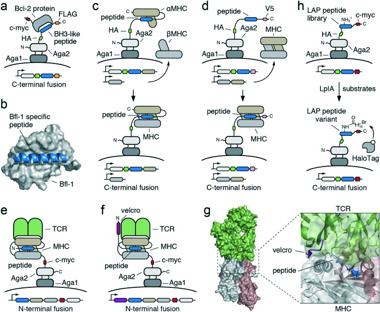
Genetically encoded peptides possess unique properties, such as a small molecular weight and ease of synthesis and modification, that make them suitable to a large variety of applications. However, despite these favorable qualities, naturally occurring peptides are often limited by intrinsic weak binding affinities, poor selectivity and low stability that ultimately restrain their final use. To overcome these limitations, a large variety of in vitro display methodologies have been developed over the past few decades to evolve genetically encoded peptide molecules with superior properties. Phage display, mRNA display, ribosome display, bacteria display, and yeast display are among the most commonly used methods to engineer peptides. While most of these in vitro methodologies have already been described in detail elsewhere, this review describes solely the yeast surface display technology and its valuable use for the evolution of a wide range of peptide formats.


Figures

Fig. 1
Fig. 1. Schematic representation of the yeast display technology based on Aga1–Aga2 system. a) The protein or peptide of interest (POI, blue) is displayed as a C-terminal fusion to the Aga2 protein (light grey), flanked by two tags for immunofluorescent detection: the hemagglutinin (HA) epitope tag at the N-terminus (green) and the c-myc epitope tag at the C-terminus (red). The Aga2 protein forms two disulfide bonds with the membrane-anchored Aga1 protein (dark grey); b) the POI (blue) is displayed as a N-terminal fusion to the Aga2 protein (light grey), flanked by the HA tag at the N-terminus (green) and the c-myc tag at the C-terminus (red); c) co-expression of two different POIs (blue and brown) fused at the N- and at the C-terminus of the Aga2 protein (light gray). The first POI (blue) is flanked by the HA tag (green) at the N-terminus and the c-myc tag at the C-terminus (red) whereas the second POI (brown) has a FLAG tag (orange) at the C-terminus; d) schematic representation for the selection of engineered POIs from a yeast display library by using fluorescence-activated cell sorting (FACS). Yeast cells, each displaying a different POI variant (light to dark blue), are incubated with a biotinylated target (light gray with white dot). Addition of fluorescently-labeled affinity reagents against the c-myc epitope (red) and the biotin (dark grey with green dot) enables the selection of dual positive yeast clones displaying full-length and properly folded POI that bind to the soluble target of interest. The FACS and two-color labeling allow the binding affinity to be normalized to cell surface expression and the affinities between clones accurately discriminated.
Fig. 2
Fig. 2. Schematic representation of linear peptides evolved using yeast display technology. a) BH3-like peptides (blue) are displayed on the surface of yeast as a C-terminus fusion of the Aga2 protein (light gray) and recognized by a Bcl-2 protein (dark gray); b) crystal structure of human Bfl-1 (gray surface) in complex with a Bfl-1-specific peptide (blue cartoon, PDB code: ; 6E3I); c) Top, the peptide (blue) located at the N-terminus of the αMHC subunit (brown), is displayed on the surface of yeast as a C-terminus fusion of the Aga2 protein (light gray) while the βMHC subunit (medium gray) is produced and secreted in a soluble form. Bottom, interaction between the displayed Aga2–peptide–αMHC fusion and the secreted βMHC leads to the assembly of a functional peptide-loaded MHC complex on the surface of yeast; d) Top, the peptide (blue), fused to the C-terminus of the Aga2 protein (light gray), is displayed on the surface of yeast while the whole dimeric MHC (brown and medium gray) is secreted in a soluble form. Bottom, formation of the peptide-loaded MHC complex on the surface of yeast is driven by non-covalent interactions between the displayed peptide and the secreted MHC molecule; e) the loaded peptide (blue), linked to the whole dimeric MHC (brown and medium gray), is encoded as a unique molecule and displayed on the surface of yeast as a N-terminus fusion of the Aga2 protein (light gray). Functional peptide-loaded MHC complex displayed on the surface of yeast is recognized by the soluble dimeric TCR receptor (green); f) an additional peptide sequence, named “velcro” (purple), is placed at the N-terminus of the loaded peptide (blue) and is displayed on the surface of yeast as a fusion of the whole dimeric MHC (brown and medium gray) and Aga2 (light gray) proteins; g) Left, crystal structure of a TCR (green surface) in complex with a peptide (blue cartoon) loaded on a MHC (brown and medium gray surface) in the presence of the affinity-enhancing “velcro” peptide (purple, PDB code: ; 6BGA). Right, close-up of the velcro-peptide–MHC–TCR complex; h) Top, LplA acceptor peptide (LAP, blue), fused to the C-terminus of Aga2 protein (light gray), is displayed on the surface of yeast and recognized by the LplA enzyme that catalyzes the conjugation of the 11-bromoundecanoic acid (bottom), an alkyl bromide that can be further specifically and covalently modified by the self-labeling enzyme HaloTag (medium grey). The white hexagon represents any probe. The epitope tags flaking the engineered peptides are colored in green (HA), red (c-myc), orange (FLAG) and pink (V5).
Fig. 3
Fig. 3. Schematic representation of cyclic peptides and peptides with a well-defined tertiary structure evolved using yeast display technology. a) The cyclic peptide “meditope” (blue) is displayed on the surface of yeast as a C-terminus fusion of the Aga2 protein (light gray) and recognized by a specific pocket located in the antigen-binding fragment (Fab) of the therapeutic antibody cetuximab. The light and heavy chains of the antibody are shown in brown and medium gray, respectively; b) Left, crystal structure of the Fab (brown and medium gray surface) in complex with meditope (blue cartoon, PDB code: ; 4GW1). Right, close-up of the Fab pocket occupied by the cyclic peptide meditope. The disulfide bridge of the peptide is shown in yellow, the antibody light and heavy chains are colored in brown and medium gray, respectively; c) the single-chain insulin analogue (blue) is displayed on the surface of yeast as a N-terminus fusion of a long and flexible stalk region (black line) anchored to the yeast cell wall. Properly folded single-chain insulin analogues retain the ability to bind the ectodomain of the insulin receptor (InsR, medium gray); the lanthipeptides (d) and the knottins (e) are displayed on the surface of yeast as a C-terminus of Aga2 subunit (light gray) and recognized by a heterodimeric integrin receptor (brown and medium gray); f) similar approach has been used to display numerous peptides with well-defined tertiary structures (blue) on the surface of yeast and detect their binding to multiple therapeutic targets (medium gray); g) crystal structure of a dimeric peptide derived from the avian pancreatic polypeptide (aPP, blue cartoon) in complex with a KRas mutant (gray surface, PDB code: ; 5WPL). The intermolecular disulfide bond is shown in yellow; h) designed peptides with well-defined tertiary structures (blue) are displayed on the surface of yeast as a C-terminus fusion of the Aga2 protein (light gray) and incubated with a protease. Proteolytic cleavage of the peptides (“truncated” forms) leads to loss of the c-myc tag at the C-terminus and consequent loss of fluorescence. Only yeast cells displaying stable structured peptides (“resistant” forms) retain fluorescence after proteolysis; i) Left, crystal structure of a designed peptide with a well-defined tertiary structure (blue cartoon) in complex with the hemagglutinin (HA, brown and medium gray) of influenza A virus PR8 (PDB code: ; 5VLI). Right, close-up of the peptide–HA complex; l) Left, crystal structure of a designed peptide with a well-defined tertiary structure (blue cartoon) in complex with the botulinum BoNT HCB neurotoxin (brown and medium gray, PDB code: ; 5VID). Right, close-up of the complex. The epitope tags flaking the engineered peptides are colored in green (HA), red (c-myc) and purple (strep).

References

    1. Koutsopoulos S., Peptide applications in biomedicine, biotechnology and bioengineering, Woodhead Publishing, 2018.
    1. Süssmuth R. D., Mainz A. Angew. Chem., Int. Ed. 2017;56:3770–3821. - PubMed
    1. Cunningham A. D., Qvit N., Mochly-Rosen D. Curr. Opin. Struct. Biol. 2017;44:59–66. - PMC - PubMed
    1. Packer M. S., Liu D. R. Nat. Rev. Genet. 2015;16:379–394. - PubMed
    1. Tizei P. A. G., Csibra E., Torres L., Pinheiro V. B. Biochem. Soc. Trans. 2016;44:1165–1175. - PMC - PubMed