. 2019 Dec 5;17(12):e3000563.
doi: 10.1371/journal.pbio.3000563. eCollection 2019 Dec.

Mediator MED23 regulates inflammatory responses and liver fibrosis

Affiliations

Mediator MED23 regulates inflammatory responses and liver fibrosis

Zhichao Wang et al. PLoS Biol. .

Abstract

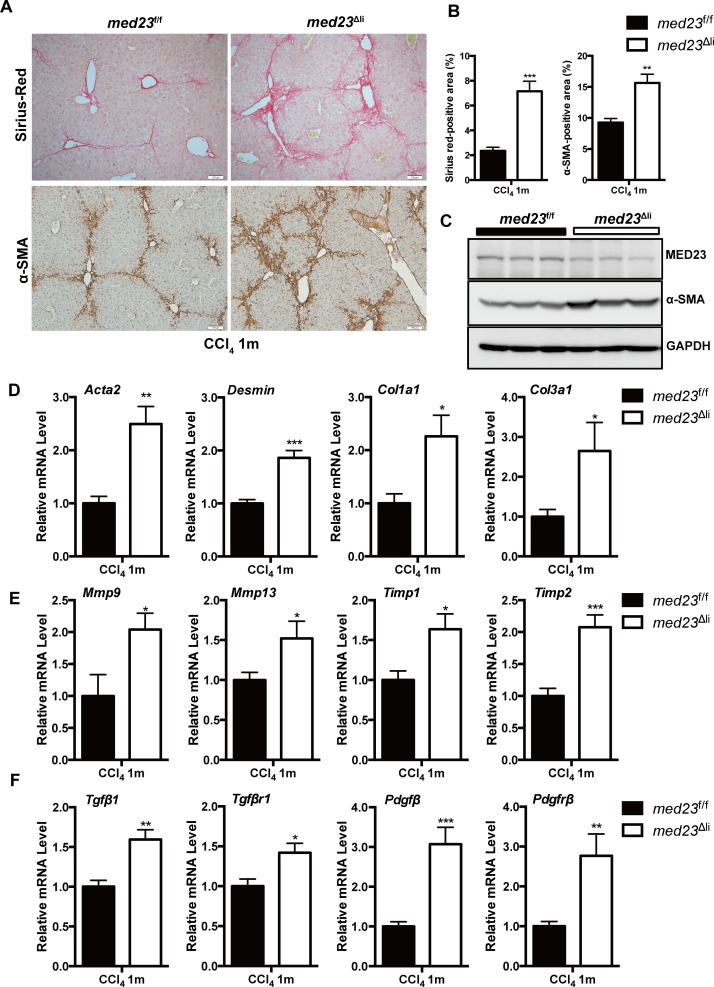
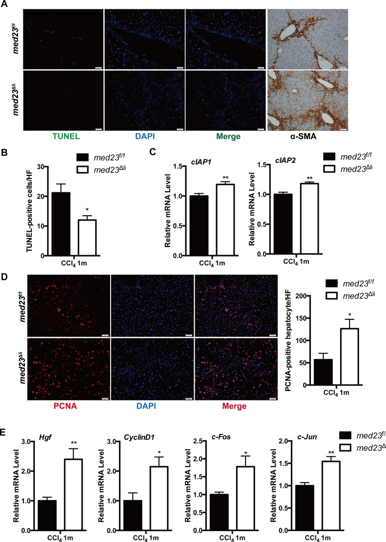
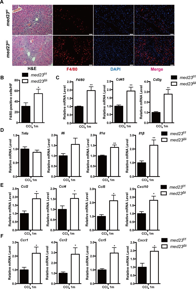
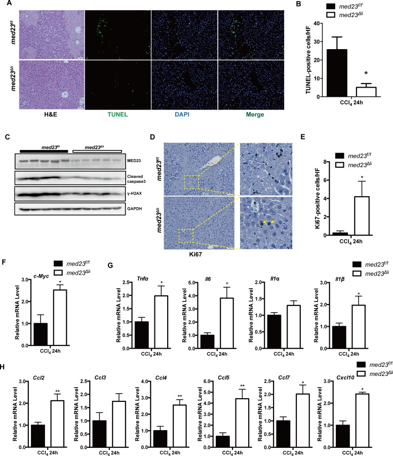
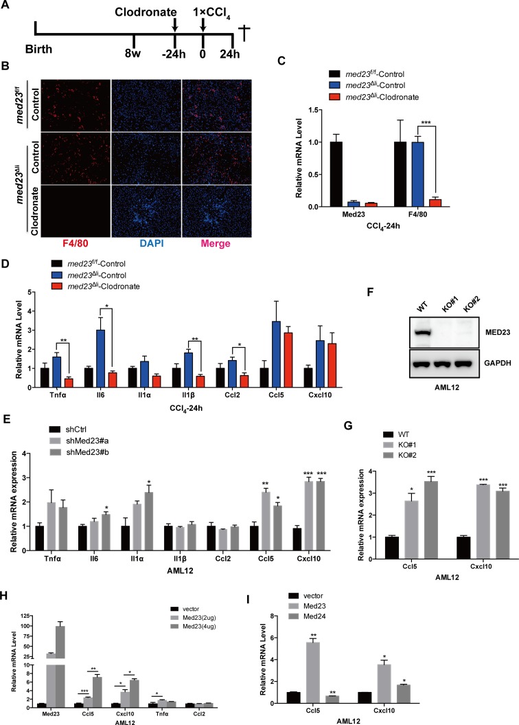
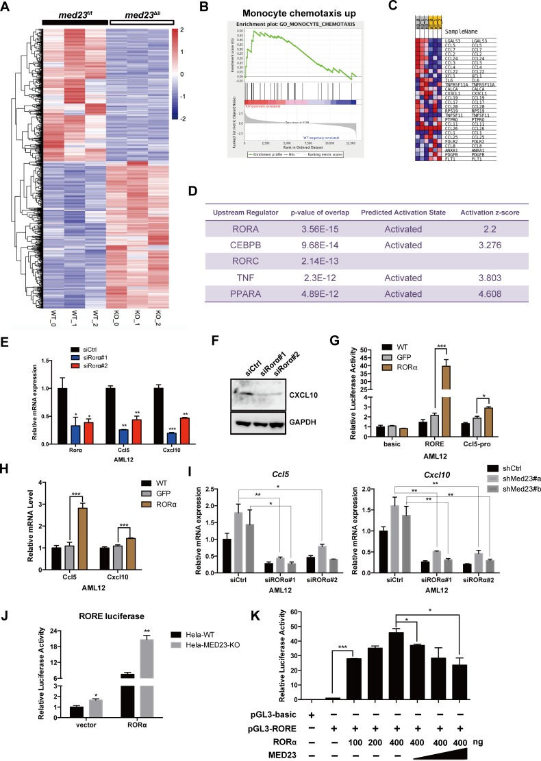
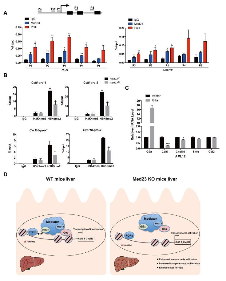
Liver fibrosis, often associated with cirrhosis and hepatocellular carcinomas, is characterized by hepatic damage, an inflammatory response, and hepatic stellate cell (HSC) activation, although the underlying mechanisms are largely unknown. Here, we show that the transcriptional Mediator complex subunit 23 (MED23) participates in the development of experimental liver fibrosis. Compared with their control littermates, mice with hepatic Med23 deletion exhibited aggravated carbon tetrachloride (CCl4)-induced liver fibrosis, with enhanced chemokine production and inflammatory infiltration as well as increased hepatocyte regeneration. Mechanistically, the orphan nuclear receptor RAR-related orphan receptor alpha (RORα) activates the expression of the liver fibrosis-related chemokines C-C motif chemokine ligand 5 (CCL5) and C-X-C motif chemokine ligand 10 (CXCL10), which is suppressed by the Mediator subunit MED23. We further found that the inhibition of Ccl5 and Cxcl10 expression by MED23 likely occurs because of G9a (also known as euchromatic histone-lysine N-methyltransferase 2 [EHMT2])-mediated H3K9 dimethylation of the target promoters. Collectively, these findings reveal hepatic MED23 as a key modulator of chemokine production and inflammatory responses and define the MED23-CCL5/CXCL10 axis as a potential target for clinical intervention in liver fibrosis.


Conflict of interest statement

The authors have declared that no competing interests exist.

Figures

Fig 1
Fig 1. Analysis of liver fibrosis in med23f/f and med23Δli mice after chronic administration of CCl4.
(A) Histology analysis of livers from med23f/f and med23Δli mice. The liver sections of med23f/f and med23Δli mice after chronic CCl4 administration were stained with Sirius red and α-SMA. Representative pictures were shown. (B) Quantification of Sirius red–positive (n = 5 per group) and α-SMA-positive areas (n = 7 per group) in the liver sections of med23f/f and med23Δli mice. (C) The total protein was extracted from whole livers of med23f/f and med23Δli mice and analyzed by western blotting using the indicated antibodies. GAPDH was used as a loading control. (D-F) The total RNA was extracted from whole livers of med23f/f and med23Δli mice and then analyzed by qRT-PCR to detect the expression of the liver fibrosis-associated genes. The expression was normalized to Gapdh (n = 7 per group). Data are presented as means ± SEM. *P < 0.05, **P < 0.01, ***P < 0.001. For underlying data, see S1 Data file. α-SMA, alpha-smooth muscle actin; CCl4, carbon tetrachloride; Col, collagen; GAPDH, glyceraldehyde 3-phosphate dehydrogenase; med23, Mediator complex subunit 23; med23Δli, liver-specific knockout of Med23; med23f/f, med23-floxed; Mmp, matrix metalloproteinase; Pdgfβ, platelet derived growth factor beta; Pdgfrβ, platelet-derived growth factor receptor beta; qRT-PCR, quantitative real-time PCR; Tgfβ1, transforming growth factor beta 1; Tgfβr1, transforming growth factor beta receptor 1; Timp, tissue inhibitor of metalloproteinase.
Fig 2
Fig 2. Reduced liver cell death and enhanced hepatocyte proliferation in med23Δli mice after chronic administration of CCl4.
(A) Representative views of TUNEL staining and α-SMA staining in the liver sections of med23f/f and med23Δli mice. (B) Quantification of TUNEL-positive cells from the liver sections of med23f/f and med23Δli mice (med23f/f, n = 6; med23Δli, n = 7). (C) The expression of antiapoptotic genes (cIAP1 and cIAP2) in whole-liver extracts was analyzed by qRT-PCR (n = 7 per group). (D) Representative views and quantification of PCNA staining in the liver sections of med23f/f and med23Δli mice (n = 7 per group). (E) qRT-PCR analysis of proliferative genes (Hgf, CyclinD1, c-Fos, and c-Jun) in whole-liver extracts of med23f/f and med23Δli mice (n = 7 per group). Data are presented as means ± SEM. *P < 0.05, **P < 0.01. For underlying data, see S1 Data file. α-SMA, alpha-smooth muscle actin; CCl4, carbon tetrachloride; cIAP, cellular inhibitor of apoptosis protein; Hgf, hepatocyte growth factor; med23, Mediator complex subunit 23; med23Δli, liver-specific knockout of Med23; med23f/f, med23-floxed; PCNA, proliferating cell nuclear antigen; qRT-PCR, quantitative real-time PCR.
Fig 3
Fig 3. Increased inflammatory infiltration in med23Δli mice after the chronic administration of CCl4.
(A) Liver sections from med23f/f and med23Δli mice were stained with HE, F4/80, and DAPI respectively. (B) Quantification of F4/80-positive cells from the liver sections of med23f/f and med23Δli mice (med23f/f, n = 5; med23Δli, n = 7). (C) The mRNA expression of immune cells markers (F4/80, Cd45, and Cd3g) in whole-liver extracts was analyzed by qRT-PCR (n = 7 per group). (D-F) qRT-PCR analysis of the expression of cytokines, chemokines, and their receptors in the liver (n = 7 per group). Data are presented as means ± SEM. *P < 0.05, **P < 0.01. For underlying data, see S1 Data file. Ccl, C-C motif chemokine ligand; CCl4, carbon tetrachloride; Ccr, C-C motif chemokine receptor; Cxcl, C-X-C motif chemokine ligand; Cxcr, C-X-C motif chemokine receptor; HE, hematoxylin–eosin; Il, interleukin; med23, Mediator complex subunit 23; med23Δli, liver-specific knockout of Med23; med23f/f, med23-floxed; qRT-PCR, quantitative real-time PCR; Tnfα, tumor necrosis factor alpha.
Fig 4
Fig 4. More proinflammatory cytokine and chemokine secretion after acute administration of CCl4.
(A) Representative views of HE, TUNEL, and DAPI staining in the liver sections of med23f/f and med23Δli mice after acute CCl4 administration for 24 hours. (B) Quantification of TUNEL-positive cells from the liver sections of med23f/f and med23Δli mice (med23f/f, n = 6; med23Δli, n = 5). (C) The total protein was extracted from whole livers of med23f/f and med23Δli mice and analyzed by western blotting using the indicated antibodies. GAPDH was used as a loading control. (D, E) Representative views of Ki67 staining (D) and quantification (E) in the liver sections of med23f/f and med23Δli mice (med23f/f, n = 5; med23Δli, n = 4). (F) qRT-PCR analysis of c-Myc in whole-liver extracts of med23f/f and med23Δli mice (med23f/f, n = 6; med23Δli, n = 5). (G, H) qRT-PCR analysis of cytokines and chemokines in whole-liver extracts of med23f/f and med23Δli mice (med23f/f, n = 6; med23Δli, n = 5). Data are presented as means ± SEM. *P < 0.05, **P < 0.01. For underlying data, see S1 Data file. γ-H2AX, phosphorylation of the histone variant H2AX; Ccl, C-C motif chemokine ligand; CCl4, carbon tetrachloride; Cxcl, C-X-C motif chemokine ligand; GAPDH, glyceraldehyde 3-phosphate dehydrogenase; HE, hematoxylin–eosin; Il, interleukin; med23, Mediator complex subunit 23; med23Δli, liver-specific knockout of Med23; med23f/f, med23-floxed; qRT-PCR, quantitative real-time PCR; Tnfα, tumor necrosis factor alpha.
Fig 5
Fig 5. Increased Ccl5 and Cxcl10 expression directly in hepatocytes with Med23 deletion in vivo and in vitro.
(A) Strategy to delete macrophages in liver and administrate with acute CCl4 for 24 hours. (B) Representative views of F4/80 staining in the liver sections of med23f/f and med23Δli mice after tail intravenous injection of control liposome and clodronate liposome and then CCl4 administration for 24 hours. (C) qRT-PCR analysis of Med23 and F4/80 in whole-liver extracts of med23f/f and med23Δli mice (med23f/f-Control, n = 4; med23Δli-Control, n = 4; med23Δli-Clodronate, n = 7). (D, E) qRT-PCR analysis of proinflammatory cytokines and chemokines in whole-liver extracts of med23f/f and med23Δli mice (med23f/f-Control, n = 4; med23Δli-Control, n = 4; med23Δli-Clodronate, n = 7) (D) and AML12 cells with Med23 knockdown (n = 3 per group) (E). (F) The protein level of MED23 was analyzed by western blotting in AML12 cells after CRISPR/Cas9 mediated Med23 KO. GAPDH was used as a loading control. (G) qRT-PCR analysis of Ccl5 and Cxcl10 in Med23 KO AML12 cells (n = 3 per group). (H, I) qRT-PCR analysis of proinflammatory cytokines and chemokines in AML12 cells after transient transfection (n = 3 per group). Data are presented as means ± SEM. *P < 0.05, **P < 0.01, ***P < 0.001. For underlying data, see S1 Data file. AML12, alpha mouse liver 12; Ccl, C-C motif chemokine ligand; CCl4, carbon tetrachloride; Cxcl, C-X-C motif chemokine ligand; GAPDH, glyceraldehyde 3-phosphate dehydrogenase; Il, interleukin; KO, knockout; MED23, Mediator complex subunit 23; med23Δli, liver-specific knockout of Med23; med23f/f, med23-floxed; qRT-PCR, quantitative real-time PCR; shCtrl, negative control vector containing scrambled shRNA; shMed23, shRNA against Med23; Tnfα, tumor necrosis factor alpha; WT, wild-type.
Fig 6
Fig 6. Hepatocyte MED23 cooperates with RORα to regulate Ccl5 and Cxcl10.
(A) Heatmap analysis of differential expression genes between med23f/f and med23Δli mouse livers after acute administration of CCl4 for 24 hours. We found that 593 genes were down-regulated (>2-fold) and 660 genes were up-regulated (>2-fold) in the med23Δli mice. (B-C) GSEA analysis of up-regulated genes (>2-fold) in med23Δli mice (B) and genes included in the panel B (C). (D) IPA was performed to predict the upstream regulator of up-regulated genes (>2-fold). The top five regulators are listed. (E) qRT-PCR analysis of RORα, Ccl5, and Cxcl10 expression in AML12 cells after RORα knockdown (n = 3 per group). (F) Western blotting analysis of CXCL10 protein expression in AML12 cells after RORα knockdown. GAPDH was used as a loading control. (G) Effect of RORα overexpression on RORE and Ccl5-pro luciferase reporter activity in AML12 cells (n = 3 per group). (H) qRT-PCR analysis of Ccl5 and Cxcl10 expression in AML12 cells after RORα overexpression (n = 4 per group). (I) qRT-PCR analysis of Ccl5 and Cxcl10 expression in shCtrl and shMed23 AML12 cells after RORα knockdown. The expression was normalized to Gapdh (n = 3 per group). (J, K) Effect of MED23 on RORE-luciferase reporter activity in HeLa cell line (n = 3 per group). Data are presented as means ± SEM. *P < 0.05, **P < 0.01, ***P < 0.001. For underlying data, see S1 Data file. AML12, alpha mouse liver 12; Ccl, C-C motif chemokine ligand; CCl4, carbon tetrachloride; CEBPB, CCAAT enhancer binding protein beta; Cxcl, C-X-C motif chemokine ligand; GAPDH, glyceraldehyde 3-phosphate dehydrogenase; GFP, green fluorescent protein; GSEA, gene set enrichment analysis; IPA, Ingenuity Pathway Analysis; KO, knockout; MED23, Mediator complex subunit 23; med23Δli, liver-specific knockout of Med23; med23f/f, med23-floxed; PPARα, peroxisome proliferator activated receptor alpha; qRT-PCR, quantitative real-time PCR; RORα, RAR-related orphan receptor alpha; RORE, RORα response element; shCtrl, negative control vector containing scrambled shRNA; shMed23, shRNA against Med23; siCtrl, negative control vector containing scrambled siRNA; siRORα, siRNA against RORα; TNFα, tumor necrosis factor alpha; WT, wild-type.
Fig 7
Fig 7. Med23 deletion causes decreased H3K9me2 occupancy among the promoter of Ccl5 and Cxcl10.
(A, B) ChIP analysis of MED23 and Pol II occupancy in AML12 cells (n = 3 per group) (A) as well as H3K4me3 and H3K9me2 occupancy in med23f/f and med23Δli mouse livers after acute administration of CCl4 (med23f/f, n = 3; med23Δli, n = 4) (B). IgG was used as a negative control. The precipitated DNA was analyzed by qRT-PCR with primers targeting the promoter regions of Ccl5 and Cxcl10. The relative binding level of each factor was calculated by normalization to the input DNA. (C) qRT-PCR analysis of proinflammatory cytokines and chemokines in AML12 cells after G9a transient transfection (n = 3 per group). (D) A proposed model for the role of MED23 in hepatocyte after CCl4 challenge. MED23 modulates RORα transcriptional activity possibly via G9a-mediated H3K9me2 modification to the target promoters for transcriptional repression. Data are presented as means ± SEM. *P < 0.05, **P < 0.01, ***P < 0.001. For underlying data, see S1 Data file. AML12, alpha mouse liver 12; Ccl, C-C motif chemokine ligand; CCl4, carbon tetrachloride; ChIP, chromatin immunoprecipitation; Cxcl, C-X-C motif chemokine ligand; IgG, immunoglobulin G; KO, knockout; MED23, Mediator complex subunit 23; med23Δli, liver-specific knockout of Med23; med23f/f, med23-floxed; Pol II, RNA polymerase II; qRT-PCR, quantitative real-time PCR; RORα, RAR-related orphan receptor alpha; WT, wild-type.

References

    1. Bataller R, Brenner DA. Liver fibrosis. The Journal of clinical investigation. 2005;115(2):209–18. 10.1172/JCI24282 - DOI - PMC - PubMed
    1. Pellicoro A, Ramachandran P, Iredale JP, Fallowfield JA. Liver fibrosis and repair: immune regulation of wound healing in a solid organ. Nat Rev Immunol. 2014;14(3):181–94. 10.1038/nri3623 - DOI - PubMed
    1. Cordero-Espinoza L, Huch M. The balancing act of the liver: tissue regeneration versus fibrosis. The Journal of clinical investigation. 2018;128(1):85–96. 10.1172/JCI93562 - DOI - PMC - PubMed
    1. Schuppan D, Kim YO. Evolving therapies for liver fibrosis. Journal of Clinical Investigation. 2013;123(5):1887–901. 10.1172/JCI66028 PubMed PMID: WOS:000319025100008. - DOI - PMC - PubMed
    1. Seki E, De Minicis S, Osterreicher CH, Kluwe J, Osawa Y, Brenner DA, et al. TLR4 enhances TGF-beta signaling and hepatic fibrosis. Nature medicine. 2007;13(11):1324–32. 10.1038/nm1663 - DOI - PubMed

Publication types