. 2019 Dec 5;14(12):e0226001.
doi: 10.1371/journal.pone.0226001. eCollection 2019.

In vivo ultrasound thermal ablation control using echo decorrelation imaging in rabbit liver and VX2 tumor

Affiliations

In vivo ultrasound thermal ablation control using echo decorrelation imaging in rabbit liver and VX2 tumor

Mohamed A Abbass et al. PLoS One. .

Abstract

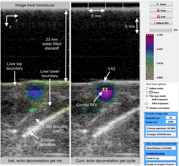
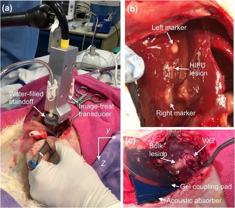
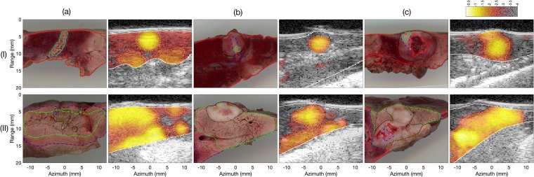
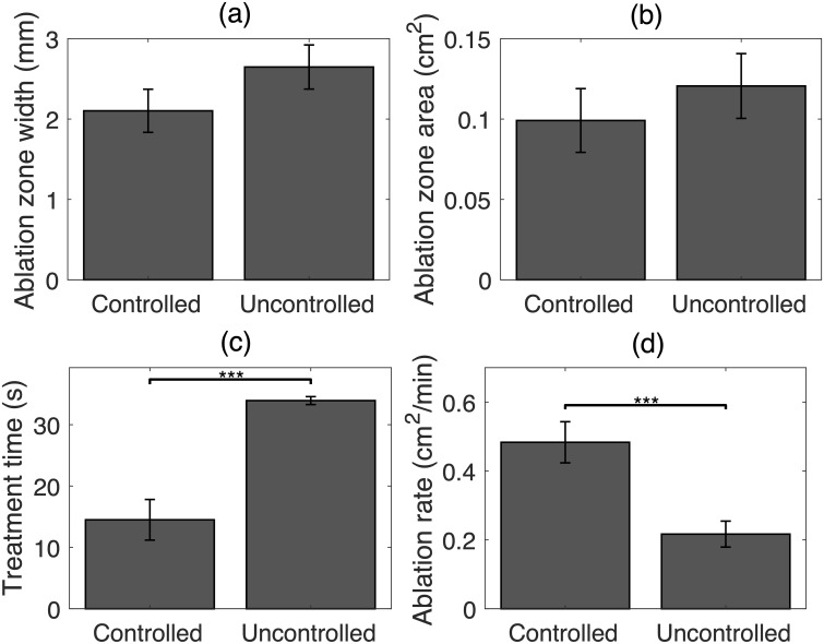
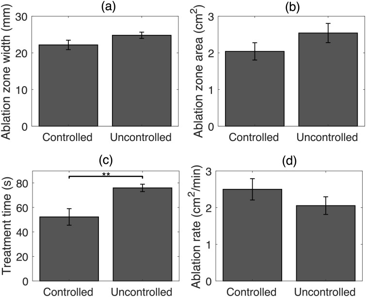
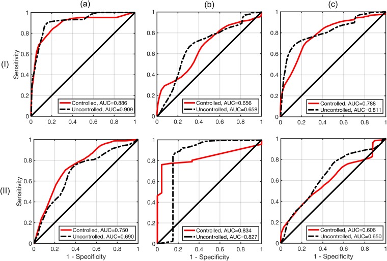
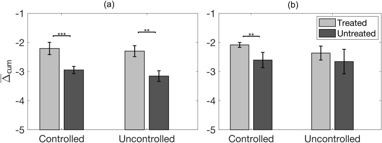
The utility of echo decorrelation imaging feedback for real-time control of in vivo ultrasound thermal ablation was assessed in rabbit liver with VX2 tumor. High-intensity focused ultrasound (HIFU) and unfocused (bulk) ablation were performed using 5 MHz linear image-ablate arrays. Treatments comprised up to nine lower-power sonications, followed by up to nine higher-power sonications, ceasing when the average cumulative echo decorrelation within a control region of interest exceeded a predefined threshold (- 2.3, log10-scaled echo decorrelation per millisecond, corresponding to 90% specificity for tumor ablation prediction in previous in vivo experiments). This threshold was exceeded in all cases for both HIFU (N = 12) and bulk (N = 8) ablation. Controlled HIFU trials achieved a significantly higher average ablation rate compared to comparable ablation trials without image-based control, reported previously. Both controlled HIFU and bulk ablation trials required significantly less treatment time than these previous uncontrolled trials. Prediction of local liver and VX2 tumor ablation using echo decorrelation was tested using receiver operator characteristic curve analysis, showing prediction capability statistically equivalent to uncontrolled trials. Compared to uncontrolled trials, controlled trials resulted in smaller thermal ablation regions and higher contrast between echo decorrelation in treated vs. untreated regions. These results indicate that control using echo decorrelation imaging may reduce treatment duration and increase treatment reliability for in vivo thermal ablation.


Conflict of interest statement

One author (PGB) was employed by Guided Therapy Systems/Ardent Sound, a company that develops and markets therapeutic ultrasound products. The research materials provided for this study are prototypes incorporating technology patented by Guided Therapy Systems/Ardent Sound, but do not comprise a commercial product, either marketed or under development. This affiliation does not alter our adherence to PLOS ONE policies on sharing data and materials.

Figures

Fig 1
Fig 1. Graphical user interface of the C++ application used for in vivo US thermal ablation imaging and control.
Left: instantaneous hybrid B-mode/echo decorrelation image. Right: cumulative echo decorrelation map for each therapy cycle, corrected in real time using decorrelation from sham ablation cycles; the control region of interest is bounded by a yellow line.
Fig 2
Fig 2. Experimental setup.
(A) Image-treat array placed on the rabbit liver capsule during open surgery. (B) HIFU thermal ablation of liver followed by left and right marking exposures. (C) Bulk thermal ablation of VX2 tumor.
Fig 3
Fig 3. Histologic and hybrid B-mode/echo decorrelation for HIFU and bulk US controlled trials.
In the tissue sections, the red, black, blue, and green boundaries indicate the segmented tissue, tumor, treated, and fully ablated regions. In the US images, the white line indicates segmented tissue boundaries and the yellow dashed line represents the optimum prediction threshold for local tissue ablation for all US exposures of both groups.
Fig 4
Fig 4. Statistical analysis of ablation results in rabbit liver and VX2 tumor for controlled and uncontrolled HIFU trials.
Means and standard errors of (A) ablation zone width, (B) ablation zone area, (C) treatment time, and (D) ablation rate. (*** p < 10−3).
Fig 5
Fig 5. Statistical analysis of ablation results for controlled and uncontrolled bulk US trials.
Means and standard errors of (A) ablation zone width, (B) ablation zone area, (C) treatment time, and (D) ablation rate. (** p < 10−2).
Fig 6
Fig 6. Assessment of prediction capability for echo decorrelation imaging.
Receiver operating characteristic curves for echo decorrelation prediction of treatment in rabbit liver (I) and VX2 tumor (II) for HIFU (A), bulk US (B), and all exposures combined (C) for both controlled and uncontrolled HIFU experiments.
Fig 7
Fig 7. Statistics of cumulative echo decorrelation in treated and untreated tissue.
(A) Means and standard errors of log10-scaled cumulative decorrelation per ms in treated and untreated rabbit liver for controlled vs. uncontrolled HIFU and bulk US exposures combined. (B) Corresponding statistics for treated and untreated VX2 tumor. (** p < 10−2 and *** p < 10−3).

References

    1. Forner A, Reig M, Bruix J. Hepatocellular carcinoma. The Lancet. 2018;391(10127):1301–1314. 10.1016/S0140-6736(18)30010-2 - DOI - PubMed
    1. Sherman M. Hepatocellular carcinoma: epidemiology, surveillance, and diagnosis. Semin Liver Dis. 2010;30(01):3–16. 10.1055/s-0030-1247128 - DOI - PubMed
    1. Ardito F, Vellone M, Cassano A, De Rose AM, Pozzo C, Coppola A, et al. Chance of cure following liver resection for initially unresectable colorectal metastases: analysis of actual 5-year survival. J Gastrointest Surg. 2013;17(2):352–359. 10.1007/s11605-012-2103-3 - DOI - PubMed
    1. Forner A, Gilabert M, Bruix J, Raoul JL. Treatment of intermediate-stage hepatocellular carcinoma. Nat Rev Clin Oncol. 2014;11:525–535. 10.1038/nrclinonc.2014.122 - DOI - PubMed
    1. Delis SG, Dervenis C. Selection criteria for liver resection in patients with hepatocellular carcinoma and chronic liver disease. World J Gastroenterol. 2008;14(22):3452–3460. 10.3748/wjg.14.3452 - DOI - PMC - PubMed

Publication types