. 2019 Dec 1;8(4):279.
doi: 10.3390/pathogens8040279.

Generation and Immunogenicity of a Recombinant Pseudorabies Virus Co-Expressing Classical Swine Fever Virus E2 Protein and Porcine Circovirus Type 2 Capsid Protein Based on Fosmid Library Platform

Affiliations

Generation and Immunogenicity of a Recombinant Pseudorabies Virus Co-Expressing Classical Swine Fever Virus E2 Protein and Porcine Circovirus Type 2 Capsid Protein Based on Fosmid Library Platform

Muhammad Abid et al. Pathogens. .

Abstract

Pseudorabies (PR), classical swine fever (CSF), and porcine circovirus type 2 (PCV2)-associated disease (PCVAD) are economically important infectious diseases of pigs. Co-infections of these diseases often occur in the field, posing significant threat to the swine industry worldwide. gE/gI/TK-gene-deleted vaccines are safe and capable of providing full protection against PR. Classical swine fever virus (CSFV) E2 glycoprotein is mainly used in the development of CSF vaccines. PCV2 capsid (Cap) protein is the major antigen targeted for developing PCV2 subunit vaccines. Multivalent vaccines, and especially virus-vectored vaccines expressing foreign proteins, are attractive strategies to fight co-infections for various swine diseases. The gene-deleted pseudorabies virus (PRV) can be used to develop promising and economical multivalent live virus-vectored vaccines. Herein, we constructed a gE/gI/TK-gene-deleted PRV co-expressing E2 of CSFV and Cap of PCV2 by fosmid library platform established for PRV, and the expression of E2 and Cap proteins was confirmed using immunofluorescence assay and western blotting. The recombinant virus propagated in porcine kidney 15 (PK-15) cells for 20 passages was genetically stable. The evaluation results in rabbits and pigs demonstrate that rPRVTJ-delgE/gI/TK-E2-Cap elicited detectable anti-PRV antibodies, but not anti-PCV2 or anti-CSFV antibodies. These findings provide insights that rPRVTJ-delgE/gI/TK-E2-Cap needs to be optimally engineered as a promising trivalent vaccine candidate against PRV, PCV2 and CSFV co-infections in future.

Keywords: E2 and Cap; classical swine fever virus; fosmid; porcine circovirus type 2; pseudorabies virus.


Conflict of interest statement

Authors declare no conflicts of interest.

Figures

Figure 1
Figure 1
Rescue and characterization of the recombinant virus rPRVTJ-delgE/gI/TK-E2-Cap. A. Cytopathic effects (CPEs) observed on Vero or porcine kidney 15 (PK-15) cells. Vero cells were transfected with different combinations fosmids, positive control (parental virus PRV TJ strain) and negative control (missing one fosmid). At 72-96 hpt, CPEs were visualized and supernatants were collected. PK-15 cells were infected with the collected supernatants and the CPEs were visualized at 24 hpi. B. Electron microscopy. Transmission electron microscopy of the rPRVTJ-delgE/gI/TK-E2-Cap and rPRVTJ-delgE/gI/TK viral particles, the parental virus PRVTJ was used as positive control. Scale bars are presented. C. PCR analysis of deleted/inserted genes. PCR analysis of deleted thymidine kinase (TK), gE/gI genes, and E2 and Cap genes inserted in the DNA of rPRVTJ-delgE/gI/TK-E2-Cap using specific primers targeting TK, gE/gI, US9 (E2 gene) and US4 (Cap gene) regions, respectively. DNA of PRVTJ was used as the negative control and non-DNA templates were used as mock.
Figure 2
Figure 2
Confirmation of E2 and Cap expression. A. Confirmation of E2 and Cap expression by immunofluorescence assay (IFA). The E2 and Cap proteins expression was verified by IFA in PK-15 cells infected with rPRVTJ-delgE/gI/TK-E2-Cap. Briefly, PK-15 cells were infected with classical swine fever virus (CSFV), porcine circovirus type 2 (PCV2) rPRVTJ-delgE/gI/TK-E2-Cap and PRV TJ at a multiplication of infectivity (MOI) of 1 or left un-infected (mock). At 48 hpi, the cells were fixed and analyzed by IFA using the anti-E2 MAb HQ06 and anti-Cap MAb 36A9 primary antibodies and fluorescein isothiocyanate (FITC)-labeled anti-mouse secondary antibody. Bars, 400 nm. The results were visualized under a fluorescent microscope (TE2000-U Nikon, Japan). B. Expression of E2 and Cap by Western blotting. To confirm E2 and Cap expression, PK-15 cells were either infected with the CSFV, rPRVTJ-delgE/gI/TK-E2-Cap, rPRVTJdelgE/gI/TK-Cap, rPRVTJ-delgE/gI/TK or mock infected. At 48 hpi, the cells were lysed and assayed by Western blotting using the anti-E2 MAb HQ06 and anti-Cap MAb 36A9 (1:300) in PBS, subsequently incubated with IRDye 800CW-labeled goat anti-mouse secondary antibody (Li-Cor). GAPDH was used as a loading control. The arrows indicate the representative bands of the CSFV E2 protein and PCV2 Cap fused with the PRV gG.
Figure 3
Figure 3
The replication kinetics and stability of recombinant viruses. A. One-step growth curve. PK-15 cells were infected with the rPRVTJ-delgE/gI/TK-E2-Cap, PRVTJ-delgE/gI/TK or PRVTJ at an MOI of 10. The cell culture supernatants were harvested at various intervals and used to determine viral titers. B. Plaque assay. The rPRVTJ-delgE/gI/TK-E2-Cap, rPRVTJ-delgE/gI/TK and PRVTJ were diluted 10-fold by serial dilution to achieve a titer required to form single plaques. The experiments were conducted in three repeats, and the corresponding results are presented. C. PCR identification. The recombinant virus propagated in PK-15 cells for 20 passages. The genome of the rPRVTJ-delgE/gI/TK-E2-Cap (F5, F10, F15 and F20) as a template for the amplification of ΔTK, ΔgE/gI, E2 and Cap genes by PCR. The PRVTJ genome was used as a negative control and non-DNA templates were used as mock. D. E2 and Cap expression by IFA. Expression of E2 and Cap proteins in PK-15 cells infected with the recombinant virus rPRVTJ-delgE/gI/TK-E2-Cap (F5, F10, F15 and F20) was confirmed by IFA using anti-E2 MAb HQ06 and anti-Cap MAb 36A9 as primary antibodies and FITC-labeled anti-mouse immunoglobulin G (IgG)as secondary antibody. Bars, 400 nm.
Figure 4
Figure 4
Gross pathological examination of various organs of immunized rabbits. Groups of rabbits (n = 5) were vaccinated with different doses (107, 106 and 105 TCID50) of rPRVTJ-delgE/gI/TK-E2-Cap, 105 TCID50 of rPRVTJ-delgE/gI/TK, or DMEM. At six weeks post-immunization, all the immunized rabbits were euthanized and various organs, that is, lungs, liver, kidneys, heart and spleen were collected for gross pathological examination.
Figure 5
Figure 5
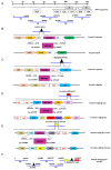
Construction of recombinant fosmids with TK/gE/gI deletions, and E2 and Cap gene insertions. A. The genomic organization of PRV, and the five fosmids generated from the PRV-TJ strain used for virus rescue. Numbers representing the fosmids names and the position of each fosmid fragment in the genome of PRV-TJ strain. B. Schematic diagram representing modification procedure for the intermediate fosmid and Fosmid-f-ΔTK. For fosmid modification, the rpsL-neo counter-selection marker cassette (rpsL-neo) harboring 50 bp left and right oligonucleotide homologous arms was inserted between 59144 and 59421 bp of PRV-TJ genome by the Red/ET-mediated recombination. Partially deleted TK (ΔTK) gene fragment with flanking homologous arms was used for replacing the rpsL-neo counter-selection marker to construct Fosmid-f-ΔTK. C. Schematic diagram representing modification procedure of the intermediate fosmid and Fosmid-s-ΔgE/gI. Partially deleted gE/gI (ΔgE/gI) genes fragment with 50 bp left and right oligonucleotide homologous arms was inserted between 122765 and 125120 bp of PRV-TJ genome as described above to generate Fosmid-f-ΔgE/gI. D. The schematic diagram representing modification procedure of the intermediate fosmid and the Fosmid-s-ΔgE/gI-US9E2. The E2 expression cassette harboring 50 bp left and right oligonucleotide homologous arms inserted between 125794 and 125795 bp (US9) of PRV-TJ genome. E. The schematic diagram representing modification procedure of the intermediate fosmid and Fosmid-s-ΔgE/gI-US9E2-US4Cap. The Cap gene flanked by 50 bp left and right oligonucleotide homologous arms was fused between the last amino acid and the stop codon of gG (US) i.e., 121084 and 121085 bp of the PRVTJ genome. F. Fosmid-f-ΔTK, s-ΔgE/gI-US9E2-US4Cap and other fosmids used to rescue rPRVTJ-delgE/gI/TK-E2-Cap.

References

    1. Mettenleiter T.C. Aujeszky’s disease (pseudorabies) virus: The virus and molecular pathogenesis-state of the art, June 1999. Vet. Res. 2000;31:99–115. doi: 10.1051/vetres:2000110. - DOI - PubMed
    1. Pomeranz L.E., Reynolds A.E., Hengartner C.J. Molecular biology of pseudorabies virus: Impact on neurovirology and veterinary medicine. Microbiol. Mol. Biol. Rev. 2005;69:462–500. doi: 10.1128/MMBR.69.3.462-500.2005. - DOI - PMC - PubMed
    1. An T.Q., Peng J.M., Tian Z.J., Zhao H.Y., Li N., Liu Y.M., Chen J.Z., Leng C.L., Sun Y., Chang D. Pseudorabies virus variant in Bartha-K61–vaccinated pigs, china, 2012. Emerg. Infect. Dis. 2013;19:1749. doi: 10.3201/eid1911.130177. - DOI - PMC - PubMed
    1. Freuling C., Müller T., Mettenleiter T. Vaccines against pseudorabies virus (PrV) Vet. Microbiol. 2017;206:3–9. doi: 10.1016/j.vetmic.2016.11.019. - DOI - PubMed
    1. Chen Y., Guo W., Xu Z., Yan Q., Luo Y., Shi Q., Chen D., Zhu L., Wang X. A novel recombinant pseudorabies virus expressing parvovirus VP2 gene: Immunogenicity and protective efficacy in swine. Virol. J. 2011;8:307. doi: 10.1186/1743-422X-8-307. - DOI - PMC - PubMed