. 2019 Dec 1;9(12):1058.
doi: 10.3390/ani9121058.

Anatomic Study of the Elbow Joint in a Bengal Tiger (Panthera tigris tigris) Using Magnetic Resonance Imaging and Gross Dissections

Affiliations

Anatomic Study of the Elbow Joint in a Bengal Tiger (Panthera tigris tigris) Using Magnetic Resonance Imaging and Gross Dissections

Mario Encinoso et al. Animals (Basel). .

Abstract

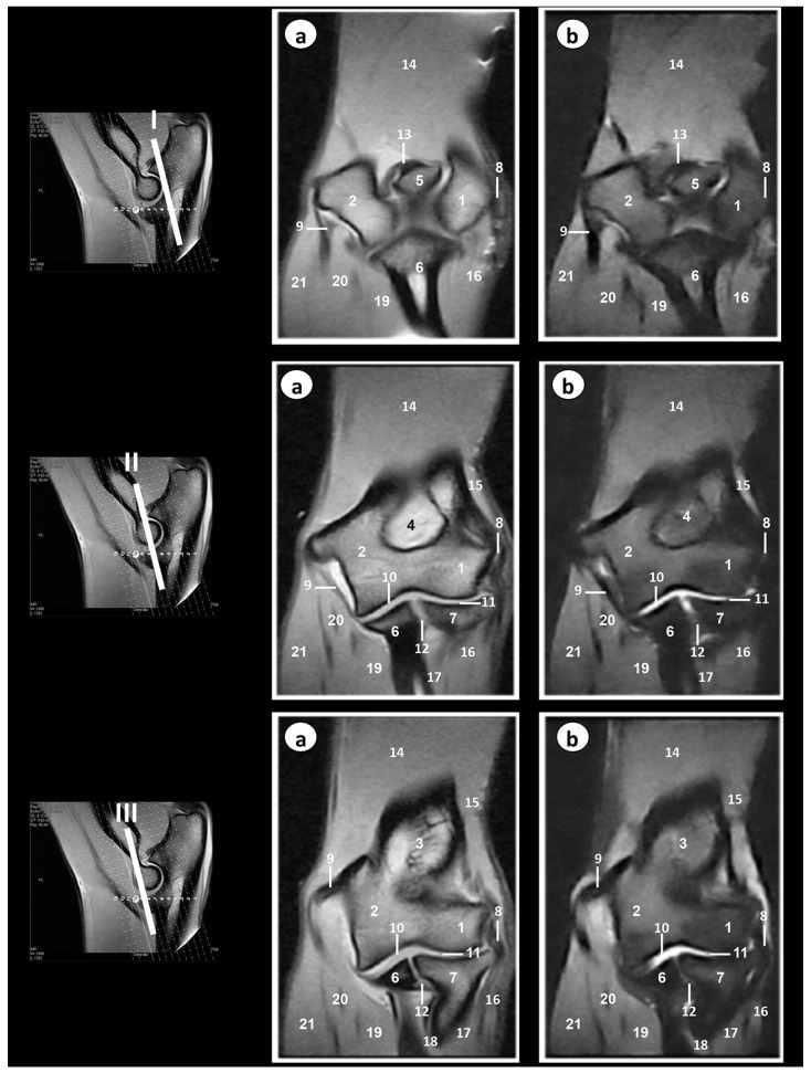
The objective of our research was to describe the normal appearance of the bony and soft tissue structures of the elbow joint in a cadaver of a male mature Bengal tiger (Panthera tigris tigris) scanned via MRI. Using a 0.2 Tesla magnet, Spin-echo (SE) T1-weighting, and Gradient-echo short tau inversion recovery (GE-STIR), T2-weighting pulse sequences were selected to generate sagittal, transverse, and dorsal planes. In addition, gross dissections of the forelimb and its elbow joint were made. On anatomic dissections, all bony, articular, and muscular structures could be identified. The MRI images allowed us to observe the bony and many soft tissues of the tiger elbow joint. The SE T1-weighted MR images provided good anatomic detail of this joint, whereas the GE-STIR T2-weighted MR pulse sequence was best for synovial cavities. Detailed information is provided that may be used as initial anatomic reference for interpretation of MR images of the Bengal tiger (Panthera tigris tigris) elbow joint and in the diagnosis of disorders of this region.

Keywords: Bengal tiger (Panthera tigris tigris); anatomy; elbow joint; magnetic resonance imaging.


Conflict of interest statement

The authors declare no conflict of interest.

Figures

Figure 1
Figure 1
Dissection images of the Bengal tiger forelimb. (a) Lateral aspect and (b) medial aspect. 1. Tensor fasciae antebrachii muscle; 2. Triceps brachii muscle (long head); 3. Triceps brachii muscle (lateral head); 4. Triceps brachii muscle (accessory head); 5. Olecranon tuberosity; 6. Brachialis muscle; 7. Biceps brachii muscle; 8. Anconeus muscle; 9. Extensor carpi radialis muscle; 10. Extensor digitorum communis muscle; 11. Extensor digitorum lateralis muscle; 12. Abductor digiti I longus muscle; 13. Extensor carpi ulnaris muscle; 14. Flexor carpi ulnaris muscle (humeral head); 15. Flexor carpi ulnaris muscle (cubital head); 16. Flexor digitorum superficialis muscle; 17. Flexor digitorum profundus muscle; 18. Flexor carpi radialis muscle; 19. Pronator teres muscle; 20. Brachial vein; 21. Brachial artery; 22. Median artery; 23. Radial artery; 24. Radial nerve; 25. Cubital nerve; 26. Median nerve.
Figure 2
Figure 2
Dissection images of the Bengal tiger right elbow joint. (a) Lateral aspect, (b) medial aspect, (c) cranial aspect, and (d) proximal aspect. 1. Humerus (body); 2. Humerus (metaphysis); 3. Humerus (lateral condyle); 4. Humerus (medial condyle); 5. Humerus (lateral epicondyle); 6. Humerus (medial epicondyle); 7. Olecranon fossa; 8. Olecranon tuberosity; 9. Ulna (body); 10. Radius (head); 11. Radius (body); 12. Lateral collateral ligament; 13. Medial collateral ligament; 14. Radial annular ligament; 15. Olecranon ligament; 16. Interosseoum antebrachii ligament; 17. Membrana interossea antebrachia; 18. Supracondylar foramen.
Figure 3
Figure 3
Sagittal MR images of the right elbow joint at the level of the lateral epicondyle of humerus (level I), lateral part of olecranon fossa (level II), medial part of olecranon fossa (level III), and medial epicondyle of humerus (level IV). The lines depict the level of section. The images are oriented so that the left is caudal and the right is cranial. (a) SE T1-weighted MR images and (b) GE-STIR T2-weighted images. 1. Humerus (body); 2. Humerus (metaphysis); 3. Humerus (lateral epicondyle); 4. Humerus (medial epicondyle); 5. Radius (head); 6. radius (body); 7. Ulna (olecranon tuberosity); 8. Ulna (anconeus process); 9. Ulna (body); 10. Humeroradial joint (articular cavity); 11. Humeroulnar joint (articular cavity); 12. Proximal radioulnar joint (articular cavity); 13. Radial annular ligament; 14. Olecranon ligament; 15. Triceps brachii muscle; 16. Biceps brachii muscle; 17. Brachialis muscle; 18. Extensor digitorum communis; 19. Extensor carpi radialis muscle; 20. Flexor carpi ulnaris muscle.
Figure 4
Figure 4
Transverse MR images of the right elbow joint at the level of olecranon tuberosity (level I), epicondyles of humerus (level II), and proximal radioulnar joint (level III). The lines depict the level of section. The images are oriented so that the left is medial, and the right is lateral. (a) SE T1-weighted MR images and (b) GE-STIR T2-weighted images. 1. Humerus (lateral epicondyle); 2. Humerus (medial epicondyle); 3. Ulna (olecranon tuberosity); 4. Ulna (body); 5. Radius head; 6. Humeroulnar joint (articular cavity); 7. Proximal radioulnar joint (articular cavity); 8. Lateral collateral ligament; 9. Medial collateral ligament; 10. Olecranon ligament; 11. Radial annular ligament; 12. Anconeus muscle; 13. Extensor carpi ulnaris muscle; 14. Extensor digitorum lateralis muscle; 15. Extensor digitorum communis muscle; 16. Extensor carpi radialis muscle; 17. Brachialis muscle; 18. Biceps brachii muscle; 19. Flexor digitorum superficialis muscle; 20. Flexor carpi radialis muscle; 21. Flexor digitorum profundus muscle; 22. fFexor carpi ulnaris muscle (humeral head); 23. Flexor carpi ulnaris muscle (ulnar head).
Figure 5
Figure 5
Dorsal MR images of the right elbow joint at the level of anconeus processus of ulna (level I), olecranon fossa (level II), and head of radius (level III). The lines depict the level of section. The images are oriented so that the right side of the image is lateral and the top is proximal. (a) SE T1-weighted MR images and (b) GE-STIR T2-weighted images. 1. Humerus (lateral epicondyle); 2. Humerus (medial epicondyle); 3. Humerus (metaphysis); 4. Olecranon fossa; 5. Anconeus process; 6. Ulna (body); 7. Radius (head); 8. Lateral collateral ligament; 9. Medial collateral ligament; 10. Humeroulnar joint (articular cavity); 11. Humerorradial joint (articular cavity); 12. Proximal radioulnar joint (articular cavity); 13. Olecranon ligament; 14. Triceps brachii muscle; 15. Anconeus muscle; 16. Extensor digitorum lateralis muscle; 17. Extensor digitorum communis muscle; 18. Extensor carpi ulnaris muscle; 19. Flexor digitorum profundus muscle; 20. Flexor carpi ulnaris muscle; 21. Flexor digitorum superficialis muscle.

References

    1. Bredella M.A., Tirman P.F., Fritz R.C., Feller J.F., Wischer T.K., Genant H.K. MR imaging findings of lateral ulnar collateral ligament abnormalities in patients with lateral epicondylitis. Am. J. Roentgenol. 1999;173:1379–1382. doi: 10.2214/ajr.173.5.10541124. - DOI - PubMed
    1. Kijowski R., Tuite M., Sanford M. Magnetic resonance imaging of the elbow. Part I: Normal anatomy, imaging technique, and osseous abnormalities. Skelet. Radiol. 2004;33:685–697. doi: 10.1007/s00256-004-0853-z. - DOI - PubMed
    1. Sampath S.C., Sampath S.C., Bredella M.A. Magnetic resonance imaging of the elbow: A structured approach. Sports Health. 2013;5:34–39. doi: 10.1177/1941738112467941. - DOI - PMC - PubMed
    1. Tnibar M.A., Auer J.A., Bakkali S. Ultrasonography of the equine elbow technique and normal appearance. J. Equine Vet. Sci. 2001;21:177–187. doi: 10.1016/S0737-0806(01)70120-7. - DOI - PubMed
    1. Snaps F.R., Saunders J.H., Park R.D., Daenen B., Balligand M.H., Dondelinger R.F. Comparison of spin echo, gradient echo and fat saturation magnetic resonance imaging sequences for imaging the canine elbow. Vet. Radiol. Ultrasound. 1998;39:518–523. doi: 10.1111/j.1740-8261.1998.tb01642.x. - DOI - PubMed

LinkOut - more resources