

Changes in specific serum biomarkers during the induction of prostatic hyperplasia in dogs
- PMID: 31805935
- PMCID: PMC6896469
- DOI: 10.1186/s12917-019-2201-5
Changes in specific serum biomarkers during the induction of prostatic hyperplasia in dogs
Abstract
Background: Prostatic hyperplasia (PH) is one of the most important disorders in intact dogs. In this study, we aimed to induce PH experimentally using the combination of testosterone and estrogen and evaluate important factors associated with this disease.
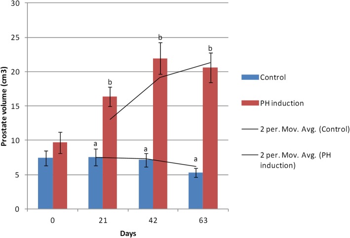
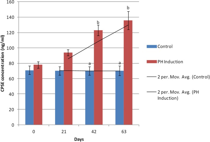
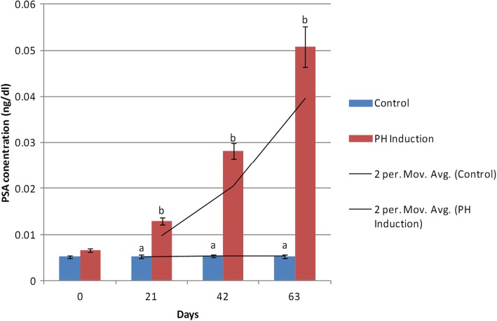
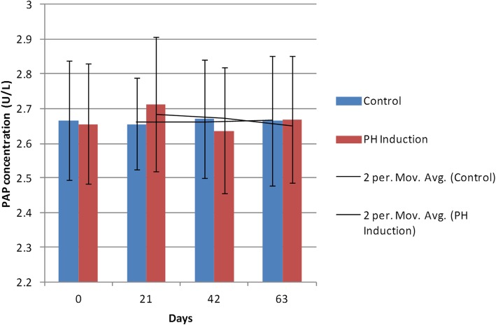
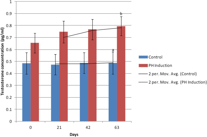
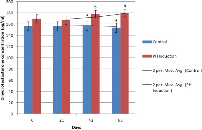
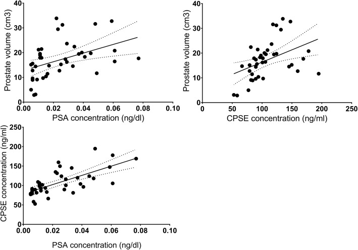
Results: The results showed that in the induction group, prostate volume and prostate specific antigen (PSA) concentration increased significantly on day 21 onwards compared to those of the control group. Canine prostatic specific esterase (CPSE) and dihydrotestosterone (DHT) concentrations increased significantly on day 42 onwards while the testosterone levels increased on day 63. In addition, prostatic acid phosphatase (PAP) concentration did not change significantly in the control and induction groups. Biochemistry profiles and hematologic factors were measured for monitoring the function of liver and kidney, and there were no adverse effects following the induction of PH.
Conclusions: It seems that testosterone and estrogen administration led to prostatic hyperplasia during 2 months. Investigating the size of the prostate, accompanied by prostate markers including CPSE, PSA, DHT, and testosterone, is helpful for the PH diagnosis. However, further studies should be carried out on PAP.
Keywords: Canine prostate specific esterase; Dog; Estrogen; Prostate specific antigen; Prostatic hyperplasia; Testosterone.
Conflict of interest statement
We would like to confirm that there are no known conflicts of interest associated with this publication.
Figures
References
-
- Atalan G, Holt P, Barr F, Brown P. Ultrasonographic estimation of prostatic size in canine cadavers. Res Vet Sci. 1999;67(1):7–15. - PubMed
-
- Berry SJ, Strandberg JD, Coffey DS, Saunders WJ. Development of canine benign prostatic hyperplasia with age. Prostate. 1986;9(4):363–373. - PubMed
-
- Barsanti J, Prasse K, Crowell W, Shotts E, Finco D. Evaluation of various techniques for diagnosis of chronic bacterial prostatitis in the dog [Escherichia coli] J Am Vet Med A. 1983;183(2):219–224. - PubMed
-
- Krawiec D, Heflin D. Study of prostatic disease in dogs: 177 cases (1981-1986) J Am Vet Med A. 1992;200(8):1809–1819. - PubMed
-
- Paclikova K, Kohout P, Vlasin M. Diagnostic possibilities in the management of canine prostatic disorders. Vet Med Praha. 2006;51(1):1.
MeSH terms
Substances
Grants and funding
LinkOut - more resources
Full Text Sources
Medical
Research Materials
Miscellaneous
