. 2020 Jan 8;147(1):dev181099.
doi: 10.1242/dev.181099.

Aurora B functions at the apical surface after specialized cytokinesis during morphogenesis in C. elegans

Affiliations

Aurora B functions at the apical surface after specialized cytokinesis during morphogenesis in C. elegans

Xiaofei Bai et al. Development. .

Abstract

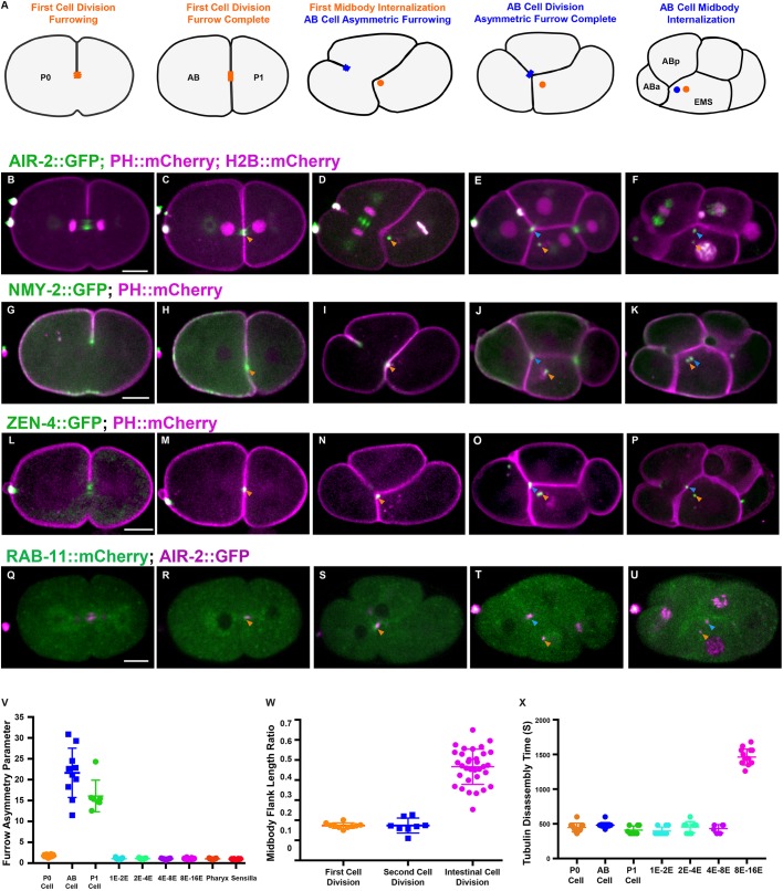
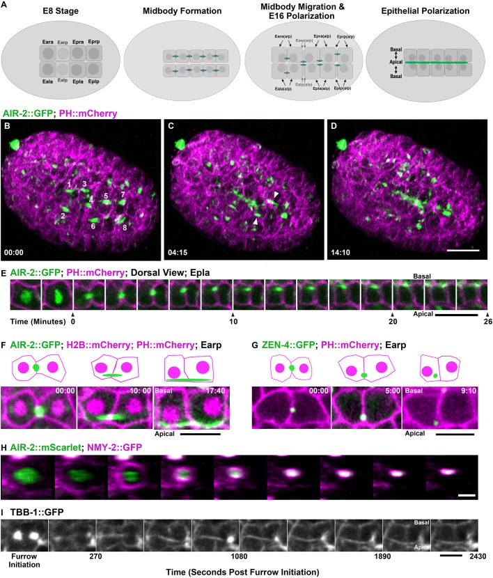
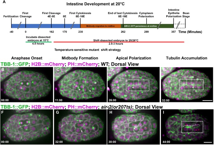
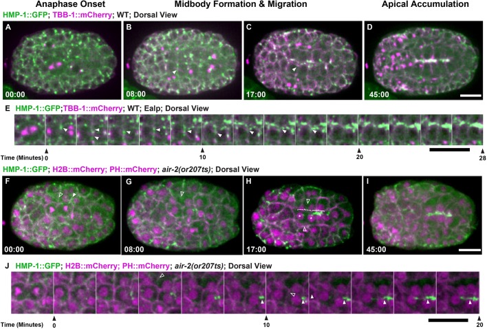
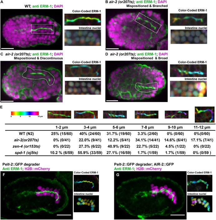
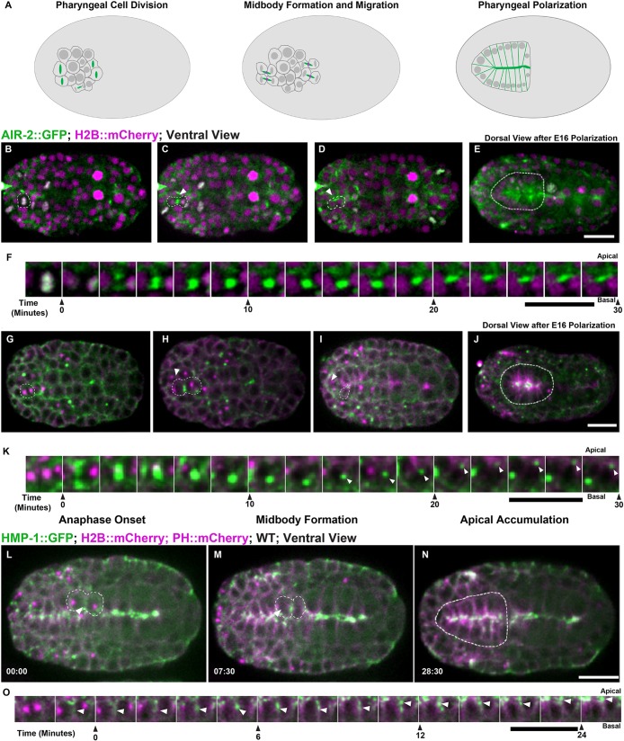
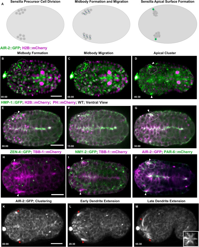
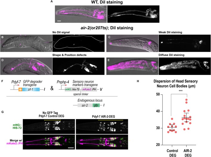
Although cytokinesis has been intensely studied, the way it is executed during development is not well understood, despite a long-standing appreciation that various aspects of cytokinesis vary across cell and tissue types. To address this, we investigated cytokinesis during the invariant Caenorhabditis elegans embryonic divisions and found several parameters that are altered at different stages in a reproducible manner. During early divisions, furrow ingression asymmetry and midbody inheritance is consistent, suggesting specific regulation of these events. During morphogenesis, we found several unexpected alterations to cytokinesis, including apical midbody migration in polarizing epithelial cells of the gut, pharynx and sensory neurons. Aurora B kinase, which is essential for several aspects of cytokinesis, remains apically localized in each of these tissues after internalization of midbody ring components. Aurora B inactivation disrupts cytokinesis and causes defects in apical structures, even if inactivated post-mitotically. Therefore, we demonstrate that cytokinesis is implemented in a specialized way during epithelial polarization and that Aurora B has a role in the formation of the apical surface.

Keywords: Apical surface; Aurora B kinase; Cytokinesis; Midbody; Morphogenesis.


Conflict of interest statement

Competing interestsThe authors declare no competing or financial interests.

Figures

Fig. 1.
Fig. 1.
Cytokinesis in the first two mitotic divisions. (A) Illustration of cytokinesis in the first two mitotic divisions. The first midbody is shown in orange, the AB midbody is blue. (B-F) Cytokinesis labeled with AIR-2::GFP (green; PH::mCherry and H2B::mCherry in magenta). AIR-2 localizes on the central spindle (B) and the midbody flank (C, orange arrowhead) and the MBR after internalization in AB (D, orange arrowhead). In the AB division, the asymmetric furrow pushes the midzone against EMS (E, blue arrowhead), which engulfs it (F, blue arrowhead). (G-K) NMY-2::GFP (green; PH::mCherry in magenta) localizes to the furrow (G) and midbody ring (H-K). (L-P) ZEN-4::GFP (green; PH::mCherry in magenta) appears on the central spindle (L) and the midbody (M-P). (Q-U) RAB-11::mCherry (green) colocalized with AIR-2::GFP (magenta) at the midbody for a short time before internalization (R-U). Arrowheads in G-U are as described for B-F. (V) Furrow asymmetry parameter is shown for different divisions. (W) Ratio of midbody microtubule length to cell length in different divisions. (X) Quantification of microtubule persistence in different cell divisions. Error bars indicate s.d. Scale bars: 10 μm.
Fig. 2.
Fig. 2.
Midbody migration and Aurora B apical localization after E8-E16 intestinal divisions. (A) Diagram of intestinal E8-E16 divisions indicating Aurora B localization (green; midbody ring in magenta). (B-D) Lattice light-sheet imaging of E8-E16 divisions with AIR-2::GFP (green) with PH::mCherry (magenta). AIR-2::GFP labels midbodies (labeled 1-8 in B) that migrate to the nascent apical surface (arrowheads, C) where it persists (D). Scale bar: 10 μm. (E) Montage of Epla division with AIR-2::GFP (green; PH::mCherry in magenta) showing midbody formation (t=0) and migration to the apical midline. Scale bar: 5 μm. (F,G) Comparison of AIR-2::GFP (F) and ZEN-4::GFP (G) localization to the apical midline. Scale bars: 5 μm. Time shown in minutes:seconds. In schematics, midbody is green, H2B::mCherry and PH::mCherry are magenta. (H) En face view of the E8-E16 contractile ring labeled with NMY-2::GFP (magenta; AIR-2::mScarlet in green; images taken at 90 second intervals) shows symmetrical furrowing. Scale bar: 2 μm. (I) Single z-plane imaging of midbody flank microtubules during Epra cell division. Scale bar: 5 μm.
Fig. 3.
Fig. 3.
Aurora B is required for E8-E16 cytokinesis and epithelial polarization. Microtubule dynamics during the E8-E16 divisions. (A) Time line of embryonic divisions and shifts for temperature-sensitive mutants. (B-E) In wild-type (WT) embryos, spindle midzone microtubules (TBB-1::GFP, green; H2B::mCherry and PH::mCherry shown in magenta) form in late anaphase (arrowheads, C) and migrate to the apical midline (D) and persist (E). (F-I) In air-2(or207) embryos, spindle midzone microtubules are diminished (G, filled arrowhead). When cytokinesis failures occur (G,H, unfilled arrowheads), nuclei fail to reach the apical midline (G-I, red asterisks) and apical microtubule accumulation is reduced (dashed bracket indicates failed divisions, solid bracket indicates successful divisions with apical accumulation). Dashed boxes indicate gut cells. Scale bars: 10 μm.
Fig. 4.
Fig. 4.
Aurora B is required for adhesion dynamics during E8-E16 cytokinesis. Adhesion dynamics during E8-E16 division and polarization. (A-D) HMP-1::GFP (green; microtubules in magenta) localizes to the furrow and midbody (B, arrowhead) during cytokinesis. HMP-1::GFP migrates with the midbody (C, arrowhead) to the apical surface where it accumulates after polarization (D). (E) Montage of HMP-1::GFP during E8-E16 division shows furrow and midbody (arrowheads) migration to the apical midline. (F-I) Aurora B mutants have reduced HMP-1::GFP on the furrow and midbody (F, unfilled arrowheads). When cells fail cytokinesis (G,H, unfilled arrowheads), HMP-1 accumulation is delayed (dashed bracket shows failed cytokinesis, solid bracket indicates successful E8 division with apical accumulation). Asterisks in H indicate nuclei. (I) HMP-1 signal eventually spreads along the midline. (J) Montage of HMP-1::GFP in Aurora B mutant E8-E16 cells that fail cytokinesis (unfilled arrowheads indicate furrow regression) and have delayed apical accumulation. Filled arrowheads indicate HMP-1 signal. Scale bars: 10 μm.
Fig. 5.
Fig. 5.
Gut morphogenesis is disrupted in cytokinesis mutants. Apical surface staining after E8-E16 division and polarization. (A) ERM-1 apical staining (dashed rectangle) in wild-type (WT) bean-stage embryos. Maximum z-projected images of ERM-1 and nuclei color-coded according to z-depth (scale shown in F) show tissue organization. (B-D) In air-2(or207) embryos, apical surfaces are mispositioned (B-D), branched (B), contain gaps (C) or have broader staining (D). (E) Quantification of the defective apical z-plane distribution in different mutants (more colors indicate greater distortion in the z-plane). (F) ERM-1 staining and distribution of nuclei in a control embryo (GFP degrader only expressed) shows normal lumen width (1.15±0.11 µm, n=10) and nuclear distribution. (G) Endogenous AIR-2::GFP degradation in the intestine results in significantly broadened ERM-1 staining (2.53±0.33 µm, n=8) and disorganized nuclei. Scale bars: 10 μm.
Fig. 6.
Fig. 6.
Cytokinesis during pharyngeal precursor cell polarization. (A) Illustration of cell division in PPCs with Aurora B (green; midbody ring in magenta). (B-E) PPC division labeled with AIR-2::GFP (green; H2B::mCherry in magenta) from both ventral (B-D, dashed line highlights one cell, arrowhead indicates midbody) and dorsal (E, dashed line highlights pharynx) views. AIR-2::GFP localizes to chromosomes in metaphase (B), moves to the central spindle in anaphase (C), and appears on the midbody which moves toward the midline (D). AIR-2 persists at the pharyngeal apical surface for an extended time (E). (F) Montage showing AIR-2::GFP migrating toward the midline. (G-K) Imaging of NMY-2::GFP (green; TBB-1::mCherry in magenta), during midbody migration to the midline (I,K). NMY-2::GFP accumulates at the midline during apical constriction (J). (L-N) During PPC cytokinesis, α-catenin (HMP-1::GFP, green; tubulin in magenta) accumulates on the furrow (arrowhead in L) and adjacent to the midbody (arrowhead, M) before accumulating at the midline (N). (O) Montage of HMP-1::GFP in PPC cell at the furrow, midbody and apical midline. Time shown in minutes:seconds. WT, wild type. Scale bars: 10 μm.
Fig. 7.
Fig. 7.
Midbody components label dendrites of sensilla neurons. (A) Diagram of SPC divisions with Aurora B (green; midbody ring in magenta). (B-D) Cytokinesis in SPCs expressing AIR-2::GFP (green; H2B::Cherry in magenta) gives rise to multiple midbodies (dashed outline, B,C) that cluster together (arrowheads, D). (E-G) HMP-1::GFP accumulates at the furrow and midbody (arrowheads, E,F), accumulates at the apical cluster, and remains at the tip (G) during dendrite extension. (H) ZEN-4::GFP (green; microtubules in magenta) is internalized and degraded before the microtubule-rich cluster forms (arrowheads). (I) NMY-2::GFP (green; microtubules in magenta) remains at the tip of the dendrite as it extends (arrowheads). (J) PAR-6::mCherry (green) and AIR-2::GFP (magenta) colocalizes to the cluster (arrowheads), indicating that this is the apical surface. (K-M) AIR-2::GFP labels the dendrites during extension. Inset in M is a rotated maximum z-projection of sensilla after dendrite extension. Time shown in minutes:seconds. Scale bars: 10 μm.
Fig. 8.
Fig. 8.
Cytokinesis mutants have disrupted sensilla neuron morphology. (A-E) Dendrite and neuron morphology revealed by DiI staining in L1 larvae. (A) In wild type (WT), two dendrite bundles and amphid and phasmid neurons are labeled. (B-E) air-2(or207ts) mutants show no DiI signal (B), weak signal (C), dendrite shape and positioning defects (D) and diffuse staining throughout the head of the animal (E). Dashed line outlines indicate animal position in B. (F) Construct used for post-mitotic degradation of AIR-2::GFP in sensory neurons. (G) Sensory neuron nuclei (green) and plasma membranes (magenta) for the indicated conditions. Scale bars: 10 μm. (H) Quantification of sensory neuron cell body distribution, measured as indicated by the yellow lines in G. Error bars are the 95% confidence interval. ***P<0.001 (two-tailed unpaired t-tests in GraphPad Prism; n=12 for control DEG and n=16 for AIR-2 DEG).

References

    1. Achilleos A., Wehman M. and Nance J. (2010). PAR-3 mediates the initial clustering and apical localization of junction and polarity proteins during C. elegans intestinal epithelial cell polarization. Development 137, 1833-1842. 10.1242/dev.047647 - DOI - PMC - PubMed
    1. Amini R., Goupil E., Labella S., Zetka M., Maddox A. S., Labbe J. C. and Chartier N. T. (2015). C. elegans Anillin proteins regulate intercellular bridge stability and germline syncytial organization (vol 206, pg 129, 2014). J. Cell Biol. 209, 467-467. 10.1083/jcb.20131011704212015c - DOI - PMC - PubMed
    1. Bastos R. N., Gandhi S. R., Baron R. D., Gruneberg U., Nigg E. A. and Barr F. A. (2013). Aurora B suppresses microtubule dynamics and limits central spindle size by locally activating KIF4A. J. Cell Biol. 202, 605-621. 10.1083/jcb.201301094 - DOI - PMC - PubMed
    1. Bembenek J. N., Verbrugghe K. J. C., Khanikar J., Csankovszki G. and Chan R. C. (2013). Condensin and the spindle midzone prevent cytokinesis failure induced by chromatin bridges in C. elegans embryos. Curr. Biol. 23, 937-946. 10.1016/j.cub.2013.04.028 - DOI - PMC - PubMed
    1. Bernabé-Rubio M., Andrés G., Casares-Arias J., Fernández-Barrera J., Rangel L., Reglero-Real N., Gershlick D. C., Fernández J. J., Millán J., Correas I. et al. (2016). Novel role for the midbody in primary ciliogenesis by polarized epithelial cells. J. Cell Biol. 214, 259-273. 10.1083/jcb.201601020 - DOI - PMC - PubMed

Publication types

Substances