. 2020 Jan 17;367(6475):301-305.
doi: 10.1126/science.aaw9544. Epub 2019 Dec 5.

Lipid-gated monovalent ion fluxes regulate endocytic traffic and support immune surveillance

Affiliations

Lipid-gated monovalent ion fluxes regulate endocytic traffic and support immune surveillance

Spencer A Freeman et al. Science. .

Abstract

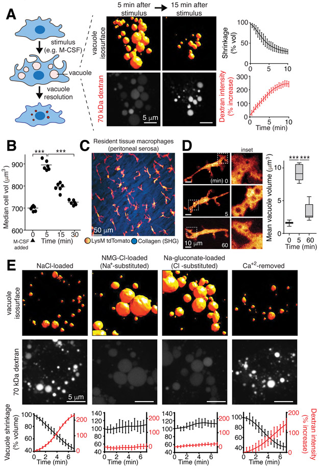
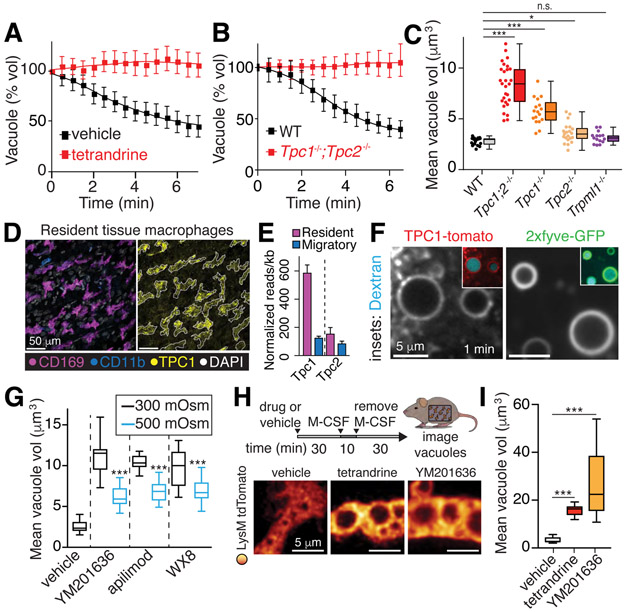
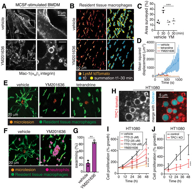
Despite ongoing (macro)pinocytosis of extracellular fluid, the volume of the endocytic pathway remains unchanged. To investigate the underlying mechanism, we used high-resolution video imaging to analyze the fate of macropinosomes formed by macrophages in vitro and in situ. Na+, the primary cationic osmolyte internalized, exited endocytic vacuoles via two-pore channels, accompanied by parallel efflux of Cl- and osmotically coupled water. The resulting shrinkage caused crenation of the membrane, which fostered recruitment of curvature-sensing proteins. These proteins stabilized tubules and promoted their elongation, driving vacuolar remodeling, receptor recycling, and resolution of the organelles. Failure to resolve internalized fluid impairs the tissue surveillance activity of resident macrophages. Thus, osmotically driven increases in the surface-to-volume ratio of endomembranes promote traffic between compartments and help to ensure tissue homeostasis.


Figures

Figure 1.
Figure 1.. Vacuolar shrinkage requires monovalent ion efflux.
A, Volume and 70 kDa rhodamine-dextran fluorescence intensity changes of macropinosomes induced in bone marrow derived macrophages (BMDM) by macrophage colony-stimulating factor (M-CSF); data are means ± SEM of >100 vacuoles from 3 independent experiments (i.e. n=3). Measurement of vacuole resolution was initiated after a 5-min stimulation with M-CSF in medium containing dextran, followed by an immediate wash. B, Cell (BMDM) volume was measured electronically before and at the indicated times after M-CSF stimulation; >104 cells per point, n=3. p values determined by unpaired, two-sided t-tests. Here and elsewhere *** indicates p<0.001, ** is p<0.01 and * is p<0.05. C, Intravital observation of td-Tomato-labeled resident tissue macrophages (RTM; pseudocolored yellow/red) of the peritoneal serosa and second harmonic imaging (SHG) of collagen (blue). See also Videos S1 and S2. D, Visualization and volume quantification of M-CSF-induced macropinosomes in RTM in vivo; means, upper and lower quartiles (boxes), and distribution (whiskers) are graphed. >50 vacuoles, n=3. p values determined by Mann-Whitney U test. E, Macropinosomes of M-CSF-stimulated BMDM in media containing indicated solutes and dextran. Representative images acquired at 5 min. See also Video S3. Bottom row: mean ± SEM macropinosomal volume and dextran intensity from 3 independent video recordings representing >150 macropinosomes. See also Fig. S1.
Figure 2.
Figure 2.. Monovalent ion efflux mechanisms.
A, Macropinosome volume changes in presence of 5 μM tetrandrine, measured in BMDM. Measurement of vacuole resolution was initiated once cells were washed after a 5-min stimulation with M-CSF in medium containing dextran; tetrandrine or vehicle were present throughout. Means ± SEM, n=3. See also Fig. S2 and Video S4. B-C, Macropinosome volume changes following stimulation with M-CSF in wt, Tpc1/Tpc2 single and double KO, and Trpml1 KO BMDM. In C, means, upper and lower quartiles (boxes), distribution (whiskers), and observations from fields each containing 3-5 cells (dots) measured 10 min after macropinosome formation; n=3. D, Staining of the peritoneal serosa. Outline of CD169 signal (left) overlaid on TPC1 signal (right). E, RNA-seq. Resident tissue macrophages were Cx3cr1/Ccr2. Migratory cells were Cx3cr1/Ccr2+. F, BMDM expressing TPC1-tomato or 2xfyve-GFP to detect PtdIns(3)P. Dextran shown in cyan. G, BMDM stimulated with M-CSF in the presence and 70 kDa rhodamine-dextran and, where indicated, the PIKfyve inhibitors YM201636, apilimod, or WX8 (all used at 500 nM). Resolution was recorded as in A. 5 min after iso-osmotic recording, a hyperosmotic solution (final 500 mOsm) was added to verify the osmotic responsiveness of the vacuoles. H-I, Visualization and volume quantification of macropinosomes in RTM treated in situ with YM201636 (500 nM) or tetrandrine (5 μM); >15 cells, n=3. All p values determined by Mann-Whitney U tests.
Figure 3.
Figure 3.. Osmotically-driven shrinkage induces tubulation.
A, BMDM were stimulated with M-CSF and the distribution of 70 kDa rhodamine-dextran and FM1-43 imaged at the indicated times after removal of the stimulus. B-C, Mean number of tubules (exceeding 1 μm in length) measured 5 min after stimulation with M-CSF; >100 macropinosomes (n=3) for each condition. D-E, Macropinosomes containing sulforhodamine B and N-methylglucamine chloride or formed in cells treated with YM201636 (500 nM) were recorded 10 min after formation, before and after being subjected to hypertonic solution. See also Video S5. >100 vacuoles, n=6. F, TEM was used to measure the diameter of tubules emerging from macropinosomes; 85 tubules quantified. G, Liposomes formed of whole brain lipid, rhodamine-labelled phosphatidylethanolamine, and PtdIns(4,5)P2 in 20 mOsm solution. The mean aspect ratio for 3-5 fields of liposomes incubated with or without recombinant human BIN1 was quantified by imaging, n=3. H, HT1080 cells expressing GFP, 2xfyve-GFP, or 5xfyve-GFP were pulsed with SRB for 10 min. Mean number of tubules (exceeding 1 μm in length); >100 macropinosomes (n=3) for each condition. All p values determined by unpaired, two-sided t-tests. I, Model of mechanism proposed to underlie macropinosomal tubulation.
Figure 4.
Figure 4.. Vacuolar resolution maintains cellular responsiveness and tissue surveillance.
A, BMDM were stimulated for 30 min with M-CSF, with or without YM201636, fixed and immunostained for Mac-1. B-E, Resident tissue macrophages (LysM-tdTomato) with or without YM201636 or tetrandrine were stimulated with M-CSF for 10 min followed by removal of the stimulus for 30 min. Cells were then imaged for 30 min. C, surveillance area measured in the absence (left) or presence (right) of YM201636 (YM) over time, n=3. In D-E, 30 min after M-CSF removal, laser-induced microlesions were generated, marked by the resultant autofluorescence (orange in E). Mean squared displacement of the macrophages is graphed in D; means ± SD, n=3. Representative images in E taken at 15 min post-injury. F-G, Experiments performed as in E. Representative images denoting the presence of neutrophils are shown. In G, the % of lesions with neutrophil swarming was quantified for 6 microlesions per animal, n=3. H, HT1080 cells expressing TPC1-tomato were incubated with 70 kDa dextran for 10 min before imaging. I-J, wildtype and TPC1 KO HT1080 cell growth measured in the absence or presence of YM201636 or tetrandrine (TTD) by cell counting. Means ± SD, n=3. All p values determined by Mann-Whitney U tests.

Comment in

References

    1. Steinman RM, Brodie SE, Cohn ZA, Membrane flow during pinocytosis. A stereologic analysis. J Cell Biol 68, 665–687 (1976). - PMC - PubMed
    1. Swanson JA, Shaping cups into phagosomes and macropinosomes. Nat Rev Mol Cell Biol 9, 639–649 (2008). - PMC - PubMed
    1. Swanson JA, Watts C, Macropinocytosis. Trends Cell Biol 5, 424–428 (1995). - PubMed
    1. Yoshida S, Hoppe AD, Araki N, Swanson JA, Sequential signaling in plasma-membrane domains during macropinosome formation in macrophages. J Cell Sci 122, 3250–3261 (2009). - PMC - PubMed
    1. Uderhardt S, Martins AJ, Tsang JS, Lammermann T, Germain RN, Resident Macrophages Cloak Tissue Microlesions to Prevent Neutrophil-Driven Inflammatory Damage. Cell 177, 541–555 e517 (2019). - PMC - PubMed

Publication types