. 2020 Jan 10;295(2):645-656.
doi: 10.1074/jbc.RA119.011929. Epub 2019 Dec 5.

DLC1 SAM domain-binding peptides inhibit cancer cell growth and migration by inactivating RhoA

Affiliations

DLC1 SAM domain-binding peptides inhibit cancer cell growth and migration by inactivating RhoA

Rakesh Joshi et al. J Biol Chem. .

Abstract

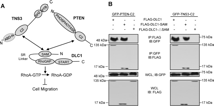
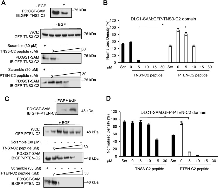
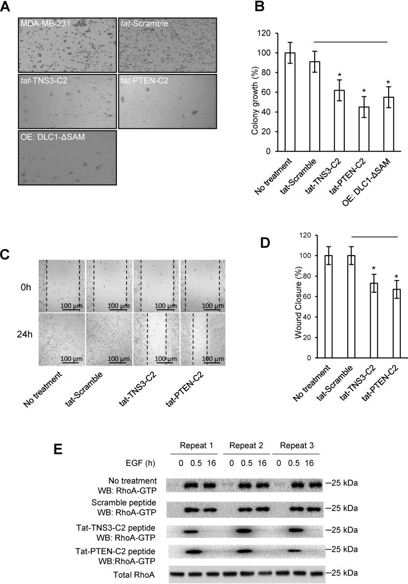
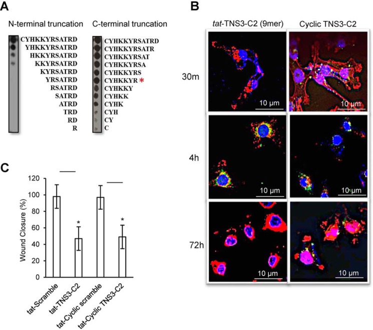
Deleted-in-liver cancer 1 (DLC1) exerts its tumor suppressive function mainly through the Rho-GTPase-activating protein (RhoGAP) domain. When activated, the domain promotes the hydrolysis of RhoA-GTP, leading to reduced cell migration. DLC1 is kept in an inactive state by an intramolecular interaction between its RhoGAP domain and the DLC1 sterile α motif (SAM) domain. We have shown previously that this autoinhibited state of DLC1 may be alleviated by tensin-3 (TNS3) or PTEN. We show here that the TNS3/PTEN-DLC1 interactions are mediated by the C2 domains of the former and the SAM domain of the latter. Intriguingly, the DLC1 SAM domain was capable of binding to specific peptide motifs within the C2 domains. Indeed, peptides containing the binding motifs were highly effective in blocking the C2-SAM domain-domain interaction. Importantly, when fused to the tat protein-transduction sequence and subsequently introduced into cells, the C2 peptides potently promoted the RhoGAP function in DLC1, leading to decreased RhoA activation and reduced tumor cell growth in soft agar and migration in response to growth factor stimulation. To facilitate the development of the C2 peptides as potential therapeutic agents, we created a cyclic version of the TNS3 C2 domain-derived peptide and showed that this peptide readily entered the MDA-MB-231 breast cancer cells and effectively inhibited their migration. Our work shows, for the first time, that the SAM domain is a peptide-binding module and establishes the framework on which to explore DLC1 SAM domain-binding peptides as potential therapeutic agents for cancer treatment.

Keywords: C2 domain; Ras homolog gene family, member A (RhoA); RhoGAP; SAM domain; breast cancer; cell migration; cell proliferation; deleted in liver cancer 1 (DLC1).


Conflict of interest statement

The authors declare that they have no conflicts of interest with the contents of this article

Figures

Figure 1.
Figure 1.
The SAM domain of DLC1 bound to its own RhoGAP domain and the C2 domain of TNS3 or PTEN. A, a schematic depicting the interactions between the DLC1 SAM domain and the TNS3 or PTEN C2 domain and how these interactions regulate cell migration through the RhoGAP-RhoA pathway. B, Western blots (WB) confirming the SAM-C2 domain interaction in cells. HEK293 cells co-expressing GFP-fused PTEN-C2 (left panel) or TNS3-C2 (right panel) and FLAG-tagged full-length DLC1, DLC1-SAM (SAM domain only), or DLC1-ΔSAM (SAM deletion mutant) were subjected to immunoprecipitation (IP) and immunoblotting (IB) using anti-GFP and anti-FLAG antibodies.
Figure 2.
Figure 2.
The DLC1 SAM domain bound to peptides derived from PTEN or TNS3 C2 domain. A, peptide-walking arrays of the PTEN-C2 (aa 174–403) or TNS3-C2 domain (aa 166–440) were probed with purified GST-SAM and the bound protein detected by anti-GST Western blotting. Peptides representing phosphorylated peptides, which showed no binding, are identified in white boxes. See Fig. S1 for the peptide array map and peptide identities. B, sequence alignment of the PTEN and TNS3 C2 domains with the SAM-binding peptides/motifs identified in A are shown in black boxes. C, SAM-binding peptides from B mapped onto the structure of the PTEN-C2 domain (PDB code 1D5R, aa 188–351; 230GPTR peptide in green, 257FFHK in red, and 334NRYF in blue). D and E, binding of the SAM domain to peptides from PTEN (230GPTRREDKFMYF) or TNS3 (244CYHKKYRSATRD) C2 domains. Shown are representative binding curves from fluorescence polarization (FP) analysis (n = 3) using the corresponding fluorescein-labeled C2 peptides. ΔFP, difference in fluorescence polarization.
Figure 3.
Figure 3.
The C2 peptides disrupted the DLC1 SAM-PTEN/TNS3-C2 domain-domain interactions in cells. A and B, effect of the C2 peptides on DLC1-SAM binding to the TNS3-C2 domain in HEK293DLC1 cells without EGF stimulation. The TNS3 C2 domain bound more tightly to the DLC1 SAM domain in the absence of EGF stimulation; accordingly, the TNS3-C2–derived peptide inhibited this interaction more effectively than the PTEN C2–derived peptide. Quantification of the Western blots indicates a significantly different effect between the two C2 peptides at 5 μm (n = 3; *, p < 0.01, Student's t test). C and D, effect of the C2 peptides on DLC1-SAM binding to the PTEN C2 domain in HEK293 cells in the presence of EGF. The PTEN-C2 domain bound to the SAM domain more tightly with EGF stimulation (for 30 min). Accordingly, the PTEN C2–derived peptide blocked this interaction more effectively than the TNS3 C2–derived peptide in HEK293 cells with EGF. Quantification of Western blots (D) indicates a significantly different effect between the two C2 peptides at 5 μm peptide (n = 3; *, p < 0.01, Student's t test). Scr: scrambled TSN3-C2 peptide control, applied at 30 μm.
Figure 4.
Figure 4.
The C2 peptides decreased RhoA activity in multiple cell lines and reduced colony formation by DLC1-expressing HEK293 cells. A, both TNS3 and PTEN C2 peptides were able to inhibit RhoA activation in the DLC1-overexpressing HEK293DLC1 cells in a concentration-dependent manner. B and C, the same RhoA-GTP inhibitory effect was observed for the C2 peptides in EGF-treated MDA-MB-231 cells (B) or HGF-treated HCC78 cells (C). D, colony-formation in soft agar for the HEK293DLC1 cells in the absence (no treatment) or presence of a C2 peptide or the scrambled TNS3-C2 peptide. E, quantify of colony-formation data in D; *, denotes a p value < 0.01 (n = 3), Student's t test; IB, immunoblotting.
Figure 5.
Figure 5.
C2 peptides reduced anchorage-independent growth and EGF-dependent migration of breast cancer cells. A, colony formation by MDA-MB-231 cells treated with a C2 peptide or scrambled control (of tat-TNS3-C2) or transfected with DLC1-ΔSAM. B, quantification of data in A to show efficiency in colony formation for the above cells compared with the parent cells (set as 100%). C, representative images of wound-healing in peptide-treated MDA-MB-231 cells. D, quantification of the would-healing data in C. For panels B and D, * denotes p < 0.01 (compared with the scramble peptide, n = 3), Student's t test. E, Western blots of total and active RhoA in peptide-treated MDA-MB-231 cells in response to EGF treatment. Peptide concentration in C–E: 18 μm.
Figure 6.
Figure 6.
Cyclization of the TNS3-C2 peptide led to increased stability without comprising its ability to inhibit MDA-MB-231 cell migration. A, identification of the minimal length for the active TNS3-C2 peptide by peptide truncation spot array. Peptide spot arrays representing N- or C-terminal truncation (or Ala-scanning analogues, Fig. S4A) of the TNS3-C2 peptides were probed with the GST-SAM domain. Based on the binding data, a 9-mer peptide (CYHKKYS-GC) was synthesized for cyclization. B, the cyclized C2 peptide showed increased cellular stability. MDA-MB-231 cells were transduced with fluorescein-labeled tat-TNS3-C2 9-mer linear peptide or the corresponding cyclized TNS3-C2 peptide (without tat label) and fixed at the indicated time points. The cells were stained with DAPI (blue for the nucleus) and phalloidin (red for actin) and imaged by confocal microscopy. The fluorescein-labeled peptides were shown in green punctate structures. C, serum-starved MDA-MB-231 cells were transduced with scrambled, linear, and cyclized TNS3 C2 peptides (each at 18 μm) and subjected to wound healing assays under EGF stimulation. Closed wound areas were measured at 18 h and are represented as percentages of wound area closed relative to the untreated cells; * denotes a p value < 0.01, n = 3; Student's t test.

References

    1. Zhou X., Thorgeirsson S. S., and Popescu N. C. (2004) Restoration of DLC-1 gene expression induces apoptosis and inhibits both cell growth and tumorigenicity in human hepatocellular carcinoma cells. Oncogene 23, 1308–1313 10.1038/sj.onc.1207246 - DOI - PubMed
    1. Wong C. C., Wong C. M., Ko F. C., Chan L. K., Ching Y. P., Yam J. W., and Ng I. O. (2008) Deleted in liver cancer 1 (DLC1) negatively regulates Rho/ROCK/MLC pathway in hepatocellular carcinoma. PLoS ONE 3, e2779 10.1371/journal.pone.0002779 - DOI - PMC - PubMed
    1. Xue W., Krasnitz A., Lucito R., Sordella R., Vanaelst L., Cordon-Cardo C., Singer S., Kuehnel F., Wigler M., Powers S., Zender L., and Lowe S. W. (2008) DLC1 is a chromosome 8p tumor suppressor whose loss promotes hepatocellular carcinoma. Genes Dev. 22, 1439–1444 10.1101/gad.1672608 - DOI - PMC - PubMed
    1. Zhou X., Zimonjic D. B., Park S. W., Yang X. Y., Durkin M. E., and Popescu N. C. (2008) DLC1 suppresses distant dissemination of human hepatocellular carcinoma cells in nude mice through reduction of RhoA GTPase activity, actin cytoskeletal disruption and down-regulation of genes involved in metastasis. Int. J. Oncol. 32, 1285–1291 - PMC - PubMed
    1. Durkin M. E., Avner M. R., Huh C. G., Yuan B. Z., Thorgeirsson S. S., and Popescu N. C. (2005) DLC-1, a Rho GTPase-activating protein with tumor suppressor function, is essential for embryonic development. FEBS Lett. 579, 1191–1196 10.1016/j.febslet.2004.12.090 - DOI - PubMed

Publication types

MeSH terms

Associated data

Grants and funding