

Differential Diagnosis Of Multiple-System Atrophy With Parkinson's Disease By External Anal- And Urethral-Sphincter Electromyography
- PMID: 31806975
- PMCID: PMC6842278
- DOI: 10.2147/NDT.S218073
Differential Diagnosis Of Multiple-System Atrophy With Parkinson's Disease By External Anal- And Urethral-Sphincter Electromyography
Abstract
Background: The differential diagnosis of Parkinson's disease (PD) with multiple-system atrophy (MSA) is difficult because of their similarity in symptoms and signs. The objective of this study was to investigate the value of external anal-sphincter electromyography (EAS-EMG) and urethral-sphincter electromyography (US-EMG) in differentiating MSA from PD.
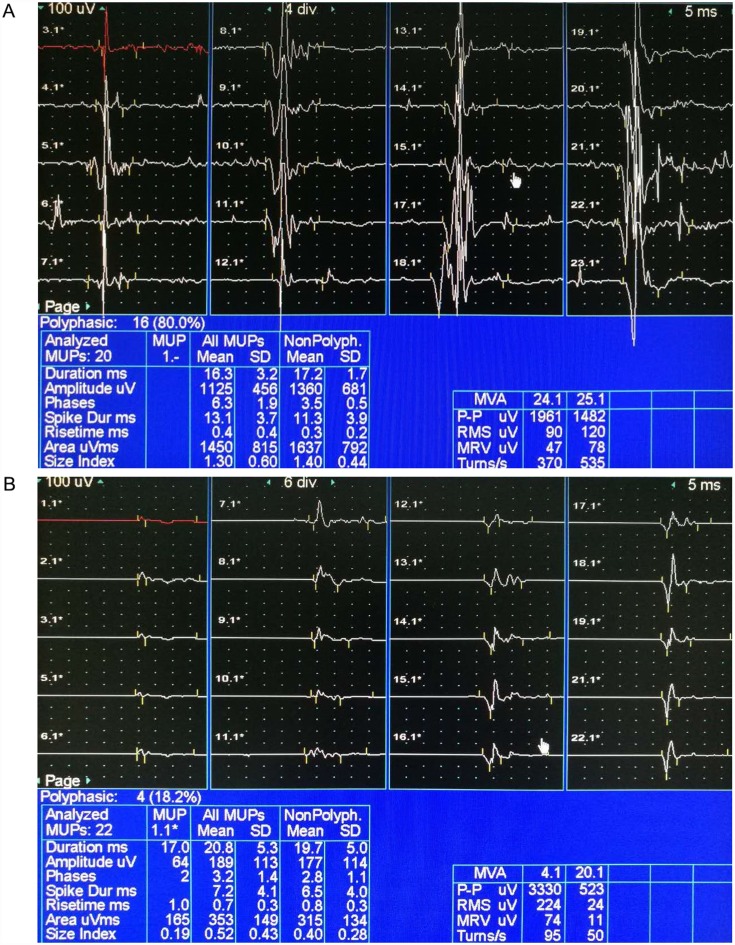
Methods: A total of 201 patients, - 101 MSA and 100 PD - were recruited in this study. Average duration and amplitude of motor unit potentials (MUPs), percentage of polyphasic MUPs, amplitude during strong contractions, and recruitment patterns during maximal voluntary contractions were recorded and analyzed to assess diagnostic efficiency of EAS-EMG and US-EMG for MSA.
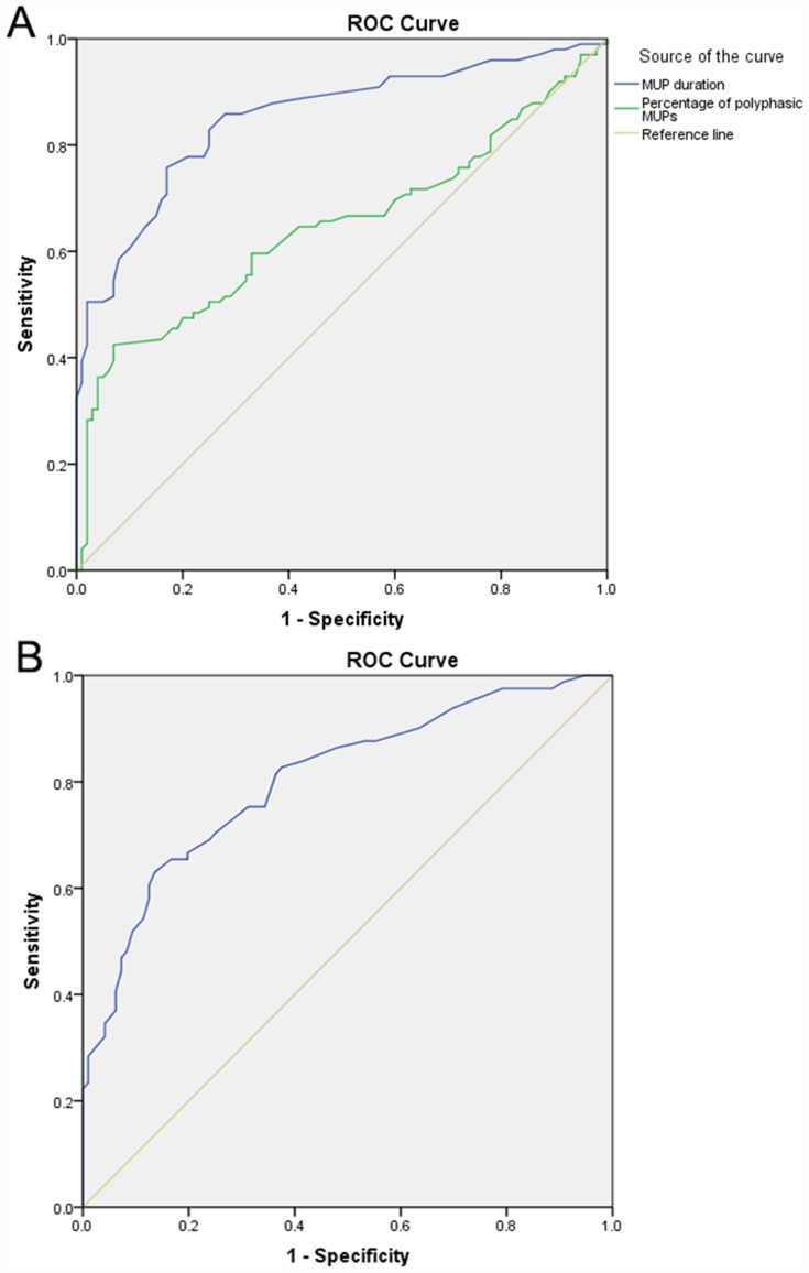
Results: Significant differences in average MUP duration and recruitment patterns during maximal voluntary contractions were found between patients with MSA and patients with PD using both EAS-EMG (P<0.001, P<0.001) and US-EMG (P<0.001, P<0.001). The percentage of polyphasic MUPs and amplitude during strong contractions showed significant differences in MSA and PD using only EAS-EMG (P<0.001, P=0.005). Cutoff points for average MUP duration in EAS-EMG and US-EMG for differential diagnosis of MSA with PD were 10.9 and 11.1 milliseconds, respectively. With average MUP duration of EAS-EMG and US-EMG being applied jointly, sensitivity and specificity in distinguishing MSA from PD were 83.2% and 71.8%, respectively.
Conclusion: EAS-EMG and US-EMG were sensitive and specific methods for the diagnosis and differential diagnosis of MSA, and the combination of both would improve the diagnostic rate of MSA compared to only one method being used.
Keywords: EAS-EMG; MSA; PD; US-EMG; differential diagnosis.
© 2019 Qiu et al.
Conflict of interest statement
The authors report no conflicts of interest in this work.
Figures
Similar articles
-
The High Value of External Anal- and Urethral-Sphincter Electromyography in Differential Diagnosis with MSA-P, PD, and PSP.Ann Indian Acad Neurol. 2023 May-Jun;26(3):241-246. doi: 10.4103/aian.aian_496_22. Epub 2023 Apr 7. Ann Indian Acad Neurol. 2023. PMID: 37538423 Free PMC article.
-
Differential value of external anal- and urethral-sphincter electromyography in multiple system atrophy cerebellar type and spinocerebellar ataxias.J Clin Neurosci. 2020 Oct;80:16-22. doi: 10.1016/j.jocn.2020.07.067. Epub 2020 Aug 15. J Clin Neurosci. 2020. PMID: 33099340
-
[Comparative study on diagnostic significance of urethral sphincter versus external anal sphincter electromyography in patients with multiple system atrophy].Zhonghua Yi Xue Za Zhi. 2013 Jul 2;93(25):1958-61. Zhonghua Yi Xue Za Zhi. 2013. PMID: 24169243 Chinese.
-
Sphincter EMG and differential diagnosis of multiple system atrophy.Mov Disord. 2001 Jul;16(4):600-7. doi: 10.1002/mds.1121. Mov Disord. 2001. PMID: 11481682 Review.
-
How to diagnose MSA early: the role of sphincter EMG.J Neural Transm (Vienna). 2005 Dec;112(12):1657-68. doi: 10.1007/s00702-005-0377-2. J Neural Transm (Vienna). 2005. PMID: 16284909 Review.
Cited by
-
A simple and effective screening strategy for early multiple system atrophy diagnosis and α-Synuclein forms in erythrocytes.Front Aging Neurosci. 2025 Feb 21;17:1533504. doi: 10.3389/fnagi.2025.1533504. eCollection 2025. Front Aging Neurosci. 2025. PMID: 40061041 Free PMC article.
-
Diagnostic and Prognostic Value of External Anal Sphincter EMG Patterns in Multiple System Atrophy.Mov Disord. 2022 May;37(5):1069-1074. doi: 10.1002/mds.28938. Epub 2022 Feb 4. Mov Disord. 2022. PMID: 35122320 Free PMC article.
-
The High Value of External Anal- and Urethral-Sphincter Electromyography in Differential Diagnosis with MSA-P, PD, and PSP.Ann Indian Acad Neurol. 2023 May-Jun;26(3):241-246. doi: 10.4103/aian.aian_496_22. Epub 2023 Apr 7. Ann Indian Acad Neurol. 2023. PMID: 37538423 Free PMC article.
-
External Anal- and Urethral-Sphincter Electromyography for Differentiating MSA-P, PD and PSP: Using a Needle to Sort the Haystack!Ann Indian Acad Neurol. 2023 May-Jun;26(3):221-222. doi: 10.4103/aian.aian_154_23. Epub 2023 Apr 28. Ann Indian Acad Neurol. 2023. PMID: 37538438 Free PMC article. No abstract available.
-
External anal sphincter electromyography in multiple system atrophy: implications for diagnosis, clinical correlations, and novel insights into prognosis.Neural Regen Res. 2023 Sep;18(9):1903-1907. doi: 10.4103/1673-5374.367833. Neural Regen Res. 2023. PMID: 36926706 Free PMC article. Review.
References
-
- Qiu F, Liu JG, Li LP, Song DD, Yao W, Qi XK. Comparative study on diagnostic significance of urethral sphincter versus external anal sphincter electromyography in patients with multiple system atrophy. Zhonghua Yi Xue Za Zhi. 2013;93(25):1958–1961. - PubMed
LinkOut - more resources
Full Text Sources
