

Sonographic assessment of the anatomy and common pathologies of clinically important bursae
- PMID: 31807327
- PMCID: PMC6856779
- DOI: 10.15557/JoU.2019.0032
Sonographic assessment of the anatomy and common pathologies of clinically important bursae
Abstract
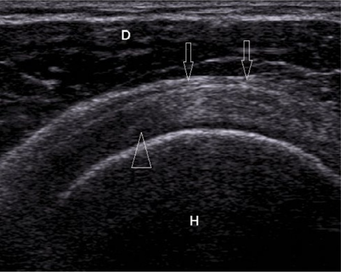
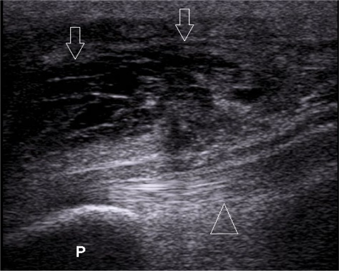
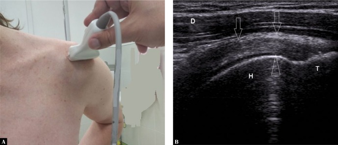
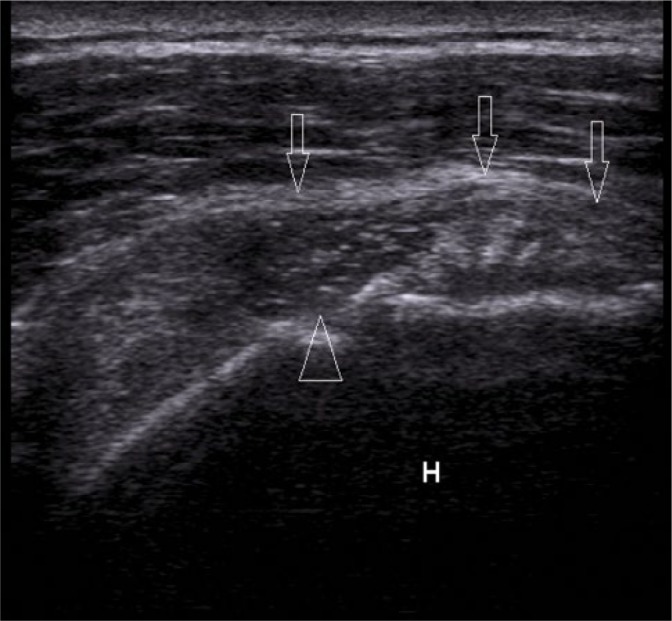
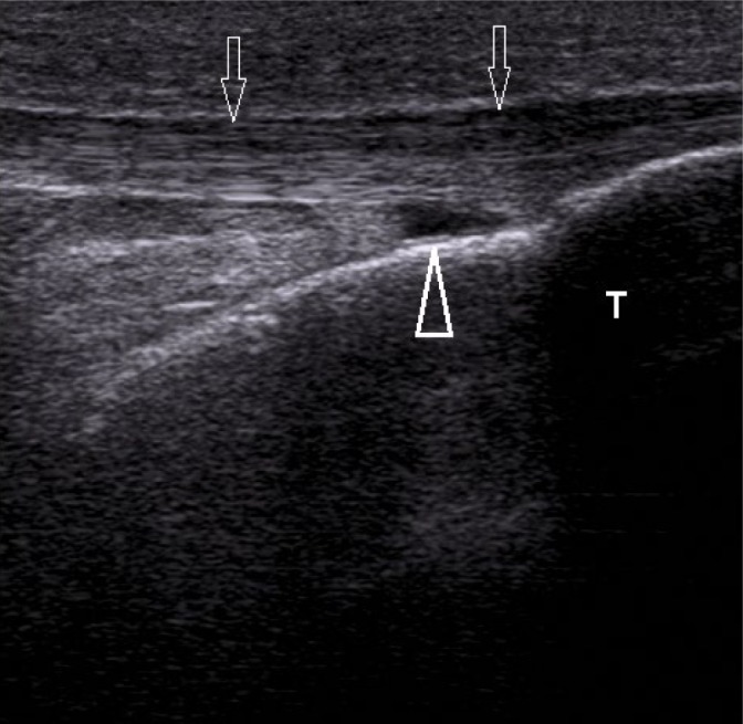
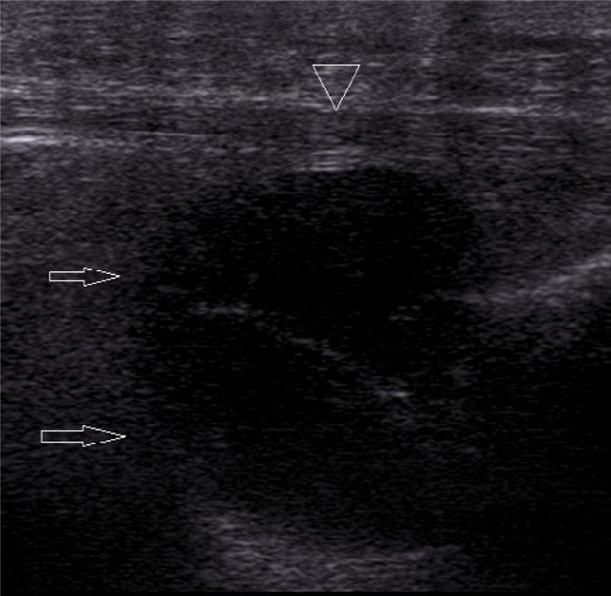
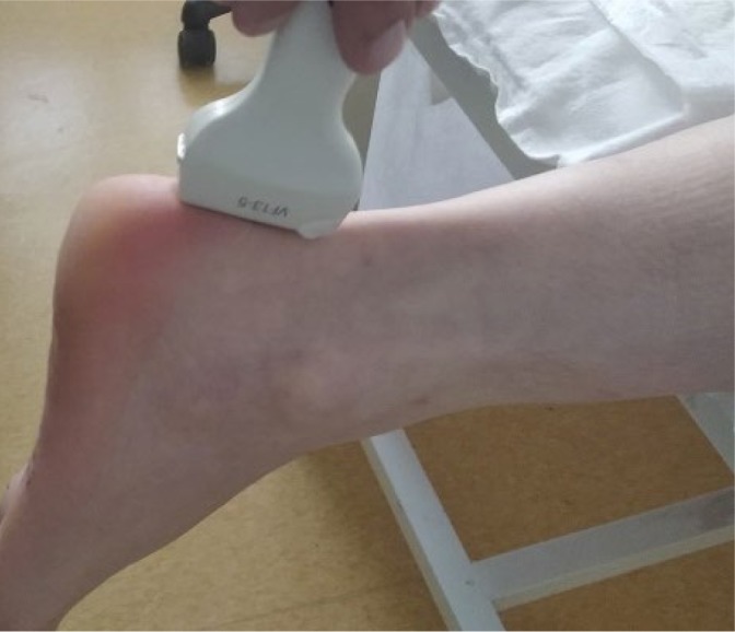
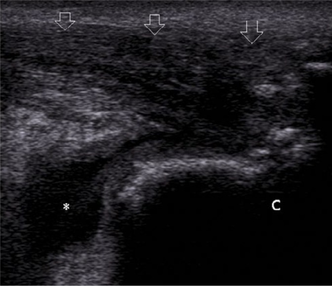
High-resolution ultrasonography has many advantages in the imaging of the musculoskeletal system, when compared to other imaging methods, particularly in superficial, easily accessible parts of the body. It is a perfect diagnostic tool for visualizing the most common pathologies of the musculoskeletal system, including the bursae. Inflammation of bursae is frequent, and it can mimic other diseases of the musculoskeletal system. Therefore, knowledge of normal ultrasound anatomy of the bursae, their exact location in the human body, and the sonographic signs of their most common pathologies is essential for establishing a quick and accurate diagnosis by ultrasound. Common conditions affecting bursae, leading to bursitis, include acute trauma, overuse syndromes, degenerative diseases, inflammatory conditions (rheumatoid arthritis, psoriatic arthritis, gout etc.), infections such as tuberculosis, synovial tumors and tumor-like conditions (pigmented villonodular synovitis, osteochondromatosis), and many more. This review article presents and explains ultrasound examples of the most frequent pathological conditions affecting bursae. Images include normal and pathological conditions of bursae around the shoulder joint, elbow, hip, knee, and ankle joint.
Keywords: anatomy; bursa; high-resolution ultrasonography; inflammation.
© Polish Ultrasound Society.
Conflict of interest statement
Conflict of interest The authors do not report any financial or personal connections with other persons or organizations which might negatively affect the contents of this publication and/or claim authorship rights to this publication.
Figures
Similar articles
-
Ultrasound evaluation of bursae: anatomy and pathological appearances.Skeletal Radiol. 2017 Apr;46(4):445-462. doi: 10.1007/s00256-017-2577-x. Epub 2017 Feb 11. Skeletal Radiol. 2017. PMID: 28190095 Review.
-
Imaging of bursae around the shoulder joint.Skeletal Radiol. 1996 Aug;25(6):513-7. doi: 10.1007/s002560050127. Skeletal Radiol. 1996. PMID: 8865483 Review.
-
Imaging of the bursae.J Clin Imaging Sci. 2011;1:22. doi: 10.4103/2156-7514.80374. Epub 2011 May 2. J Clin Imaging Sci. 2011. PMID: 21966619 Free PMC article.
-
Clinical anatomy of the subacromial and related shoulder bursae: A review of the literature.Clin Anat. 2017 Mar;30(2):213-226. doi: 10.1002/ca.22823. Epub 2017 Feb 9. Clin Anat. 2017. PMID: 28033656 Review.
-
Applications of musculoskeletal sonography.J Clin Ultrasound. 1999 Jul-Aug;27(6):293-318. doi: 10.1002/(sici)1097-0096(199907/08)27:6<293::aid-jcu1>3.0.co;2-c. J Clin Ultrasound. 1999. PMID: 10395126 Review.
Cited by
-
High-Resolution Imaging Insights into Shoulder Joint Pain: A Comprehensive Review of Ultrasound and Magnetic Resonance Imaging (MRI).Cureus. 2023 Nov 17;15(11):e48974. doi: 10.7759/cureus.48974. eCollection 2023 Nov. Cureus. 2023. PMID: 38111406 Free PMC article. Review.
-
Shoulder ultrasound: current concepts and future perspectives.J Ultrason. 2021 Jun 7;21(85):e154-e161. doi: 10.15557/JoU.2021.0025. Epub 2021 Jun 18. J Ultrason. 2021. PMID: 34258041 Free PMC article.
-
Diagnostic Accuracy of Dynamic High-Resolution Ultrasonography in Assessing Anterior Disc Displacement in Temporomandibular Joint Disorders: A Prospective Observational Study.Healthcare (Basel). 2024 Nov 25;12(23):2355. doi: 10.3390/healthcare12232355. Healthcare (Basel). 2024. PMID: 39684977 Free PMC article.
-
Sonographic pathoanatomy of greater trochanteric pain syndrome.J Ultrasound. 2024 Sep;27(3):501-510. doi: 10.1007/s40477-023-00836-x. Epub 2023 Dec 12. J Ultrasound. 2024. PMID: 38082193
-
Application of mechanical quantitative techniques in postoperative rehabilitation assessment of anterior cruciate ligament reconstruction: A study protocol.PLoS One. 2025 Aug 6;20(8):e0324663. doi: 10.1371/journal.pone.0324663. eCollection 2025. PLoS One. 2025. PMID: 40768443 Free PMC article.
References
-
- Lohr KM: Bursitis. Medscape Drugs and Diseases. New York, NY: 2016. Available from: http://emedicine.medscape.com/article/2145588-overview.
-
- DeLee JC, Drez D: Imaging effusions, cysts, and ganglia In: DeLee JC, Drez D, Miller MD (eds): DeLee and Drez’s Orthopaedic Sports Medicine: Principles and Practice. WB Saunders, Philadelphia: 2003: 1646–1648.
-
- Ruangchaijatuporn T, Gaetke-Udager K, Jacobson JA, Yablon CM, Morag Y: Ultrasound evaluation of bursae: anatomy and pathological appearances. Skeletal Radiol 2017; 46: 445–462. - PubMed
Publication types
LinkOut - more resources
Full Text Sources