

Ewing's sarcoma/primitive neuroectodermal tumor of the kidney: a case report and literature review
- PMID: 31807433
- PMCID: PMC6842790
- DOI: 10.21037/tau.2019.09.46
Ewing's sarcoma/primitive neuroectodermal tumor of the kidney: a case report and literature review
Abstract
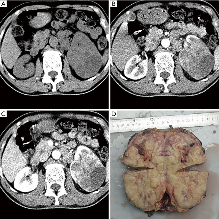
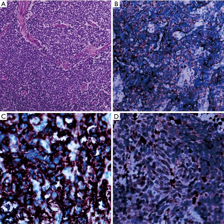
Ewing sarcoma/primitive neuroectodermal tumor (ES/PNET) is a malignant bone, and soft-tissue tumor derived from neuroectoderm. It occurs in pediatric adolescents with the histopathological features of invasiveness. Renal involvement is extremely rare, which is limited to case reports and small case series. Most patients showed non-specific symptoms, such as abdominal pain and severe hematuria. The corresponding diagnosis was based on pathological features and immunohistochemical detection. So far, the characteristics of computed tomography (CT) have been rarely described in these cases. We report an 18-year-old man diagnosed with renal ES/PNET, who suffered from a sudden left flank pain associated with gross hematuria. The CT images showed an irregular soft tissue mass with a size of 7.3 cm × 7.0 cm × 9.0 cm. The patient underwent laparoscopic nephrectomy of the left kidney. The final diagnosis of renal ES/PNET was confirmed by immunohistochemical detection and fluorescence in situ hybridization of the nephrectomy specimen. We want to point out that CT scanning is still a useful method for preliminary assessment in preoperative diagnosis.
Keywords: Ewing sarcoma (ES); computed tomography (CT); kidney; nephrectomy; primitive neuroectodermal tumor (PNET).
2019 Translational Andrology and Urology. All rights reserved.
Conflict of interest statement
Conflicts of Interest: The authors have no conflicts of interest to declare.
Figures
Similar articles
-
Renal Ewing sarcoma/primitive neuroectodermal tumor in a pregnant woman who underwent robot-assisted laparoscopic nephrectomy: a case report and literature review.Onco Targets Ther. 2018 Oct 11;11:6839-6843. doi: 10.2147/OTT.S155523. eCollection 2018. Onco Targets Ther. 2018. PMID: 30349316 Free PMC article.
-
Renal Ewing's sarcoma/primitive neuroectodermal tumor: a case report and literature review.Beijing Da Xue Xue Bao Yi Xue Ban. 2017 Oct 18;49(5):919-923. Beijing Da Xue Xue Bao Yi Xue Ban. 2017. PMID: 29045981 Review.
-
Primary Ewing's sarcoma/primitive neuroectodermal tumor of the kidney: a clinicopathologic and immunohistochemical analysis of 11 cases.Am J Surg Pathol. 2002 Mar;26(3):320-7. doi: 10.1097/00000478-200203000-00005. Am J Surg Pathol. 2002. PMID: 11859203
-
Ewing sarcoma/primitive neuroectodermal tumor of the kidney: A report of three cases.Int J Surg Case Rep. 2016;28:330-334. doi: 10.1016/j.ijscr.2016.10.014. Epub 2016 Oct 18. Int J Surg Case Rep. 2016. PMID: 27776324 Free PMC article.
-
Primary Ewing's sarcoma/primitive neuroectodermal tumor of the ileum: case report of a 16-year-old Chinese female and literature review.Diagn Pathol. 2017 May 4;12(1):37. doi: 10.1186/s13000-017-0626-3. Diagn Pathol. 2017. PMID: 28472972 Free PMC article. Review.
Cited by
-
Primary Ewing sarcoma of the kidney mimicking cystic papillary renal cell carcinoma in an older patient: A case report.World J Clin Cases. 2024 May 26;12(15):2606-2613. doi: 10.12998/wjcc.v12.i15.2606. World J Clin Cases. 2024. PMID: 38817223 Free PMC article.
-
A rare entity of Primary Ewing sarcoma in kidney.BMC Surg. 2020 Nov 11;20(1):280. doi: 10.1186/s12893-020-00948-9. BMC Surg. 2020. PMID: 33176766 Free PMC article.
-
A rare case of calcified metastasis in a patient with primary osseous Ewing's sarcoma.Pediatr Radiol. 2025 May;55(6):1313-1318. doi: 10.1007/s00247-025-06237-y. Epub 2025 Apr 15. Pediatr Radiol. 2025. PMID: 40232393
-
A unique surgical case report of Ewing sarcoma of the kidney in a 54-year-old female: Diagnostic and therapeutic challenges.Int J Surg Case Rep. 2025 Aug;133:111615. doi: 10.1016/j.ijscr.2025.111615. Epub 2025 Jul 7. Int J Surg Case Rep. 2025. PMID: 40663964 Free PMC article.
-
A primary Ewing's sarcoma of the kidney: A case report and review of literature.Urol Ann. 2023 Jul-Sep;15(3):334-336. doi: 10.4103/ua.ua_2_23. Epub 2023 Jul 17. Urol Ann. 2023. PMID: 37664090 Free PMC article.
References
Publication types
LinkOut - more resources
Full Text Sources