. 2019 Nov 27;5(11):1768-1776.
doi: 10.1021/acscentsci.9b00567. Epub 2019 Sep 24.

Development of a Split Esterase for Protein-Protein Interaction-Dependent Small-Molecule Activation

Affiliations

Development of a Split Esterase for Protein-Protein Interaction-Dependent Small-Molecule Activation

Krysten A Jones et al. ACS Cent Sci. .

Abstract

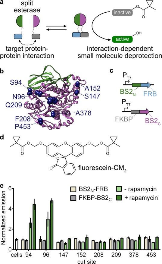
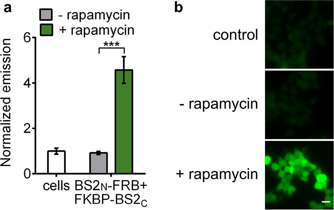
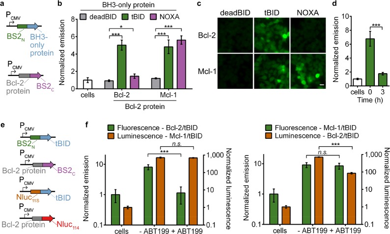
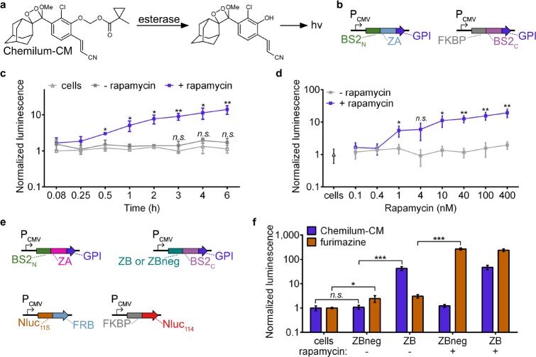
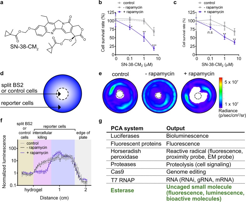
Split reporters based on fluorescent proteins and luciferases have emerged as valuable tools for measuring interactions in biological systems. Relatedly, biosensors that transduce measured input signals into outputs that influence the host system are key components of engineered gene circuits for synthetic biology applications. While small-molecule-based imaging agents are widely used in biological studies, and small-molecule-based drugs and chemical probes can target a range of biological processes, a general method for generating a target small molecule in a biological system based on a measured input signal is lacking. Here, we develop a proximity-dependent split esterase that selectively unmasks ester-protected small molecules in an interaction-dependent manner. Exploiting the versatility of an ester-protected small-molecule output, we demonstrate fluorescent, chemiluminescent, and pharmacological probe generation, each created by masking key alcohol functional groups on a target small molecule. We show that the split esterase system can be used in combination with ester-masked fluorescent or luminescent probes to measure protein-protein interactions and protein-protein interaction inhibitor engagement. We demonstrate that the esterase-based reporter system is compatible with other commonly used split reporter imaging systems for the simultaneous detection of multiple protein-protein interactions. Finally, we develop a system for selective small-molecule-dependent cell killing by unmasking a cytotoxic molecule using an inducible split esterase. Presaging utility in future synthetic biology-based therapeutic applications, we also show that the system can be used for intercellular cell killing via a bystander effect, where one activated cell unmasks a cytotoxic molecule and kills cells physically adjacent to the activated cells. Collectively, this work illustrates that the split esterase system is a valuable new addition to the split protein toolbox, with particularly exciting potential in synthetic biology applications.


Conflict of interest statement

The authors declare the following competing financial interest(s): A.R.L. declares a financial stake in BioLum Sciences, LLC.

Figures

Figure 1
Figure 1
Development of a split esterase sensor. (a) Schematic of a PPI-driven split esterase assembly to unmask 1-methylcyclopropyl (CM)-masked molecules. Interacting proteins are fused to inactive fragments of BS2 esterase. Interaction between the fusion proteins results in assembly of the esterase which cleaves an inactive small molecule to generate an active molecule and output signal. (b) Mapping the cut sites onto a homologous B. subtilis esterase structure (PDB 1QE3). The BS2N fragment (green) and BS2C (magenta) from the lead split site, S94, are shown. Split sites occur after the designated amino acids. (c) Vector system to identify PPI-dependent esterase fragments in E. coli. (d) Chemical structure of masked fluorophore fluorescein-CM2. (e) Fluorescence output of split esterase fragments. E. coli expressing BS2N-fused FRB (tan), FKBP-fused BS2C (gray), or both in the absence (light green) or presence (green) of rapamycin were incubated with fluorescein-CM2 for 4 h and then analyzed for fluorescence. Error bars are the standard deviation for n = 3 replicates.
Figure 2
Figure 2
Split BS2 can detect small-molecule-activated PPIs. (a) HEK293T cells cotransfected with BS2N-fused FRB and FKBP-fused BS2C or HEK293T control cells (white) were incubated with rapamycin (green) or a DMSO control (gray). After 24 h, fluorescein-CM2 was applied, and the cells were analyzed by a plate reader for fluorescence. (b) HEK293T cells were treated identically to conditions in part a and analyzed by fluorescence microscopy. Error bars are the standard deviation for n = 4 replicates. Unpaired t test; ***P < 0.0001. Scale bars shown are 20 μm.
Figure 3
Figure 3
Split BS2 can detect Bcl-2 family PPIs and inhibitor engagement. (a) Vector system to test Bcl-2 split esterase PPI detection. (b) HEK293T cells cotransfected with the plasmids shown in part a were incubated with fluorescein-CM2 and analyzed for fluorescence by a plate reader. The normalized emission for interactions between deadBID/Bcl-2 protein (gray), tBID/Bcl-2 protein (green), and NOXA/Bcl-2 protein is shown. HEK293T control cells (white) were similarly analyzed. (c) HEK293T cells cotransfected and incubated with fluorescein-CM2 as in part b were analyzed by fluorescence microscopy. (d) HEK293T cells cotransfected with BS2N-fused tBID and Bcl-2-fused BS2C were treated with DMSO (time = 0) or ABT199 for 0–3 h (green) followed by incubation with fluorescein-CM2 and analyzed for fluorescence by a plate reader. HEK293T control cells (white) were similarly analyzed. (e) Vector system to simultaneously detect two PPIs and Bcl-2 inhibition. (f) HEK293T cells were cotransfected with the split esterase plasmids or split Nluc plasmids shown in part e. The two cell populations were mixed and treated with ABT-199 or a DMSO control. After 24 h, the cells were incubated with fluorescein-CM2 and analyzed (green). Immediately after analysis, furimazine was added, and the cells were reimaged (orange). The Bcl-2/tBID interaction was selectively blocked and detected with the esterase reporter (left) or Nluc reporter (right). HEK293T control cells were similarly analyzed. Error bars are the standard deviation for n = 4 (b), n = 6 replicates (d), and n = 8 replicates (f). Unpaired t test; *P < 0.01, ***P < 0.0001. Scale bars shown are 20 μm.
Figure 4
Figure 4
Multiplexed PPI analysis with split BS2. (a) Chemilum-CM is unmasked by esterase activity and generates a photon. (b) Vector system to monitor extracellular PPIs. (c) HEK293T cells were cotransfected with plasmids shown in part b. Rapamycin (blue) or a DMSO control (gray) were added to cells for 0–6 h. Media was replaced with Chemilum-CM and analyzed for luminescence. HEK293T control cells (white) were similarly analyzed. (d) HEK293T cells cotransfected as in part c or HEK293T control cells (triangle) were incubated with rapamycin (blue) or a DMSO control (gray). After 6 h, the cells were analyzed with Chemilum-CM as in part c. (e) Vector system to simultaneously monitor extracellular and intracellular PPIs. (f) HEK293T cells were transfected with all four plasmids shown in part d. Rapamycin or a DMSO control was added to the cells for 24 h. Media was replaced with Chemilum-CM (10 μM) and analyzed for luminescence (blue). The cells were then rinsed, incubated with furimazine, and analyzed for bioluminescence (orange). Error bars are the standard deviation for n = 4 replicates. Unpaired t test; *P < 0.01, **P < 0.001, ***P < 0.0001.
Figure 5
Figure 5
Small-molecule-induced intra- and intercellular cell death. (a) Chemical structure of masked chemotherapeutic SN-38-CM2. (b) MDA-MB-231 luciferase cells were cotransfected with GPI-anchored BS2N-fused FRB and FKBP-fused BS2C. Rapamycin (blue square) or a DMSO control (gray square) was added to cells for 12 h prior to addition of SN-38-CM2. After 6 h, the cells were rinsed, cultured for 40 h, and imaged with d-luciferin. MDA-MB-231 luciferase control cells (gray dashed circle) were similarly analyzed. (c) MDA-MB-231 cells were cotransfected with GPI-anchored BS2N-fused FRB and FKBP-fused BS2C and then coplated with MDA-MB-453 luciferase cells. Rapamycin (blue square) or a DMSO control (gray square) and SN-38-CM2 were added to cells and imaged with d-luciferin as in part b. (d) Schematic of the coculture cytotoxicity assay. Split esterase or control MDA-MB-231 cells were plated in the center of a 3.5 cm2 dish (dashed center circle). MDA-MB-453 luciferase cells were plated around the split BS2 or control cells. PPI-mediated cleavage of SN-38-CM2 by split BS2 results in an active molecule to induce killing of neighboring cells. (e) Representative bioluminescence images of cocultures after treatment as in part d. (f) Quantification of light emission from bioluminescence images of cocultures. Photon counts along a 2 cm line in the direction of most observed cell killing from the hydrogel embedded cells (hydrogel) to the edge of each dish are plotted. (g) Commonly used PCA systems and their corresponding outputs. Error bars are the standard deviation for n = 4 replicates (b, c) and standard error of the mean for n = 8 replicates. Unpaired t test; *P < 0.01, **P < 0.001, ***P < 0.0001.

References

    1. Braun P. Interactome mapping for analysis of complex phenotypes: Insights from benchmarking binary interaction assays. Proteomics 2012, 12 (10), 1499–1518. 10.1002/pmic.201100598. - DOI - PubMed
    1. Jones S.; Thornton J. M. Principles of protein-protein interactions. Proc. Natl. Acad. Sci. U. S. A. 1996, 93 (1), 13–20. 10.1073/pnas.93.1.13. - DOI - PMC - PubMed
    1. Sudhof T. C. The synaptic cycle - A cascade of protein-protein interactions. Nature 1995, 375 (6533), 645–653. 10.1038/375645a0. - DOI - PubMed
    1. Kolch W. Meaningful relationships: The regulation of the Ras/Raf/MEK/ERK pathway by protein interactions. Biochem. J. 2000, 351, 289–305. 10.1042/0264-6021:3510289. - DOI - PMC - PubMed
    1. Arkin M. R.; Tang Y. Y.; Wells J. A. Small-molecule inhibitors of protein-protein interactions: Progressing toward the reality. Chem. Biol. 2014, 21 (9), 1102–1114. 10.1016/j.chembiol.2014.09.001. - DOI - PMC - PubMed