. 2020 Feb 20;48(3):1531-1550.
doi: 10.1093/nar/gkz1153.

AtFKBP53: a chimeric histone chaperone with functional nucleoplasmin and PPIase domains

Affiliations

AtFKBP53: a chimeric histone chaperone with functional nucleoplasmin and PPIase domains

Ajit Kumar Singh et al. Nucleic Acids Res. .

Abstract

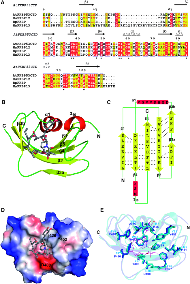
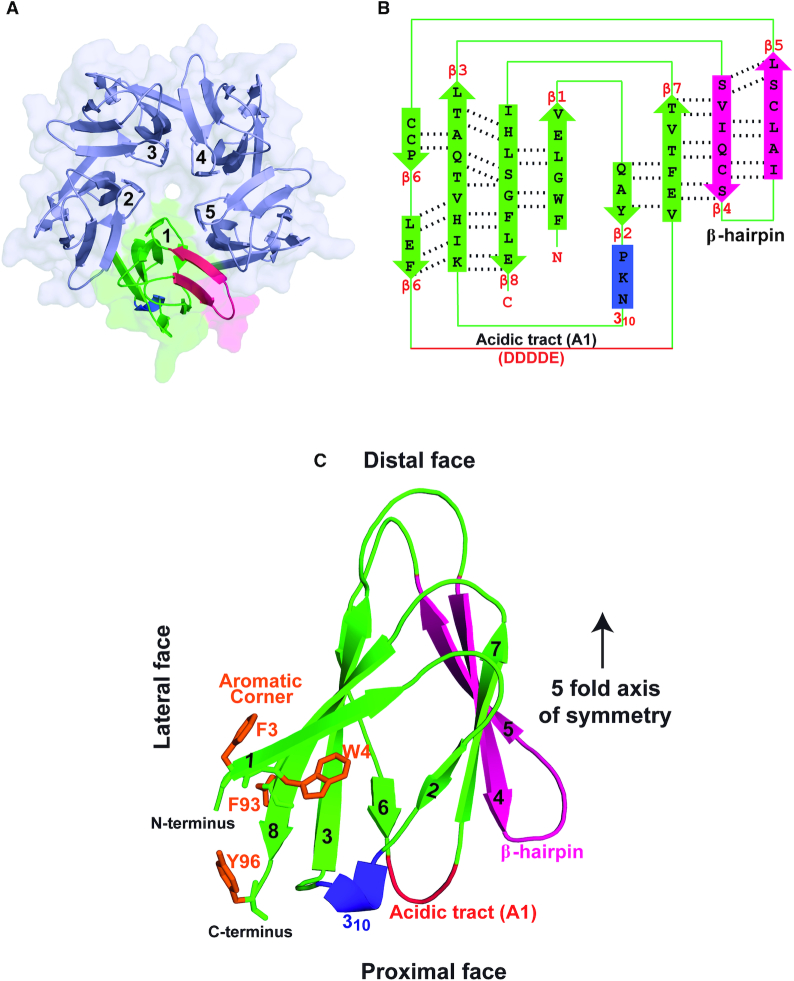
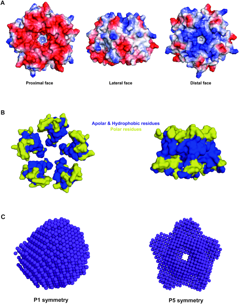
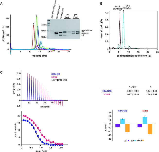
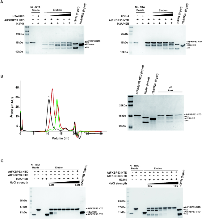
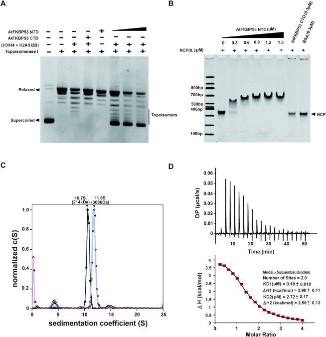
FKBP53 is one of the seven multi-domain FK506-binding proteins present in Arabidopsis thaliana, and it is known to get targeted to the nucleus. It has a conserved PPIase domain at the C-terminus and a highly charged N-terminal stretch, which has been reported to bind to histone H3 and perform the function of a histone chaperone. To better understand the molecular details of this PPIase with histone chaperoning activity, we have solved the crystal structures of its terminal domains and functionally characterized them. The C-terminal domain showed strong PPIase activity, no role in histone chaperoning and revealed a monomeric five-beta palm-like fold that wrapped over a helix, typical of an FK506-binding domain. The N-terminal domain had a pentameric nucleoplasmin-fold; making this the first report of a plant nucleoplasmin structure. Further characterization revealed the N-terminal nucleoplasmin domain to interact with H2A/H2B and H3/H4 histone oligomers, individually, as well as simultaneously, suggesting two different binding sites for H2A/H2B and H3/H4. The pentameric domain assists nucleosome assembly and forms a discrete complex with pre-formed nucleosomes; wherein two pentamers bind to a nucleosome.


Figures

Figure 1.
Figure 1.
Sequence alignment and crystal structure of AtFKBP53 CTD reveals a canonical FKBD fold. (A) Multiple sequence alignment of AtFKBP53 FKBD with human FKBP12 (HsFKBP12; UniProtKB – P62942), human FKBP13 (HsFKBP13; UniProtKB – P26885) and Burkholderia pseudomallei FKBP (BpFKBP; UniProtKB – Q63J95) was done using the T-Coffee program. The indicated residue numbering is based on hFKBP12, and secondary structures are based on AtFKBP53 CTD. The red coloured box highlights strictly conserved residues and the yellow coloured box highlights residues that are semi-conserved. The strictly conserved eleven residues known to play a role in PPIase activity and FK506-binding are marked by *. (B) Cartoon representation of AtFKBP53 FKBD in complex with FK506, showing the α-helices (red), β-strands (yellow), and loops (green). FK506 compound is shown as a stick model with elements highlighted in different colours (grey, C; blue, N; and red, O). (C) 2D topology diagram of AtFKBP53 FKBD with β-strands shown as arrows (yellow), α-helices as boxes (red), loops as lines (green) and the hydrogen bonding patterns between β-strands marked with dotted lines. (D) Electrostatic surface charge distribution of AtFKBP53 FKBD with a linear colour ramp of red (–kT) to blue (+kT) where the residues in the binding pocket involved in direct hydrogen bonding with FK506 are highlighted. FK506 compound is shown as a stick model with the elements highlighted in different colours (grey, C; blue, N; and red, O). (E) Superposition of crystal structure of AtFKBP53 FKBD (blue) with FKBP12 from H. sapiens (cyan) (HsFKBP12; PDB ID: 1FKJ); the 11 conserved residues involved in FK506-binding are represented as sticks and FK506 compound from 1FKJ as lines with elements highlighted in different colours (magenta, C; blue, N; and red, O).
Figure 2.
Figure 2.
Crystal structure of AtFKBP53 NTD reveals a nucleoplasmin fold. (A) Cartoon and surface representation of the crystal structure of AtFKBP53 NTD pentamer, wherein one monomer is highlighted in different colours. (B) 2D jelly-roll topology diagram of the monomer, showing the arrangement of the β strands, with the hydrogen bonding patterns marked with dotted lines. The acidic tract A1 formed of residues DDDDE, the 310 helix, and the β-hairpin motif are also labelled. (C) Cartoon representation of AtFKBP53 NTD monomer having the characteristic teardrop shape, labelled for the distal, lateral and proximal faces and the five-fold axis of symmetry (with a black arrow). The N and the C-terminal ends go in and come out respectively, from the proximal face. The aromatic corner made up of F3, W4, F93, Y96 (stick representation, with side-chains in orange), the 310 helix (in purple), and the β-hairpin motif formed by β4 and β5 (in pink) are also highlighted.
Figure 3.
Figure 3.
Surface charge, apolar and hydrophobic residue distribution and oligomeric status of AtFKBP53 NTD pentamer. (A) The electrostatic surface potential of AtFKBP53 NTD pentamer in three different orientations shows its proximal, lateral and distal faces. The figures were generated using PyMOL and coloured linearly from red (–kT) to blue (+kT). The proximal and distal faces reveal a relative abundance of acidic and basic charges, respectively. (B) The beta-sheets forming the inner and the outer cores of AtFKBP53 NTD pentamer in different orientations have been depicted in surface representation in yellow, showing the distribution of apolar and hydrophobic residues in blue, forming a concentric ring-like network, facing each other and thereby stabilizing the pentamer structure. (C) AtFKBP53 NTD envelope generated from the SAXS scattering profile using the ATSAS software DAMMIF and DAMAVER in P1 and P5 symmetries.
Figure 4.
Figure 4.
AtFKBP53 NTD pentamer interacts with H2A/H2B and H3/H4. (A). Analytical size-exclusion chromatography profile of AtFKBP53 NTD (purple), H2A/H2B (light blue), H3/H4 (violet), AtFKBP53 NTD/H2A/H2B mixture (green) and AtFKBP53 NTD/H3/H4 mixture (red) indicating stable complex formation. 18% SDS-PAGE gel showing the peak fractions obtained from size-exclusion chromatography, confirming complex formation (inset). (B) Analytical ultracentrifugation distance distribution c(S) versus sedimentation coefficient (S) plot obtained from SEDFIT software for AtFKBP53 NTD/H2A/H2B complex (dark red) and AtFKBP53 NTD/H3/H4 complex (sky blue). (C) Calorimetric titration of AtFKBP53 NTD with H2A/H2B and H3/H4 via ITC at 25°C. In the left upper panel heat changes of injections of AtFKBP53 NTD to H2A/H2B (blue) and H3/H4 (red) are shown. The resulting isotherms fitted with one-set of site binding model were shown in the left lower panel. The upper right panel shows the best fit values and fitting errors while the lower-left panel shows the graphical representation of thermodynamic parameters from the titration experiments. The data were acquired in a buffer containing 20 mM PIPES (pH 7.4) and 300 mM NaCl.
Figure 5.
Figure 5.
AtFKBP53 NTD can bind to both H2A/H2B and H3/H4, simultaneously. (A) The sequential binding experiment, wherein the left panel shows eluted fractions of saturated AtFKBP53 NTD–H3/H4 complex titrated with an increasing gradient of H2A/H2B, run on an 18% SDS PAGE gel. The right panel shows the 18% SDS PAGE gel image for the eluted fractions of saturated AtFKBP53 NTD–H2A/H2B complex, titrated with an increasing gradient of H3/H4. (B) Analytical size-exclusion chromatogram of AtFKBP53 NTD–H2A/H2B complex (green), AtFKBP53 NTD–H3/H4 complex (red) and AtFKBP53 NTD–H2A/H2B/H3/H4 complex (black). Collected peak fraction for AtFKBP53 NTD–H2A/H2B-H3/H4 complex analysed on an 18% SDS-PAGE gel (right panel) shows individual bands for all the components in the complex and confirms simultaneous binding of AtFKBP53 NTD to H2A/H2B and H3/H4. (C) Eluted fractions from separate pull-down experiments of AtFKBP53 NTD–H2A/H2B complex (left panel) and AtFKBP53 NTD–H3/H4 complex (right panel) performed with increasing salt concentrations (0.3–1.0 M) analysed on an 18% SDS-PAGE gel. The presence of both H2A/H2B and H3/H4 bound to AtFKBP53 NTD even at higher ionic strength conditions suggest a major role for hydrophobic interactions in stabilizing both the complexes. The intensity of the H2A/H2B bands at 0.5 M ionic strength suggests a role also for ionic interactions in stabilizing the complexes.
Figure 6.
Figure 6.
AtFKBP53 NTD is a functional histone chaperone and it interacts with NCP. (A) Supercoiling assay using relaxed closed circular (rcc) pUC19 plasmid (0.5 μg) incubated with histone H2A/H2B, H3/H4 and increasing amounts of AtFKBP53 NTD (1.0–3.0 μM). AtFKBP53 NTD shows supercoiling activity in the presence of histones and (rcc) pUC19 plasmid DNA, whereas AtFKBP53 FKBD did not show any supercoiling activity in the presence of histones and (rcc) pUC19. AtFKBP53 NTD and histones alone were also not able to induce supercoiling in rccPU19 DNA. (B) Electrophoretic mobility shift assay (EMSA) of NCP with AtFKBP53 NTD pentamer in incremental ratios ranging from 1:1 to 1:5 on a 6% Native-PAGE stained with ethidium bromide. NCP with AtFKBP53 CTD and BSA separately in 1:1 ratio were run as controls. The gel showed that AtFKBP53 NTD pentamer binds to NCP, and the reaction appears to reach saturation at about 1:2 ratio, whereas AtFKBP53 FKBD and BSA did not show any specific interaction with NCP. (C) SV-AUC distance distribution c(S) versus sedimentation coefficient (S) plot obtained from SEDFIT software for NCP (black), a mixture of NCP and AtFKBP53 NTD pentamer in 1:2 ratio (sky blue) and a mixture of NCP and AtFKBP53 NTD pentamer in 1:3 ratio (light pink). (D) Calorimetric titration of AtFKBP53 NTD pentamer with NCP at 25°C. In the right upper panel, heat changes from injections of AtFKBP53 NTD into NCP (black) are shown. In the lower panel, the resulting isotherms (fitted with a sequential binding model) are shown. The actual experimental positions are represented as a black line and the fitted positions are represented as red dots. The dissociation constant value (Kd) and thermodynamic parameters are shown as an inset in the lower panel. Data were acquired in 20 mM Tris (pH 7.5), 50 mM NaCl and 1.0 mM β-mercaptoethanol.

Similar articles

Cited by

References

    1. Galat A. Peptidylprolyl cis/trans isomerases (immunophilins): biological diversity–targets–functions. Curr. Top. Med. Chem. 2003; 3:1315–1347. - PubMed
    1. Gothel S.F., Marahiel M.A.. Peptidyl-prolyl cis-trans isomerases, a superfamily of ubiquitous folding catalysts. Cell Mol. Life Sci. 1999; 55:423–436. - PMC - PubMed
    1. Hamilton G.S., Steiner J.P.. Immunophilins: beyond immunosuppression. J. Med. Chem. 1998; 41:5119–5143. - PubMed
    1. Siekierka J.J., Wiederrecht G., Greulich H., Boulton D., Hung S.H., Cryan J., Hodges P.J., Sigal N.H.. The cytosolic-binding protein for the immunosuppressant FK-506 is both a ubiquitous and highly conserved peptidyl-prolyl cis-trans isomerase. J. Biol. Chem. 1990; 265:21011–21015. - PubMed
    1. Kang C.B., Hong Y., Dhe-Paganon S., Yoon H.S.. FKBP family proteins: immunophilins with versatile biological functions. Neurosignals. 2008; 16:318–325. - PubMed

Publication types

MeSH terms