. 2020 Apr 15;31(4):1093-1106.
doi: 10.1021/acs.bioconjchem.9b00790. Epub 2019 Dec 20.

Programmable Assembly of Adeno-Associated Virus-Antibody Composites for Receptor-Mediated Gene Delivery

Programmable Assembly of Adeno-Associated Virus-Antibody Composites for Receptor-Mediated Gene Delivery

Alina C Zdechlik et al. Bioconjug Chem. .

Abstract

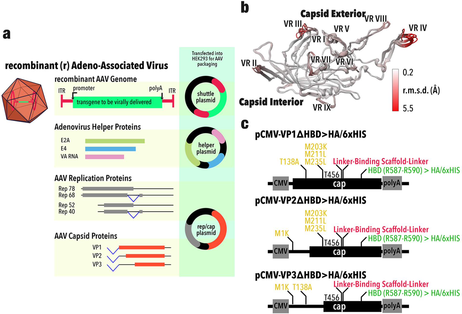
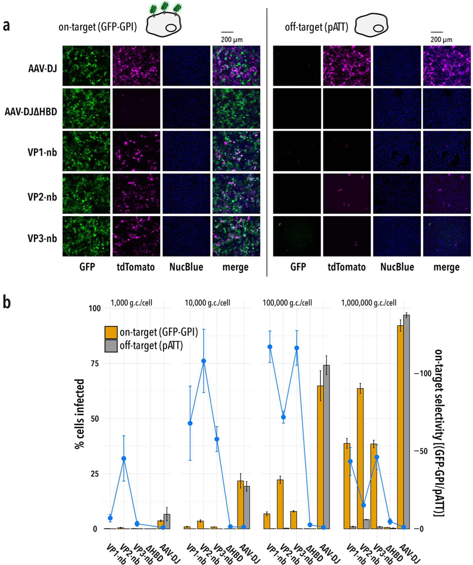
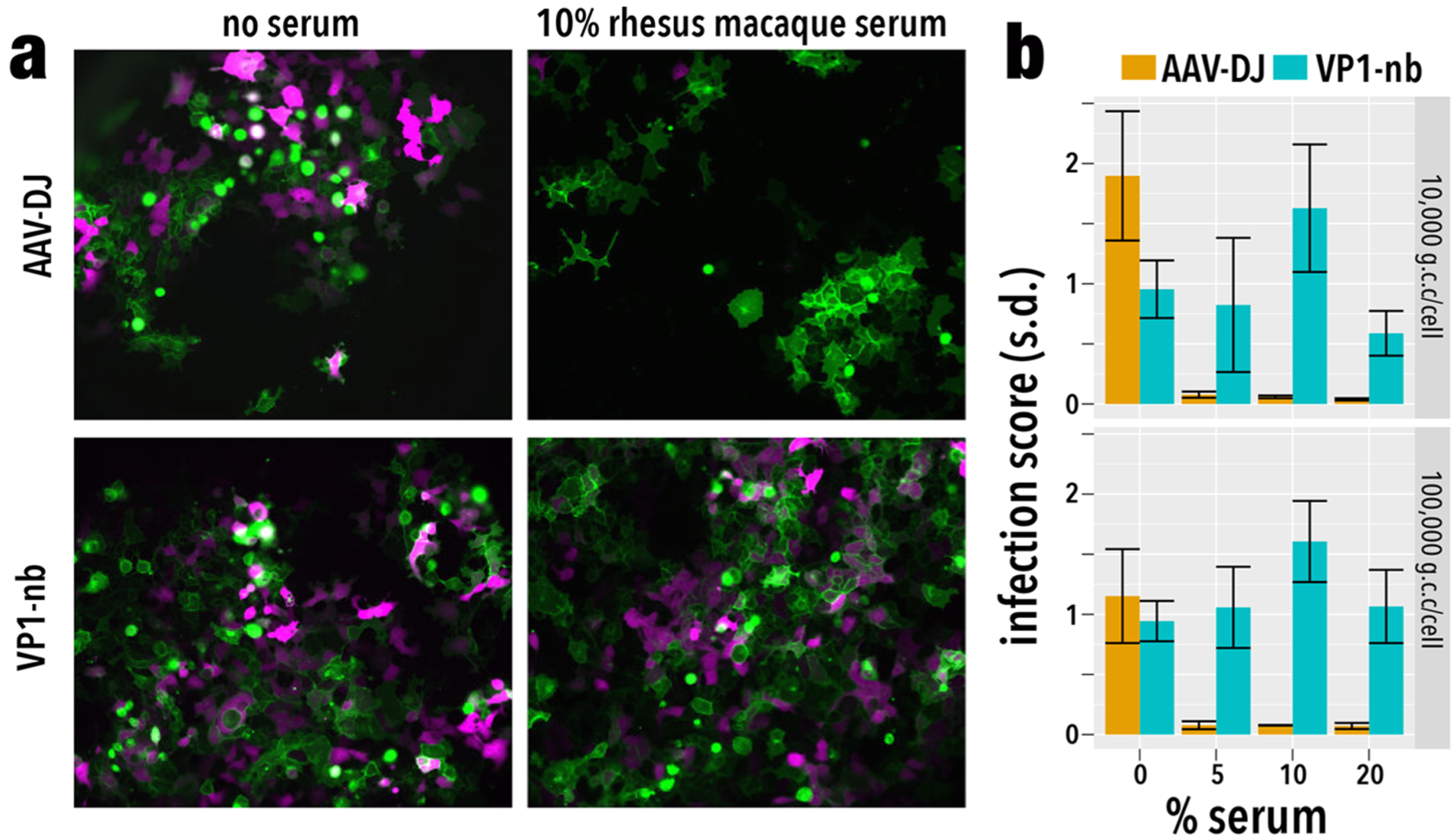
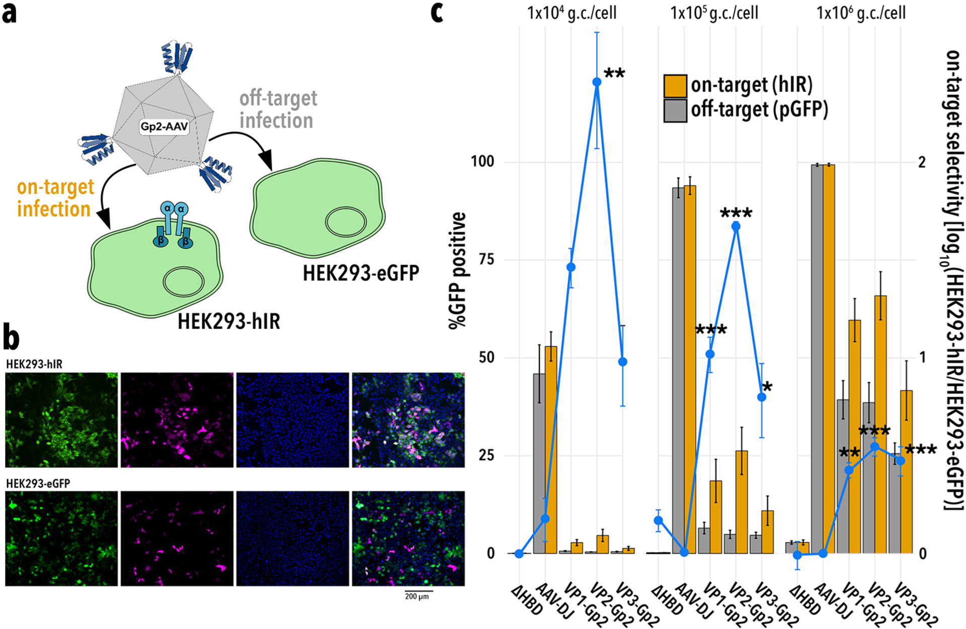
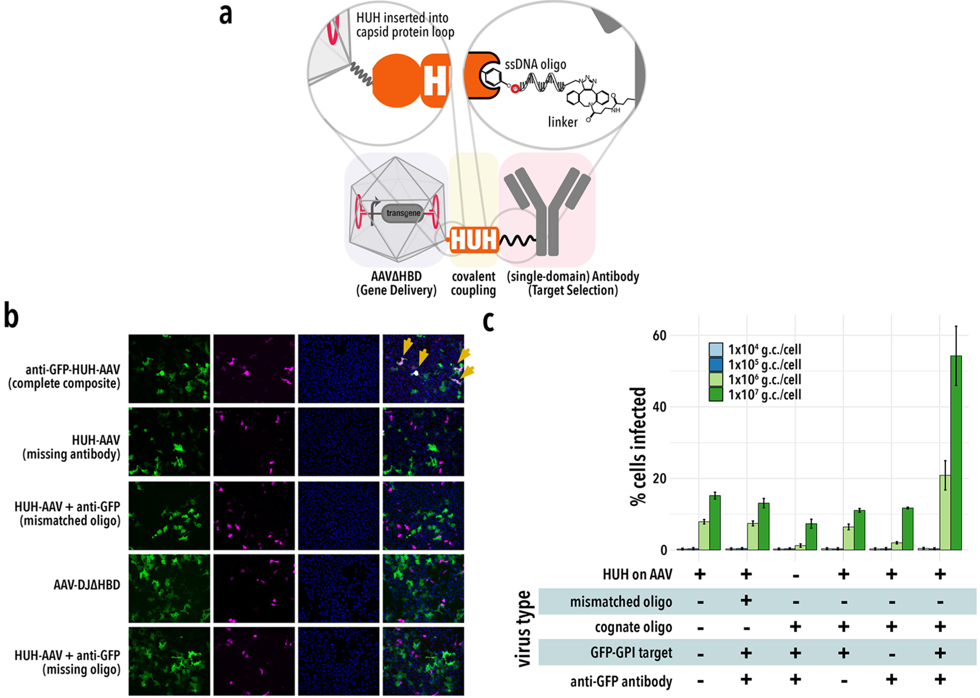
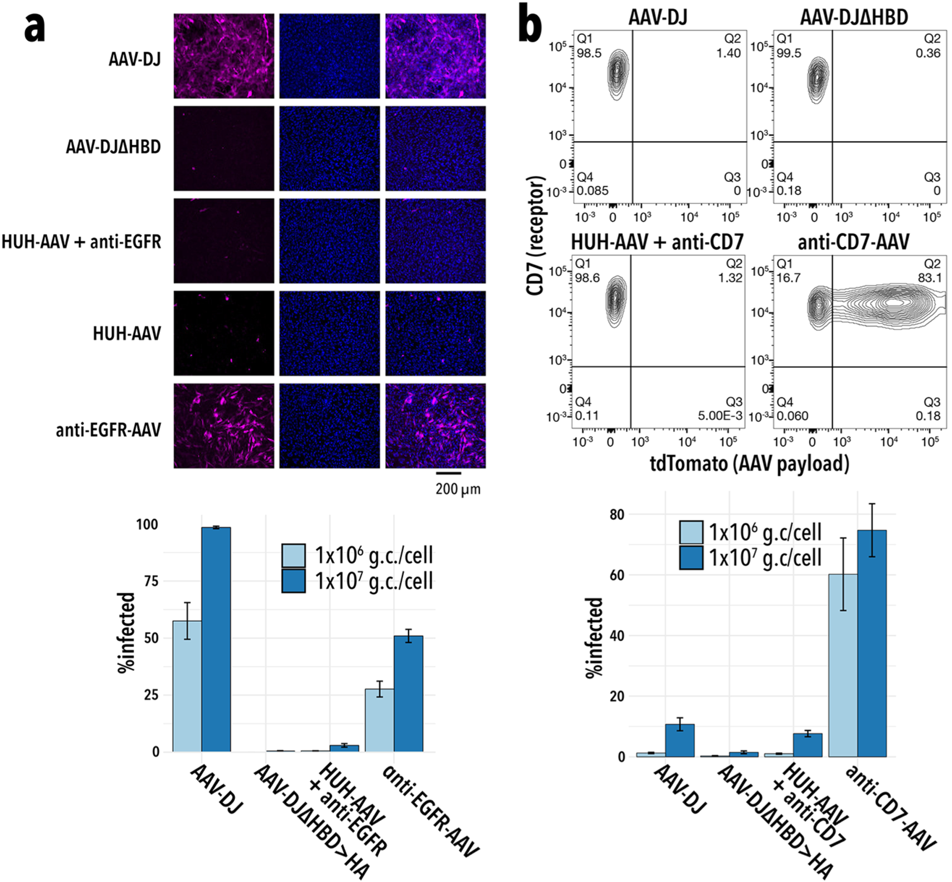
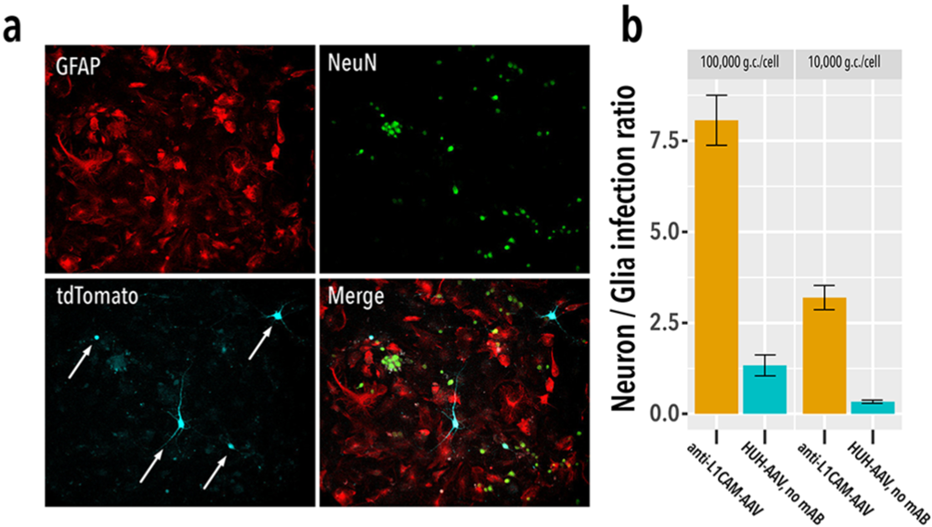
Adeno-associated virus (AAV) has emerged as a viral gene delivery vector that is safe in humans, able to infect both dividing and arrested cells and drive long-term expression (>6 months). Unfortunately, the naturally evolved properties of many AAV serotypes-including low cell type specificity and largely overlapping tropism-are mismatched to applications that require cell type-specific infection, such as neural circuit mapping or precision gene therapy. A variety of approaches to redirect AAV tropism exist, but there is still the need for a universal solution for directing AAV tropism toward user-defined cellular receptors that does not require extensive case-by-case optimization and works with readily available components. Here, we report AAV engineering approaches that enable programmable receptor-mediated gene delivery. First, we genetically encode small targeting scaffolds into a variable region of an AAV capsid and show that this redirects tropism toward the receptor recognized by these targeting scaffolds and also renders this AAV variant resistant to neutralizing antibodies present in nonhuman primate serum. We then simplify retargeting of tropism by engineering the same variable loop to encode a HUH tag, which forms a covalent bond to single-stranded DNA oligos conjugated to store-bought antibodies. We demonstrate that retargeting this HUH-AAVs toward different receptors is as simple as "arming" a premade noninfective AAV template with a different antibody in a conjugation process that uses widely available reagents and requires no optimization or extensive purification. Composite antibody-AAV nanoparticles structurally separate tropism and payload encapsulation, allowing each to be engineered independently.


Conflict of interest statement

The authors declare no competing financial interest.

Figures

Figure 1.
Figure 1.
Engineering of AAV capsid protein. a, Recombinant AAV is packaged in HEK293 cells after transfection of three plasmids: the shuttle plasmid (contains the AAV payload, green) flanked by AAV2 ITRs, red); a helper plasmid encoding Adenovirus proteins (E2A, E4) plus VA RNA required for AAV replication and packaging; a plasmid that encodes AAV rep and cap genes. While the former expresses replication factors (e.g., Rep78), the latter gives rise to three capsid proteins (VP1–3) through alternative splicing (blue lines) and start codons (ORFs shown in orange). b, Superimposed crystal structures of VP from different AAV serotypes (AAV1 PDB 3NG9, 5EGC; AAV2 PDB 1LP3; AAV3 PDB 3KIC; AAV6 PDB 3SHM, 4V86, 30AH; AAV8 2QA0; AAV9 3UX1). Root mean square deviation (rmsd) is mapped onto the structures (white to red). Variable regions (VR) and loops are annotated. c, Expression of individual modified capsid proteins is achieved by mutating (alternative) start codons (gold), inserting the targeting scaffold into position T456 (red), and replacing the endogenous heparan binding domain (HBD) with an HA or 6xHIS tag (green).
Figure 2.
Figure 2.
Receptor-mediated infection in a synthetic system. a, Wide-field fluorescent imaging of cells transiently transfected with surface-anchored GFP (left panels) or mock-transfected (right panels) 48 h after viral transduction with 1 × 10⁶ g.c./cell. Individual channels are pseudocolored green (GFP), magenta (tdTomato, the viral payload), and blue (Hoechst 33342). While wt AAV-DJ infects nonspecifically, and AAV-DJΔHBD > HA infects neither, AAV-nb composites, in which the anti-GFP nanobody is incorporated into VP1, VP2, or VP3, infects only GFP positive cells. b, On-target infection (GFP-GPI transfected, yellow) and off-target infection (mock transfected with plasmid pATT, gray) as a function of multiplicity of infection (MOI). On-target selectivity expressed as the ratio of on-target vs off-target infection (blue dots, no selectivity = 1). All error bars indicate standard error (n = 3).
Figure 3.
Figure 3.
Nanobody-AAV are resistant to neutralizing antibodies. a, HEK293 expressing GFP (green) as a synthetic receptor on the extracellular face of the cell membrane was infected—in the absence or presence of 10% pooled, mixed gender rhesus macaque serum (BioIVT)—with either 100,000 genome copies (g.c.)/cell AAV-DJ or VPl-nb(anti-GFP) AAV composites, both delivering tdTomato (magenta). b, Infection scores for AAV-DJ (gold) and VPl-nb-AAV (teal) at different serum concentrations were determined by flow cytometry at two different multiplicities of infection.
Figure 4.
Figure 4.
Receptor-mediated transduction of a breast cancer model cell line. a, AAV engineered to express a human insulin receptor (hIR) specific Gp2 targeting scaffold (Gp2IR, blue cartoon representation) is expected to infect hIR-expressing HEK293 cells (on-target; also express GFP) and not control HEK293-EGFP cells (off-target). b, Wide-field fluorescent imaging 48 h after viral transduction with 1 × 10⁶ genome copies (g.c.)/cell VP2-Gp2IR-AAV. Individual channels are pseudocolored green (GFP), magenta (tdTomato, the viral payload), and blue (Hoechst 33342). Gp2 IR-AAV preferentially infects hIR-positive cells. c, Comparing AAV tropism for HEK293-hIR (orange), and HEK293-EGFP (gray). Bar height represents % infected cells at a specific multiplicity of infection (MOI, g.c./cell). While AAV-DJ is infecting cells indiscriminately, and AAV-ΔHBD is noninfective, Gp2IR-AAV preferentially infects hIR over-expressing cells. The observed off-target infection likely is due to basal expression of endogenous hIR in control cells. Importantly, while off-target transduction is observed at high MOI, at lower MOI better specificity can be achieved. For example, at a MOI of 1 × 10⁴ g.c./cell, the ratio of on-target vs off-target infection is >11-fold for VP2-Gp2IR-AAV. Significance of the difference in on-target vs off-target infection is tested by a two-sided Dunnett’s test for multiple comparison with AAV-DJ as the control. Significance levels: * p < 0.05, ** p < 0.01, *** p < 0.001, n.s. otherwise. All error bars indicate standard error (n = 3).
Figure 5.
Figure 5.
Generalized receptor-mediated infection using antibody-AAV composites. a, Covalently linked composite structures contain an AAV particle (packaging the delivered transgene) and an antibody that selectively binds cell-type specific surface markers. A HUH domain (here, mMobA) bridges the two components and confers programmable assembly. Insets provide further detail on HUH domain display on the capsid surface and HUH/antibody linkage (formation of a phosphotyrosine adduct with conjugated ssDNA-mAb). b, Wide-field fluorescent imaging of cells transiently transfected with surface-anchored GFP 48 h after viral transduction with 1 × 10⁶ g.c./cell of the indicated virus. Individual channels are pseudocolored green (GFP), magenta (tdTomato, the viral payload), and blue (Hoechst 33342). Only complete anti-GFP-HUH-AAV composites mediate specific infection of GFP positive cells (yellow arrows). With any component missing (mMobA HUH, oligo, or antibody), only basal off-target infection is observed, which is not specific to GFP positive cells (i.e., there is no colocalization of tdTomato and GFP signals). c, Infection efficiency is quantified at different multiplicities of infection (MOI) for the indicated virus. Bar height represents % infected cells at a specific multiplicity of infection (MOI, g.c./cell). All error bars indicate standard error (n = 3).
Figure 6.
Figure 6.
Efficient retargeting of premade AAV. a, Wide-field fluorescent imaging 48 h after viral transduction of U251 MG cells with 1 × 10⁶ genome copies (g.c.)/cell of the indicated virus. Individual channels are pseudocolored magenta (tdTomato, the viral payload) and blue (Hoechst 33342). While AAV-DJ and complete anti-EGFR-AAV composites infect strongly, AAV-DJΔHBD > HA and composites formed omitting either oligo or antibody are noninfective. Infection efficiency is quantified at different multiplicities of infection (MOI) for the indicated virus (bottom panel). b, Anti-CD7-AAV composites infect Jurkat cells. Shown are representative flow analysis examples from Jurkat cells infected with the AAV-DJ, AAV-DJΔHBD > HA virus, HUH-AAV composites formed with nonoligo labeled anti-CD7, and complete anti-CD7-AAV composites. Cells were infected with each virus at a MOI of 1 × 10⁷ g.c./cell. Cells are counterstained for surface CD7 expression. Percentage of cells in each quadrant are indicated in each corner. Only complete anti-CD7-AAV composites transduce Jurkat cells efficiently. Infection efficiency is further quantified at different MOIs (bottom panel). All error bars indicate standard error (n = 3).
Figure 7.
Figure 7.
Receptor-mediated infection in primary neuron culture. a, Immunohistochemistry of primary neuron/glia coculture infected with L1CAM-conjugated AAV delivering tdTomato. IHC identifies neurons (NeuN, green), glia (GFAP, red), and infected neuron (cyan, white arrows). b, Infection ratio of neuron vs glia. While HUH-AAV without conjugated L1CAM antibody (HUH-AAV, no mAB) infects both neurons and glia (teal), L1CAM-conjugated AAV predominantly infects neurons (yellow). All error bars indicate standard error (n = 7–9). g.c., genome copies; mAB, monoclonal antibody.

Similar articles

Cited by

References

    1. Balakrishnan B, and Jayandharan GR (2014) Basic biology of adeno-associated virus (AAV) vectors used in gene therapy. Curr. Gene Ther 14, 86–100. - PubMed
    1. Smith RH (2008) Adeno-associated virus integration: virus versus vector. Gene Ther. 15, 817–822. - PubMed
    1. Vandamme C, Adjali O, and Mingozzi F (2017) Unraveling the Complex Story of Immune Responses to AAV Vectors Trial After Trial. Hum. Gene Ther 28, 1061–1074. - PMC - PubMed
    1. Dismuke DJ, Tenenbaum L, and Samulski RJ (2014) Biosafety of recombinant adeno-associated virus vectors. Curr. Gene Ther 13, 434–452. - PubMed
    1. Betley JN, and Sternson SM (2011) Adeno-associated viral vectors for mapping, monitoring, and manipulating neural circuits. Hum. Gene Ther 22, 669–677. - PMC - PubMed

Publication types