

Bilateral Gait 6 and 12 Months Post-Anterior Cruciate Ligament Reconstruction Compared with Controls
- PMID: 31809411
- PMCID: PMC7078064
- DOI: 10.1249/MSS.0000000000002208
Bilateral Gait 6 and 12 Months Post-Anterior Cruciate Ligament Reconstruction Compared with Controls
Abstract
Purpose: To compare gait biomechanics throughout stance phase 6 and 12 months after unilateral anterior cruciate ligament reconstruction (ACLR) between ACLR and contralateral limbs and compared with controls.
Methods: Vertical ground reaction force (vGRF), knee flexion angle (KFA), and internal knee extension moment (KEM) were collected bilaterally 6 and 12 months post-ACLR in 30 individuals (50% female, 22 ± 3 yr, body mass index = 23.8 ± 2.2 kg·m) and at a single time point in 30 matched uninjured controls (50% female, 22 ± 4 yr, body mass index = 23.6 ± 2.1 kg·m). Functional analyses of variance were used to evaluate the effects of limb (ACLR, contralateral, and control) and time (6 and 12 months) on biomechanical outcomes throughout stance.
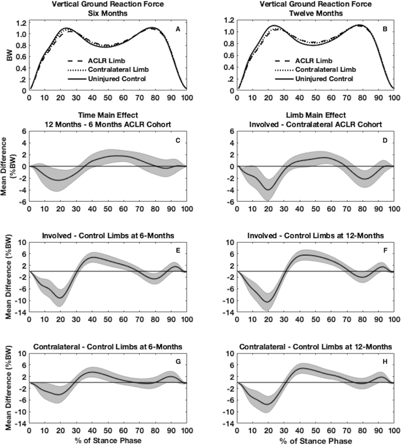
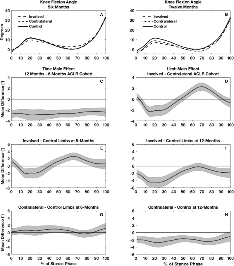
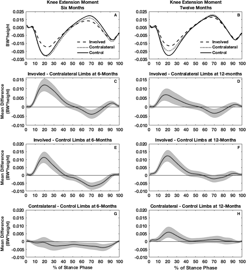
Results: Compared with the uninjured controls, the ACLR group demonstrated bilaterally lesser vGRF (ACLR, 9% body weight [BW]; contralateral, 4%BW) during early stance and greater vGRF during midstance (ACLR, 5%BW; contralateral, 4%BW) 6 months post-ACLR. Compared to the uninjured controls, the ACLR group demonstrated bilaterally lesser vGRF (ACLR, 10%BW; contralateral, 8%BW) during early stance and greater vGRF during midstance (ACLR, 5%BW; contralateral, 5%BW) 12 months post-ACLR. Compared with controls, the ACLR limb demonstrated lesser KFA during early stance at 6 (2.3°) and 12 months post-ACLR (2.0°), and the contralateral limb demonstrated lesser KFA during early stance at 12 months post-ACLR (2.8°). Compared with controls, the ACLR limb demonstrated lesser KEM during early stance at both 6 months (0.011BW × height) and 12 months (0.007BW × height) post-ACLR, and the contralateral limb demonstrated lesser KEM during early stance only at 12 months (0.006BW × height).
Conclusions: Walking biomechanics are altered bilaterally after ACLR. During the first 12 months post-ACLR, both the ACLR and contralateral limbs demonstrate biomechanical differences compared with control limbs. Differences between the contralateral and control limbs increase from 6 to 12 months post-ACLR.
Conflict of interest statement
Figures
References
-
- Hart HF, Culvenor AG, Collins NJ et al. Knee kinematics and joint moments during gait following anterior cruciate ligament reconstruction: a systematic review and meta-analysis. British journal of sports medicine. 2016;50(10):597–612. - PubMed
-
- Kaur M, Ribeiro DC, Theis JC, Webster KE and Sole G. Movement Patterns of the Knee During Gait Following ACL Reconstruction: A Systematic Review and Meta-Analysis. Sports medicine (Auckland, N.Z.). 2016;46(12):1869–95. - PubMed
-
- Erhart-Hledik JC, Favre J and Andriacchi TP. New insight in the relationship between regional patterns of knee cartilage thickness, osteoarthritis disease severity, and gait mechanics. Journal of biomechanics. 2015;48(14):3868–75. - PubMed
Publication types
MeSH terms
Grants and funding
LinkOut - more resources
Full Text Sources
Medical
