. 2019 Dec 6;14(12):e0226145.
doi: 10.1371/journal.pone.0226145. eCollection 2019.

Ameliorating effects of Gö6976, a pharmacological agent that inhibits protein kinase D, on collagen-induced arthritis

Affiliations

Ameliorating effects of Gö6976, a pharmacological agent that inhibits protein kinase D, on collagen-induced arthritis

Tae Won Yoon et al. PLoS One. .

Abstract

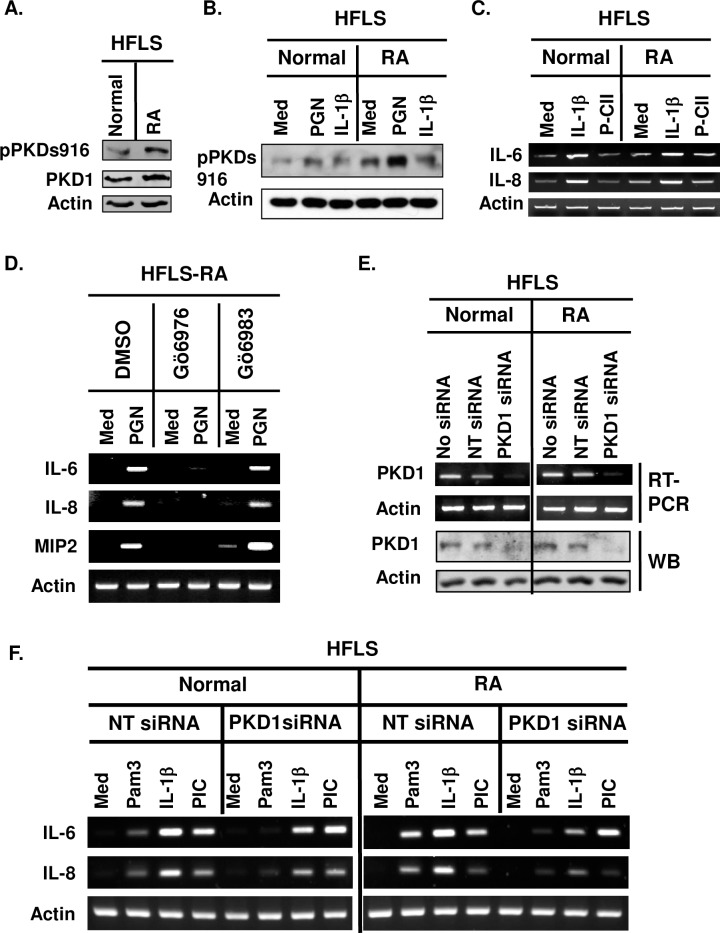
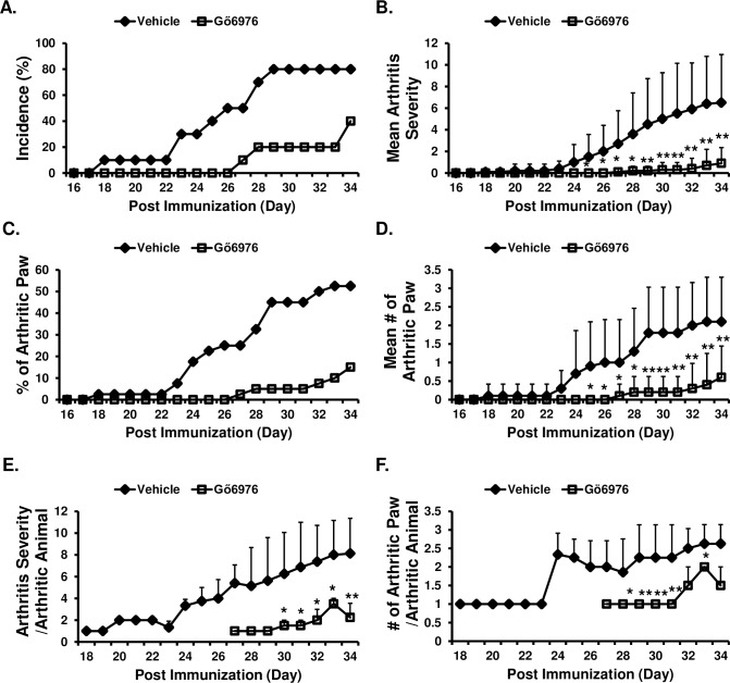
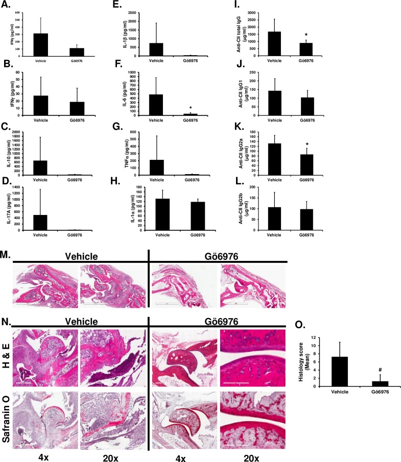
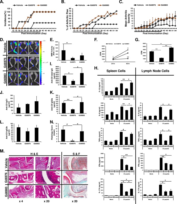
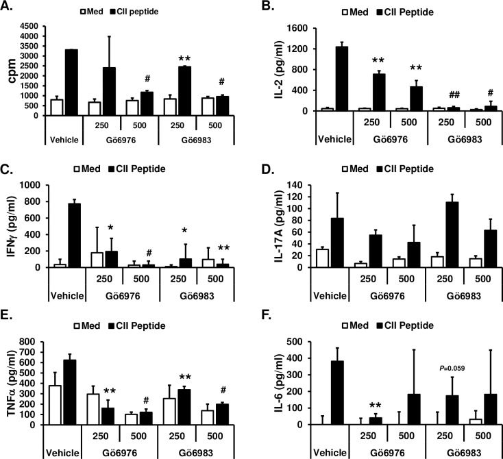
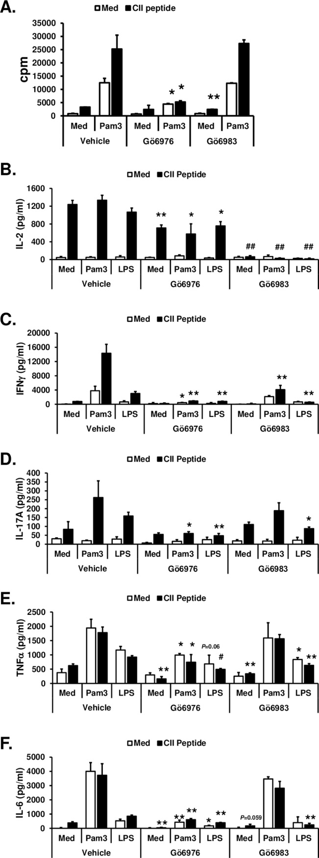
Toll-like receptor (TLR) signaling can contribute to the pathogenesis of arthritis. Disruption of TLR signaling at early stages of arthritis might thereby provide an opportunity to halt the disease progression and ameliorate outcomes. We previously found that Gö6976 inhibits TLR-mediated cytokine production in human and mouse macrophages by inhibiting TLR-dependent activation of protein kinase D1 (PKD1), and that PKD1 is essential for proinflammatory responses mediated by MyD88-dependent TLRs. In this study, we investigated whether PKD1 contributes to TLR-mediated proinflammatory responses in human synovial cells, and whether Gö6976 treatment can suppress the development and progression of type II collagen (CII)-induced arthritis (CIA) in mouse. We found that TLR/IL-1R ligands induced activation of PKD1 in human fibroblast-like synoviocytes (HFLS). TLR/IL-1R-induced expression of cytokines/chemokines was substantially inhibited in Gö6976-treated HFLS and PKD1-knockdown HFLS. In addition, serum levels of anti-CII IgG antibodies, and the incidence and severity of arthritis after CII immunization were significantly reduced in mice treated daily with Gö6976. Synergistic effects of T-cell receptor and TLR, as well as TLR alone, on spleen cell proliferation and cytokine production were significantly inhibited in the presence of Gö6976. Our results suggest a possibility that ameliorating effects of Gö6976 on CIA may be due to its ability to inhibit TLR/IL-1R-activated PKD1, which might play an important role in proinflammatory responses in arthritis, and that PKD1 could be a therapeutic target for inflammatory arthritis.


Conflict of interest statement

The authors have declared that no competing interests exist.

Figures

Fig 1
Fig 1. PKD1 can be activated and plays an essential role in both spontaneous and TLR/IL-1R-induced expression of proinflammatory mediators in human synoviocytes.
(A) Protein levels and phosphorylation status of PKD in human primary fibroblast-like synoviocytes (HFLS) from a normal donor (HFLS-N) and from a patient with RA (HFLS-RA) were detected by Western blot. (B) HFLS-N and HFLS-RA were stimulated with media (med), TLR2 ligand peptidoglycan (PGN; 50 μg/ml), or human recombinant IL-1β (10 ng/ml) for 45 min. Protein levels and phosphorylation status of PKD were detected by Western blot. (C) HFLS-N and HFLS-RA were stimulated with media (med), IL-1β (10 ng/ml), or synthetic CII peptide (P-CII; 50 μg/ml) for 4 hr. Levels of IL-6 and IL-8 mRNA were analyzed by RT-PCR. (D) HFLS-RA were pretreated with vehicle (1% v/v DMSO), Gö6976 (500 ng/ml) or Gö6983 (500 ng/ml) for 1hr and then stimulated with PGN (10 μg/ml) for 4 hr. Levels of the indicated cytokine and chemokine mRNA were analyzed by RT-PCR. (E, F) HFLS-N and HFLS-RA were transiently transfected with non-target siRNA (NT siRNA; control) or PKD1-specific siRNA (PKD1 siRNA; PKD1-knockdown). mRNA levels (E-top panel) and protein levels (E-bottom panel) of PKD1 were detected by RT-PCR and Western blot, respectively. Control and PKD1-knockdown HFLS were stimulated with Pam3CSK4 (Pam3; 500 ng/ml), IL-1β (10 ng/ml), or TLR3 ligand PIC (50 μg/ml) for 4 hr. Messenger RNA levels of the indicated gene were analyzed by RT-PCR. All experiments were repeated two to three times with similar results. S1 Fig shows uncropped blot and gel scans.
Fig 2
Fig 2. Effects of Gö6976 on the development and progression of CIA.
HLA-DR1 mice were immunized with bovine CII and treated daily with vehicle (n = 10) or Gö6976 (n = 10) for 34 days starting at the day of CII immunization. Mice were observed daily and development of arthritis was assessed. (A) Incidence of arthritis. (B) Mean arthritis severity. Data represent the mean ± SD. *, p < 0.05; **, p < 0.005. (C) The % of arthritic paws. (D) Mean number of arthritic paws in a mouse. Data represent the mean ± SD. *, p < 0.05; **, p < 0.005. (E) Arthritis severity score in an arthritic mouse. Data represent the mean ± SD. *, p < 0.05; **, p < 0.005. (F) Number of arthritic paws in an arthritic mouse. Data represent the mean ± SD. *, p < 0.05; **, p < 0.005. Experiments were repeated four times with similar results.
Fig 3
Fig 3. Effects of Gö6976 treatments on the anti-CII IgG antibody production, joint inflammation and bone destruction in HLA-DR1 mice immunized with CII.
HLA-DR1 mice were immunized with bovine CII and treated daily with vehicle (n = 4) or Gö6976 (n = 5) for 34 days starting at the day of CII immunization. Blood (at day 11 and day 35) and joints (at day 35) were harvested. (A—H) Levels of the selected cytokines in serum isolated at day 11 (A) and at day 35 (B—H) were analyzed by multiplex sandwich immunoassay. Data represent the mean concentration (pg/ml) ± SD. *, p < 0.05. (I–L) Levels of total anti-CII IgG antibodies (I), anti-CII IgG1 antibodies (J), anti-CII IgG2a antibodies (K) and anti-CII IgG2b antibodies (L) in serum isolated at day 35 were analyzed by isotype-specific anti-CII IgG ELISA. Data represent the mean concentration (μg/ml) ± SD. *, p < 0.05. (M) Representative H&E stained joint tissue section (1.5 x of the original objective; white scale bar = 2 mm). (N) Representative H&E (top) or safranin O (bottom) stained joint tissue sections. White scale bar = 600 μm at 4 x of the original objective. White scale bar = 200 μm at 20 x of the original objective. (O) Histology score (vehicle: n = 8 paws, Gö6976: n = 10 paws). Data represent the mean ± SD. #, p < 0.0005. Experiments were repeated two to four times with similar results.
Fig 4
Fig 4. Differential effects of Gö6976 and Gö6983 on CIA.
HLA-DR1 mice were immunized with bovine CII and treated daily with vehicle (n = 5), Gö6976 (n = 3), Gö6983 (n = 3) for 39 days (indicated as underline) starting at the day of CII immunization. Mice were observed daily and development of arthritis was assessed up to 54 days post immunization. (A) Incidence of arthritis. (B) The % of arthritic paws. (C) Mean arthritis severity score. Data represent the mean ± SD. *, p < 0.05. (D—E) At day 54, mice were injected i.v. with MMPSense® 750 FAST Fluorescent Imaging Agent (750F). Twenty four hours later, mice were anesthetized and then scanned using IVIS® Lumina XR System (D). Fluorescence intensities were quantified within ROI by using Living Image 4.0 software (E). Calculations are represented graphically as radiant efficiency (photons/s/cm2/str)/(μW/cm2). Data represent the mean of the group ± SD. *, p < 0.05. (F-N) At day 55, inguinal lymph nodes, spleens, blood, and hind paws were harvested. (F, G) Lymph node cells were incubated in 96-well plate (8 x 10⁵ cells/200 μl/well) for 43 hrs. One μCi of [3H]thymidine was added to each well, and the cells were harvested after an additional 5 hr of culture. Data present the mean (cpm) ± SD. *, p < 0.05; **, p < 0.005. (H) Spleen cells and lymph node cells were stimulated with media or synthetic CII peptide (10 μg/ml) for 24 hr (for IL-6 in spleen cell culture), 48 hr (for IFNγ and IL-17A in both spleen cell and lymph node cell culture), or 72 hr (for IL-1β and TNFα in both spleen cell and lymph node cell culture, and IL-6 on lymph node cell culture). The levels of cytokines in the culture supernatants were analyzed by multiplex sandwich immunoassay. Data represent the mean concentration (pg/ml) ± SD. *, p < 0.05; **. (I–L) Levels of total serum anti-CII IgG antibodies (I), anti-CII IgG1 antibodies (J), anti-CII IgG2a antibodies (K) and anti-CII IgG2b antibodies (L) were analyzed by isotype-specific anti-CII IgG ELISA. Data represent the mean concentration (μg/ml) ± SD. *, p < 0.05; **, p < 0.005. (M) Representative H&E or S&F stained joint tissue sections at day 55 post immunization. White scale bar = 600 μm at 4 x of the original objective. White scale bar = 200 μm at 20 x of the original objective. (N) Histology score (vehicle: n = 10 paws, Gö6976: n = 6 paws, Gö6983: n = 6 paws). Data represent the mean ± SD. *, p < 0.05; #, p < 0.0005.
Fig 5
Fig 5. Inhibitory effects of Gö6976 and Gö6983 on TCR-mediated T cell activation.
Spleen cells were isolated from humanized CII-specific TCR/HLA-DR1 double transgenic mice. (A) Cells (1x10⁵ cells/300 μl/well in 96-well plate) were pretreated with vehicle (1% v/v DMSO), Gö6976 (250 ng/ml or 500 ng/ml), or Gö6983 (250 ng/ml or 500 ng/ml) for 30 min and then stimulated with media or synthetic CII peptide (10 μg/ml) for 48 hrs. One μCi of [3H]thymidine was added to each well, and the cells were harvested after an additional 16 hr of culture. Data present the mean (cpm) ± SD of triplicates. (B-F) Cells (4x10⁵ cells/200 μl/well in 96-well plate) were pretreated with vehicle (1% v/v DMSO), Gö6976 (250 ng/ml or 500 ng/ml), or Gö6983 (250 ng/ml or 500 ng/ml) for 30 min and then stimulated with media or synthetic CII peptide (50 μg/ml) for 48 hrs. The levels of the indicated cytokine in the culture supernatants were measured by ELISA. Data represent the mean concentration (pg/ml) ± SD of triplicates. The differences between the control (vehicle pretreated group) and experimental group (Gö6976 or Gö6983 pretreated group) were evaluated using two-tailed Student’s t-test. *, p < 0.05; **, p < 0.005; #, p < 0.0005; ##, p < 0.00005. Experiments were repeated three to five times with similar results.
Fig 6
Fig 6. Differential effects of Gö6976 and Gö6983 on TLR-mediated spleen cell proliferation and cytokine production.
Spleen cells were isolated from humanized CII-specific TCR/HLA-DR1 double transgenic mice. (A) Cells (1x10⁵ cells/300 μl/well in 96-well plate) were pretreated with vehicle (1% v/v DMSO), Gö6976 (250 ng/ml), or Gö6983 (250 ng/ml) for 30 min and then stimulated with media or Pam3CSK4 (Pam3; 500 ng/ml) in the presence or absence of synthetic CII peptide (10 μg/ml) for 48 hrs. One μCi of [3H]thymidine was added to each well, and the cells were harvested after an additional 16 hr of culture. Data present the mean (cpm) ± SD of triplicates. (B-F) Cells (4x10⁵ cells/200 μl/well in 96-well plate) were pretreated with vehicle (1% v/v DMSO), Gö6976 (250 ng/ml), or Gö6983 (250 ng/ml) for 30 min and then stimulated with media, Pam3CSK4 (Pam3; 500 ng/ml), or LPS (25 ng/ml) in the presence or absence of synthetic CII peptide (50 μg/ml) for 48 hrs. The levels of the indicated cytokine in the culture supernatants were measured by ELISA. Data represent the mean concentration (pg/ml) ± SD of triplicates. The differences between the control (vehicle pretreated group) and experimental group (Gö6976 or Gö6983 pretreated group) were evaluated using two-tailed Student’s t-test. *, p < 0.05; **, p < 0.005; #, p < 0.0005; ##, p < 0.00005. Experiments were repeated three to five times with similar results.

Similar articles

Cited by

References

    1. Firestein GS. Evolving concepts of rheumatoid arthritis. Nature. 2003;423(6937):356–61. 10.1038/nature01661 . - DOI - PubMed
    1. van der Heijden IM, Wilbrink B, Tchetverikov I, Schrijver IA, Schouls LM, Hazenberg MP, et al. Presence of bacterial DNA and bacterial peptidoglycans in joints of patients with rheumatoid arthritis and other arthritides. Arthritis Rheum. 2000;43(3):593–8. 10.1002/1529-0131(200003)43:3<593::AID-ANR16>3.0.CO;2-1 . - DOI - PubMed
    1. Takeda K, Kaisho T, Akira S. Toll-like receptors. Annu Rev Immunol. 2003;21:335–76. 10.1146/annurev.immunol.21.120601.141126 . - DOI - PubMed
    1. Johnson GB, Brunn GJ, Kodaira Y, Platt JL. Receptor-mediated monitoring of tissue well-being via detection of soluble heparan sulfate by Toll-like receptor 4. J Immunol. 2002;168(10):5233–9. 10.4049/jimmunol.168.10.5233 . - DOI - PubMed
    1. Midwood K, Sacre S, Piccinini AM, Inglis J, Trebaul A, Chan E, et al. Tenascin-C is an endogenous activator of Toll-like receptor 4 that is essential for maintaining inflammation in arthritic joint disease. Nat Med. 2009;15(7):774–80. Epub 2009/06/30. nm.1987 [pii] 10.1038/nm.1987 . - DOI - PubMed

Publication types

MeSH terms

LinkOut - more resources