. 2019 Dec 2;8(12):2117.
doi: 10.3390/jcm8122117.

FAD/NADH Dependent Oxidoreductases: From Different Amino Acid Sequences to Similar Protein Shapes for Playing an Ancient Function

Affiliations

FAD/NADH Dependent Oxidoreductases: From Different Amino Acid Sequences to Similar Protein Shapes for Playing an Ancient Function

Lucia Trisolini et al. J Clin Med. .

Abstract

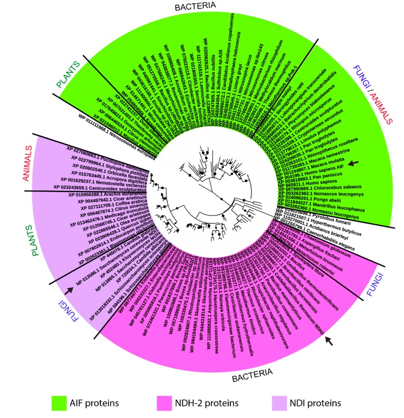
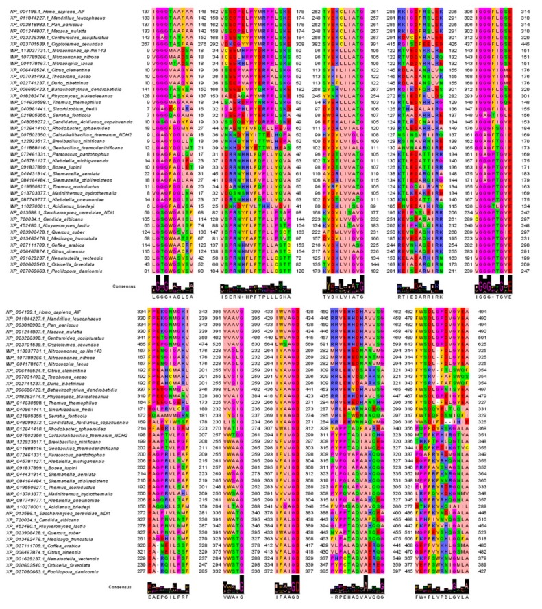
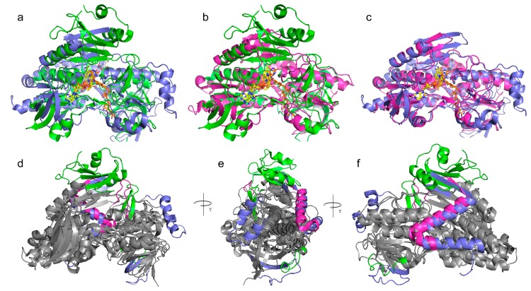
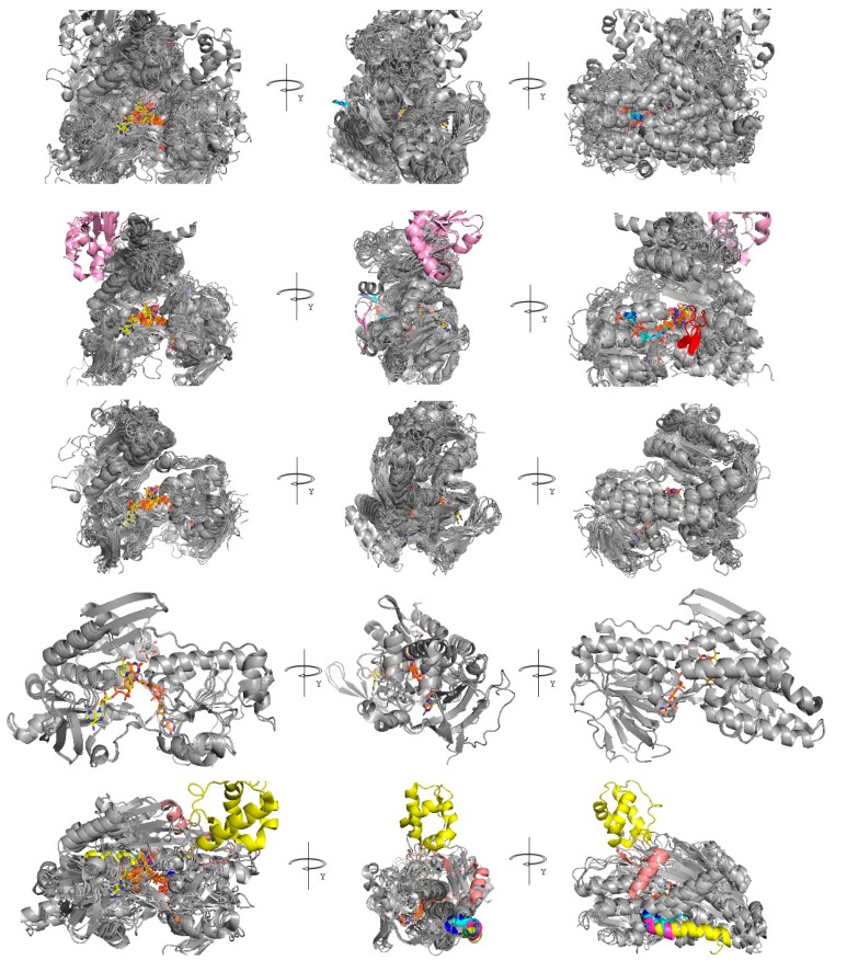
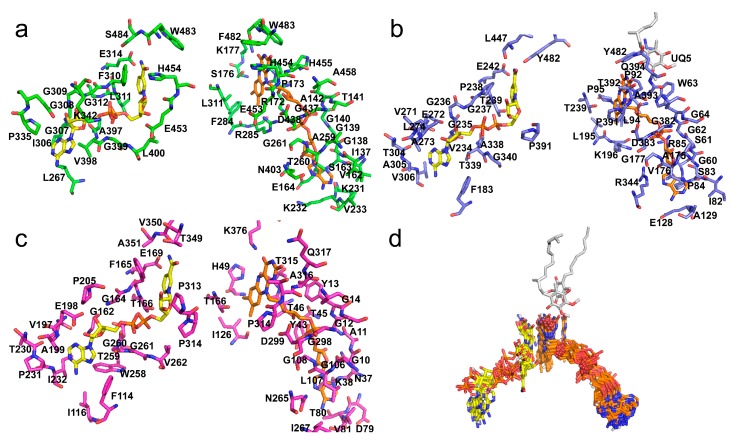
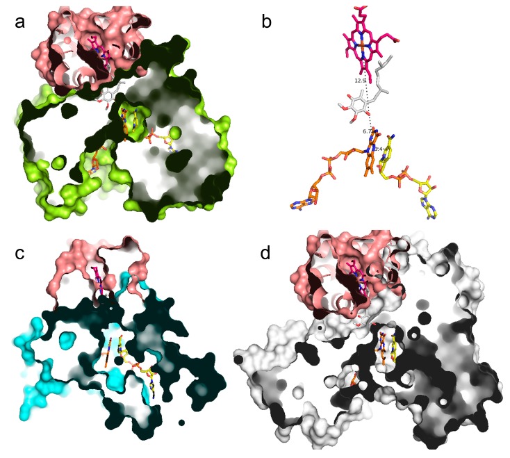
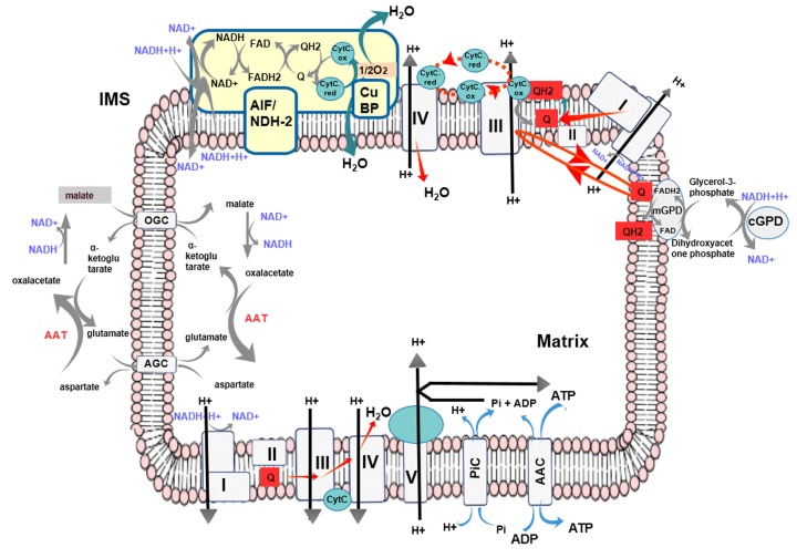
Flavoprotein oxidoreductases are members of a large protein family of specialized dehydrogenases, which include type II NADH dehydrogenase, pyridine nucleotide-disulphide oxidoreductases, ferredoxin-NAD+ reductases, NADH oxidases, and NADH peroxidases, playing a crucial role in the metabolism of several prokaryotes and eukaryotes. Although several studies have been performed on single members or protein subgroups of flavoprotein oxidoreductases, a comprehensive analysis on structure-function relationships among the different members and subgroups of this great dehydrogenase family is still missing. Here, we present a structural comparative analysis showing that the investigated flavoprotein oxidoreductases have a highly similar overall structure, although the investigated dehydrogenases are quite different in functional annotations and global amino acid composition. The different functional annotation is ascribed to their participation in species-specific metabolic pathways based on the same biochemical reaction, i.e., the oxidation of specific cofactors, like NADH and FADH2. Notably, the performed comparative analysis sheds light on conserved sequence features that reflect very similar oxidation mechanisms, conserved among flavoprotein oxidoreductases belonging to phylogenetically distant species, as the bacterial type II NADH dehydrogenases and the mammalian apoptosis-inducing factor protein, until now retained as unique protein entities in Bacteria/Fungi or Animals, respectively. Furthermore, the presented computational analyses will allow consideration of FAD/NADH oxidoreductases as a possible target of new small molecules to be used as modulators of mitochondrial respiration for patients affected by rare diseases or cancer showing mitochondrial dysfunction, or antibiotics for treating bacterial/fungal/protista infections.

Keywords: antibiotics; apoptosis-inducing factor (AIF); dihydrolipoamide dehydrogenase (DLD); flavoprotein oxidoreductases; mitochondrial respiration; molecular modeling; protein shape; thioredoxin reductase (TrxR1); type II NADH dehydrogenase (NDH-2); ubiquinone.


Conflict of interest statement

The authors declare no conflict of interest. The funders had no role in the design of the study; in the collection, analyses, or interpretation of data; in the writing of the manuscript, or in the decision to publish the results.

Figures

Figure 1
Figure 1
Phylogenetic tree of AIF/NDI/NDH-2-homologous sequences. Maximum likelihood phylogenetic tree of AIF/NDH2/NDI-homologous protein sequences selected from representative species of bacteria, Fungi, Plants, and animals. Each one of the tree leaves reports the corresponding organism and RefSeq protein accession number. Arrows indicate the query-protein sequences used for sampling all the other homologous sequences. Nodes supported by bootstrap values greater than 0.7 are indicated as black dots.
Figure 2
Figure 2
AIF/NDI/NDH-2-conserved sequence motifs. Sequence motifs detected by comparing the sampled AIF/NDI/NDH-2 homologous sequences sampled by blastp. The highlighted motifs reveal crucial protein regions involved in cofactor binding and/or the protein function mechanism. The jalviewzappo color style was used for coloring amino acids. Logo representation is also reported to highlight the most conserved residues.
Figure 3
Figure 3
Comparative analysis of AIF/NDI/NDH-2 3D structure. Panels (a,b,c) Lateral view of superposition of H. sapiens AIF (in green cartoon, 4bur.pdb) and S. cerevisae NDI (in blue cartoon, 4g73.pdb) that gives a root mean square deviation(RMSD) of 2.891 Å; H. sapiens AIF (4bur.pdb) and C. thermarum NDH2 (in magenta cartoon, 5kmr.pdb) that gives an RMSD of 1.891 Å; S. cerevisae NDI (4g73.pdb) and C. thermarum NDH2 (5kmr.pdb) that gives an RMSD of 1.384 Å. Panel (d,e,f) Three 90-degree rotation of the alignment view of 4bur, 4g74, and 5kmr. In the grey cartoon representation, the common features highlighted by the superposition of the three structures. Specific AIF/NDI/NDH-2 structural features highlighted by the proposed superposition are reported in green, blue, and magenta cartoon representations. All the three aligned proteins are represented in complex with the FAD (orange sticks, panels ac), NAD+ (yellow sticks, panels ac), and UQ (white sticks, panels a and c) cofactors, when the cited cofactors were present in the reported crystallized structures.
Figure 4
Figure 4
Comparative analysis of AIF/NDI/NDH-2/DLD structures. First row: Superimposition of all the sampled 49 crystallized structures (see Table 1). Second to fifth row: superimposition of all the AIF-like proteins, DLD-like proteins, NDI-like proteins, and NDH-2-like proteins, respectively. All the sampled proteins are reported as grey cartoon representations. Specific protein features are colored according to what is reported in the main text at each row. FAD, NADH, UQ, and CoA are reported as orange, yellow, white, and cyan sticks, respectively, where available at the reported crystallized structures, according to Table 1.
Figure 5
Figure 5
AIF/NDI/NDH-2 cofactor-binding regions. Panels (ac) Residues within 4 Å from NADH (yellow sticks, corresponding to NADHa according to NADH cofactor nomenclature reported in [26]) or FAD (orange sticks) for AIF (green sticks from 4bur.pdb), NDI (blue sticks from 4g73.pdb), and NDH-2 (magenta sticks from 5kmr.pdb) are reported and labeled. Panel (d) Zoomed-in view of cofactor coordinates from all the investigated proteins obtained by superimposing the sampled crystallized structures (see Table 1). UQ (from 4g73.pdb, corresponding to UQI, according to UQ cofactor nomenclature reported in [25]) and DCQ (from 3hyw.pdb) are reported as white sticks. See Supplementary Figure S7 for visualizing the other cofactors observed along the superimposition of the investigated crystallized FAD/NADH oxidoreductases (i.e., heme C, CoA, O2, and H2S).
Figure 6
Figure 6
AIF/NDI/NDH-2 possible protein–protein interaction surfaces. Panel (a) Zoomed-in views of the crystallized heterodimeric structure of flavoprotein dehydrogenase (yellow surf representation) and CytC (pink surf representation) from T. paradoxus (5n1t.pdb) is reported in complex with FAD, (orange sticks), the heme C center in magenta sticks and NADH and UQ (yellow and white sticks, respectively, obtained by superimposition with 4g73 (see methods)). Panel (b) FAD, NADH, heme C center (from 5n1t.pdb), and UQ (superimposed from 4g73.pdb) are reported in stick representations. Intermolecular distances are reported by dashed lines and are labeled. Panel (c,d) 3D models of a putative heterodimeric structure of AIF (4bur.pdb) or NDI (4g73) proteins (cyan or white cartoon, respectively) in complex with CytC from T. paradoxus (5n1t.pdb). FAD, NADH, and UQ are reported in stick representations (see previous panels for colors).
Figure 7
Figure 7
Scheme representation describing the putative participation of AIF to mitochondrial respiration. Cofactors involved in the reaction NAD+ to NADH, FAD to FADH2, a quinone derivative (Q) to the corresponding quinol derivative (QH2), and CytC.ox to CytC.red are indicated by labels. Respiratory chain complexes (I, II, III, and IV) and the ATP-synthase, ADP/ATP Carrier (AAC), phosphate carrier (PiC), malate/aspartate shuttle protein members (with the two carriers OGC and AGC) and the glycerol-3-phosphate dehydrogenase (GPD) shuttle members are reported to show a putative context of action in mitochondrial redox pathways for AIF. A putative copper-binding protein (CuBP) is also indicated. IMS = Intermembrane Space.

References

    1. Verdin E. NAD+ in aging, metabolism, and neurodegeneration. Science. 2015;350:1208–1213. doi: 10.1126/science.aac4854. - DOI - PubMed
    1. Bogachev A.V., Baykov A.A., Bertsova Y.V. Flavin transferase: The maturation factor of flavin-containing oxidoreductases. Biochem. Soc. Trans. 2018;46:1161–1169. doi: 10.1042/BST20180524. - DOI - PubMed
    1. Harold L.K., Antoney J., Ahmed F.H., Hards K., Carr P.D., Rapson T., Greening C., Jackson C.J., Cook G.M. FAD-sequestering proteins protect mycobacteria against hypoxic and oxidative stress. J. Biol. Chem. 2019;294:2903–2912. doi: 10.1074/jbc.RA118.006237. - DOI - PMC - PubMed
    1. Eggink G., Engel H., Vriend G., Terpstra P., Witholt B. Rubredoxin reductase of Pseudomonas oleovorans. Structural relationship to other flavoprotein oxidoreductases based on one NAD and two FAD fingerprints. J. Mol. Biol. 1990;212:135–142. doi: 10.1016/0022-2836(90)90310-I. - DOI - PubMed
    1. Ross R.P., Claiborne A. Molecular cloning and analysis of the gene encoding the NADH oxidase from Streptococcus faecalis 10C1. Comparison with NADH peroxidase and the flavoprotein disulfide reductases. J. Mol. Biol. 1992;227:658–671. doi: 10.1016/0022-2836(92)90215-6. - DOI - PubMed

LinkOut - more resources