. 2020 Jan 7;118(1):207-218.
doi: 10.1016/j.bpj.2019.11.004. Epub 2019 Nov 14.

Irregular Chromatin: Packing Density, Fiber Width, and Occurrence of Heterogeneous Clusters

Affiliations

Irregular Chromatin: Packing Density, Fiber Width, and Occurrence of Heterogeneous Clusters

Gaurav Bajpai et al. Biophys J. .

Abstract

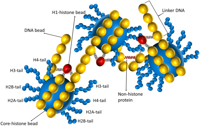
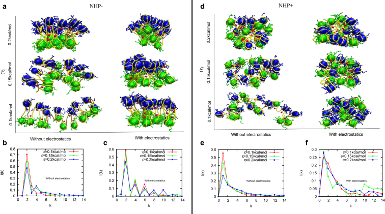
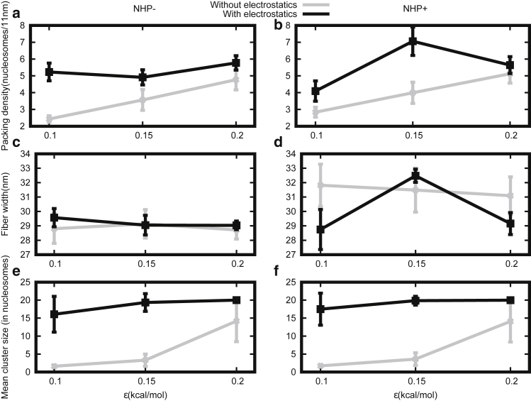
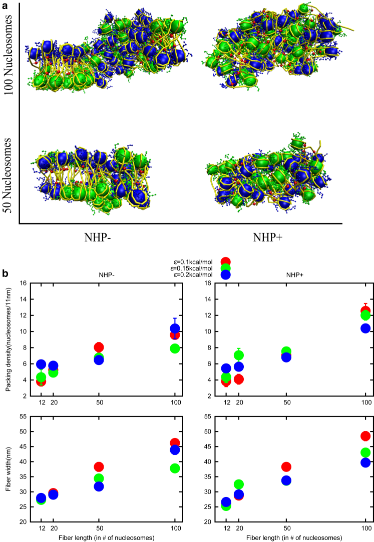
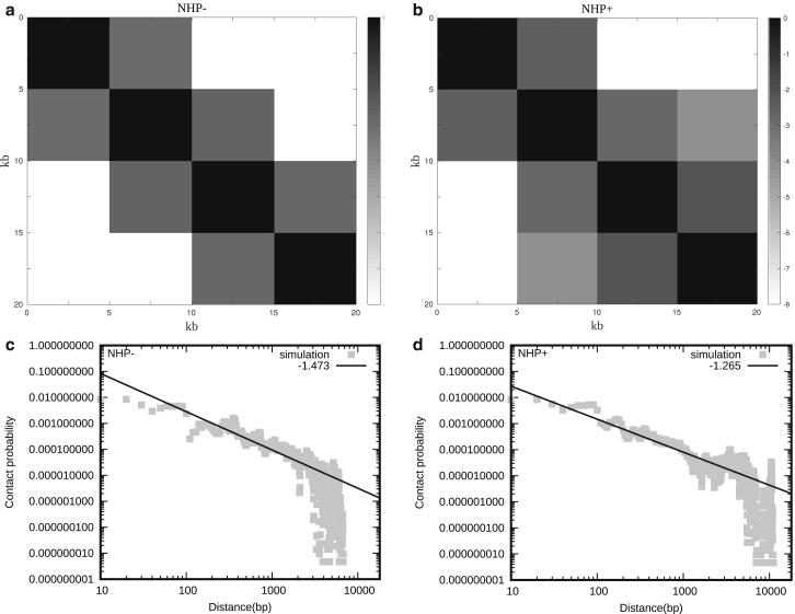
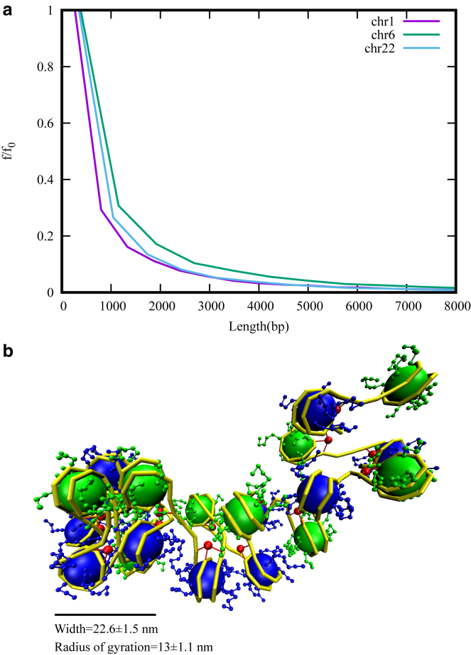
How chromatin is folded on the length scale of a gene is an open question. Recent experiments have suggested that, in vivo, chromatin is folded in an irregular manner and not as an ordered fiber with a width of 30 nm that is expected from theories of higher order packaging. Using computational methods, we examine how the interplay between DNA-bending nonhistone proteins, histone tails, intrachromatin electrostatic, and other interactions decide the nature of the packaging of chromatin. We show that although the DNA-bending nonhistone proteins make the chromatin irregular, they may not alter the packing density and size of the fiber. We find that the length of the interacting region and intrachromatin electrostatic interactions influence the packing density, clustering of nucleosomes, and the width of the chromatin fiber. Our results suggest that the heterogeneity in the interaction pattern will play an important role in deciding the nature of the packaging of chromatin.


Figures

Figure 1
Figure 1
Schematic diagram describing the model. DNA is modeled as a polymer made of type 1 beads (yellow) with a diameter of 3.4 nm. 14 DNA beads are wrapped around the core histone bead (type 2, big blue bead, 5.25 nm in diameter). Histone tails are modeled as flexible bead-spring polymer chains in which each histone-tail bead (type 3, small blue bead) is 1.56 nm in diameter and have the following lengths: tail 1 (H2A) = 4 beads, tail 2 (H2B) = 5 beads, tail 3 (H3) = 8 beads, and tail 4 (H4) = 5 beads. H1 histone (red bead, type 4) is connected to three DNA beads—the entry bead, exit bead, and the central bead of the DNA wrapped around the core histone bead. DNA-bending NHP is modeled as a spring (red) connecting two beads in the linker region.
Figure 2
Figure 2
Snapshots of chromatin configurations and the corresponding contact probabilities between kth neighbor nucleosomes (I(k)) in the absence (NHP−) and presence (NHP+) of DNA-bending nonhistone proteins (NHPs) that bind along linker DNA regions. The results are presented for four cases, as follows: case 1: simulations with no NHPs and no electrostatic interactions (left column of (a), and (b)); case 2: no NHPs but with electrostatic interactions (right column of (a), and (c)); case 3: with NHPs and no electrostatic interactions (left column of (d), and (e)); case 4: with NHPs and with electrostatic interactions (right column of (d), and (f)). All the results are presented for three different strengths of LJ (ɛ) interaction potentials (see text for details).
Figure 3
Figure 3
Packing density, fiber width, and mean cluster size are plotted for different LJ strengths (ε), in the absence of NHPs (NHP−, left side) and the presence of NHPs (NHP+, right side). (a and b) Without electrostatic interaction, the packing density increases with ɛ (gray curves). With electrostatic interaction, packing density is ∼6 nucleosomes/11 nm (black curve). (c and d) Chromatin fiber diameter (width) for different parameters is shown. With (black curve) and without (gray curve) electrostatic interaction, the fiber diameter is ∼30 nm (constant) for all cases. (e and f) Without electrostatic interaction, mean cluster size increases on increasing the LJ (ε) parameter (gray curve), but with electrostatic interaction, it remains constant (black curve). In all the subfigures, the vertical bars represent SD.
Figure 4
Figure 4
(a) Snapshots of simulation results of 50 nucleosomes and 100 nucleosomes in the absence and presence of NHPs (left to right) for ε = 0.15. Neighboring nucleosomes (odd, even) are shown in different colors so that the regular/irregular organization is more visible to the eye. (b) Shown is the packing density and fiber width on varying chromatin fiber length in the absence and presence of NHPs for different LJ interaction strengths (ε). Packing densities and fiber widths increase as we increase the chromatin length. Colors red, green, and blue represent different LJ interaction strengths ε = 0.1, 0.15, and 0.2 kcal/mol, respectively.
Figure 5
Figure 5
(a and b) Contact maps (Pij) from our simulations, between different 5-kb segments (bins), in the absence (NHP−) and presence of NHP (NHP+). The color scheme varies from black to white, representing high to low contact counts (log(Pij)). (c and d) Shown is contact probability as a function of contour distance (distance along the DNA backbone) in the absence and presence of NHP, calculated from the simulations (gray curve). The black line is a guide to the eye indicating power-law behavior, suggesting the fractal nature of the self-organization.
Figure 6
Figure 6
(a) Frequency (f) distribution of lengths of contiguous patches having H3K9me3 modifications, for three different chromosomes, scaled with maximal frequency (f0). See Fig. S6 for a similar plot for all chromosomes. Most of the patches are smaller in length; very long patches are rare. (b) A snapshot of the chromatin structure with NHPs and heterogeneous interactions is shown; the system was simulated with electrostatic interactions in the first eight nucleosomes and with no electrostatic interaction in the remaining 12 nucleosomes. Note that the radius of gyration or “width” of the dense cluster indicate a length scale less than 30 nm. To see this figure in color, go online.

References

    1. Alberts B. Sixth Edition. Garland Science, Taylor and Francis Group; New York: 2014. Molecular Biology of the Cell.
    1. Kornberg R.D. Chromatin structure: a repeating unit of histones and DNA. Science. 1974;184:868–871. - PubMed
    1. Luger K., Mäder A.W., Richmond T.J. Crystal structure of the nucleosome core particle at 2.8 A resolution. Nature. 1997;389:251–260. - PubMed
    1. Luger K., Dechassa M.L., Tremethick D.J. New insights into nucleosome and chromatin structure: an ordered state or a disordered affair? Nat. Rev. Mol. Cell Biol. 2012;13:436–447. - PMC - PubMed
    1. Sun J., Zhang Q., Schlick T. Electrostatic mechanism of nucleosomal array folding revealed by computer simulation. Proc. Natl. Acad. Sci. USA. 2005;102:8180–8185. - PMC - PubMed

Publication types

LinkOut - more resources