. 2019 Dec 6;294(49):18545-18546.
doi: 10.1074/jbc.H119.011803.

Cutting antigenic peptides down to size

Affiliations

Cutting antigenic peptides down to size

Kannan Natarajan et al. J Biol Chem. .

Abstract

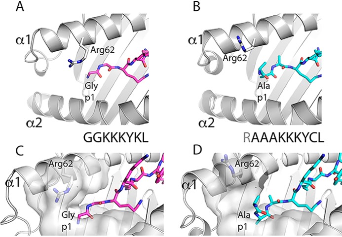
A critical step in antigen presentation is the degradative processing of peptides by aminopeptidases in the endoplasmic reticulum. It is unclear whether these enzymes act only on free peptides or on those bound to their major histocompatibility complex (MHC)-I-presenting molecules. A recent study examined the structure and biophysics of N-terminally extended peptides in complex with MHC-I, revealing the conformational adjustment of MHC to permit both binding of the peptide core and exposure of the peptide N terminus. These data suggest a mechanism by which aminopeptidase access is determined and offer an explanation for how longer peptides may be displayed at the cell surface.


Conflict of interest statement

The authors declare that they have no conflicts of interest with the contents of this article

Figures

Figure 1.
Figure 1.
Comparison of the structure of pMHC-I complex containing N-terminally extended peptide with canonical octamer-peptide/MHC-I complex reveals reorientation of p1 amine of peptide and side chain of MHC-I residue Arg-62. A ribbon diagram of the previously determined structure of HLA-B*08:01 complexed with an octamer peptide (GGKKKYKL) (PDB entry 1AGD) (A and C) is compared with that of HLA-B*08:01E76C complexed with a 10-mer (RAAAKKKYCL) (PDB entry 6P2S) (B and D) as described in detail by Li et al. (4). Attention is focused on the position of the side chain of Arg-62 of the α1 helix and the orientation of p1 nitrogen (in blue). In C and D, a surface representation of the residues lining the A pocket is shown.

References

    1. Hammer G. E., Gonzalez F., Champsaur M., Cado D., and Shastri N. (2006) The aminopeptidase ERAAP shapes the peptide repertoire displayed by major histocompatibility complex class I molecules. Nat. Immunol. 7, 103–112 10.1038/ni1286 - DOI - PubMed
    1. Saveanu L., Carroll O., Lindo V., Del Val M., Lopez D., Lepelletier Y., Greer F., Schomburg L., Fruci D., Niedermann G., and van Endert P. M. (2005) Concerted peptide trimming by human ERAP1 and ERAP2 aminopeptidase complexes in the endoplasmic reticulum. Nat. Immunol. 6, 689–697 10.1038/ni1208 - DOI - PubMed
    1. López de Castro J. A. (2018) How ERAP1 and ERAP2 shape the peptidomes of disease-associated MHC-I proteins. Front. Immunol. 9, 2463 10.3389/fimmu.2018.02463 - DOI - PMC - PubMed
    1. Li L., Batliwala M., and Bouvier M. (2019) ERAP1 enzyme-mediated trimming and structural analyses of MHC I–bound precursor peptides yield novel insights into antigen processing and presentation. J. Biol. Chem. 294, 18534–18544 10.1074/jbc.RA119.010102 - DOI - PMC - PubMed
    1. Saric T., Chang S. C., Hattori A., York I. A., Markant S., Rock K. L., Tsujimoto M., and Goldberg A. L. (2002) An IFN-γ-induced aminopeptidase in the ER, ERAP1, trims precursors to MHC class I-presented peptides. Nat. Immunol. 3, 1169–1176 10.1038/ni859 - DOI - PubMed

Publication types

LinkOut - more resources