. 2020 Mar 20;295(12):3749-3756.
doi: 10.1074/jbc.RA119.011227. Epub 2019 Dec 6.

The regulatory protein 14-3-3β binds to the IQ motifs of myosin-IC independent of phosphorylation

Affiliations

The regulatory protein 14-3-3β binds to the IQ motifs of myosin-IC independent of phosphorylation

Huan-Hong Ji et al. J Biol Chem. .

Abstract

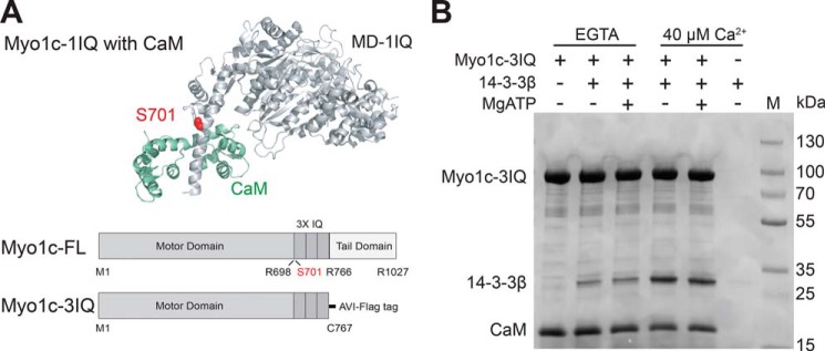
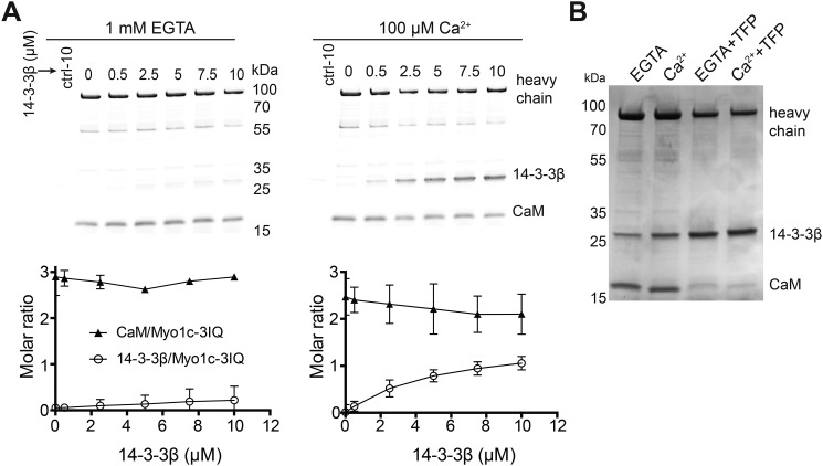
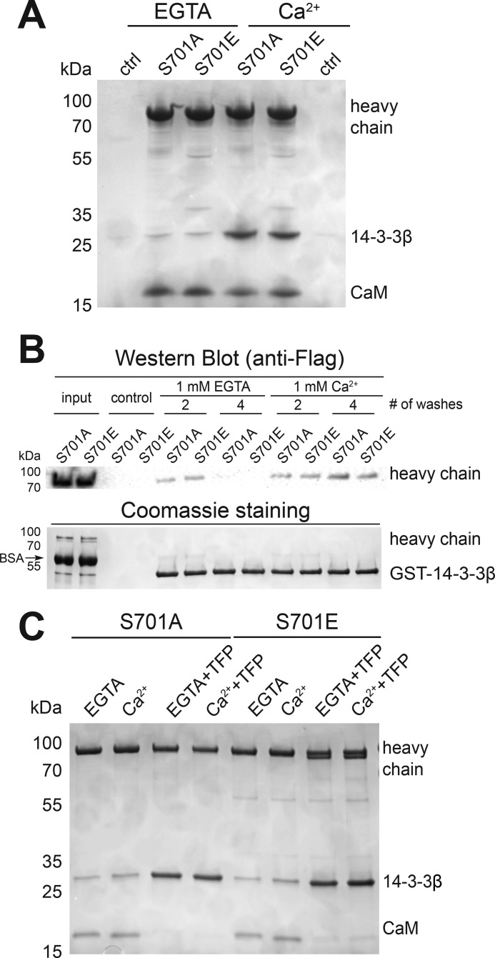
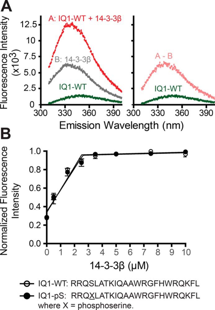
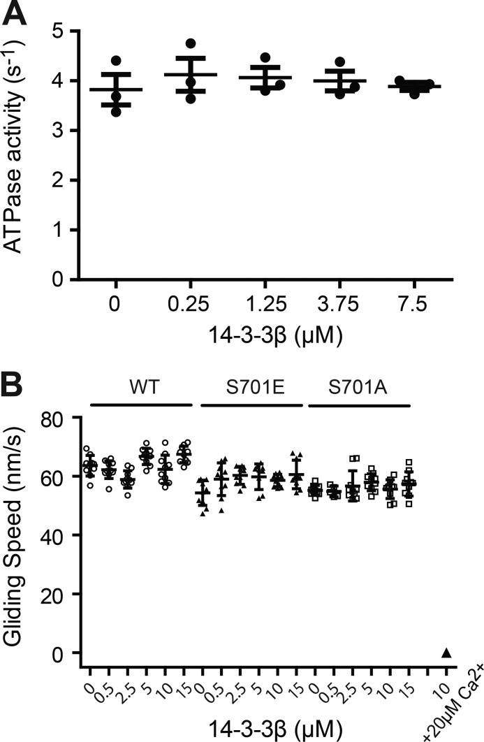
Myosin-IC (Myo1c) has been proposed to function in delivery of glucose transporter type 4 (GLUT4)-containing vesicles to the plasma membrane in response to insulin stimulation. Current evidence suggests that, upon insulin stimulation, Myo1c is phosphorylated at Ser701, leading to binding of the signaling protein 14-3-3β. Biochemical and functional details of the Myo1c-14-3-3β interaction have yet to be described. Using recombinantly expressed proteins and mass spectrometry-based analyses to monitor Myo1c phosphorylation, along with pulldown, fluorescence binding, and additional biochemical assays, we show here that 14-3-3β is a dimer and, consistent with previous work, that it binds to Myo1c in the presence of calcium. This interaction was associated with dissociation of calmodulin (CaM) from the IQ motif in Myo1c. Surprisingly, we found that 14-3-3β binds to Myo1c independent of Ser701 phosphorylation in vitro Additionally, in contrast to previous reports, we did not observe Myo1c Ser701 phosphorylation by Ca2+/CaM-dependent protein kinase II (CaMKII), although CaMKII phosphorylated four other Myo1c sites. The presence of 14-3-3β had little effect on the actin-activated ATPase or motile activities of Myo1c. Given these results, it is unlikely that 14-3-3β acts as a cargo adaptor for Myo1c-powered transport; rather, we propose that 14-3-3β binds Myo1c in the presence of calcium and stabilizes the calmodulin-dissociated, nonmotile myosin.

Keywords: 14-3-3 protein; 14-3-3β; calmodulin (CaM); cell motility; cell signaling; cytoskeleton; molecular motor; myo1c; myosin; myosin-IC.


Conflict of interest statement

The authors declare that they have no conflicts of interest with the contents of this article. The content is solely the responsibility of the authors and does not necessarily represent the official views of the National Institutes of Health

Figures

Figure 1.
Figure 1.
Effects of calcium on binding of 14-3-3β to Myo1c. A, top panel, cartoon representation of the crystal structure (PDB code 4BYF) of the Myo1c motor domain with 1-IQ (light gray) with bound CaM (green). The proposed phosphorylation site, Ser710, is highlighted in red. Bottom panel, schematic of the primary structure of full-length Myo1c and Myo1c-3IQ constructs. B, Coomassie-stained gel showing results of streptavidin bead–mediated pulldown of biotinylated 0.8 μm Myo1c-3IQ–WT and 2.0 μm CaM in the presence or absence of 2.5 μm 14-3-3β, 40 μm free calcium, and 1 mm MgATP. Lane M shows molecular weight markers.
Figure 2.
Figure 2.
Association of 14-3-3β with Myo1c-3IQ. A, top panels, SYPRO-stained gels showing results of streptavidin bead–mediated pulldown of biotinylated Myo1c-3IQ with various concentrations of 14-3-3β in the absence (1 mm EGTA) and presence of 100 μm free calcium. Bottom panels, the stoichiometry of 14-3-3β and CaM to pulled down Myo1c-3IQ as determined from quantification of gel bands as described under “Experimental procedures.” Plotted values are mean ± S.D. from three to four independent pulldown assays. B, Coomassie-stained gel showing pulldown of biotinylated Myo1c-3IQ with 1.25 μm 14-3-3β under EGTA and Ca2+ conditions. Myo1c-3IQ was treated with or without 0.5 mm TFP.
Figure 3.
Figure 3.
Effects of phosphorylation of Ser701 on binding of 14-3-3β to Myo1c. A, Coomassie-stained gel of pulldown of 0.8 μm biotinylated Myo1c-3IQ S701A and S701E in the presence of 2.5 μm 14-3-3β in the absence (EGTA) and presence of 40 μm free calcium. ctrl, control. B, GSH–agarose bead–mediated pulldown of GST-14-3-3β in the presence of 1 μm Myo1c-3IQ–S701A or –S701E. # of washes indicates number of times the agarose beads were washed after pulldown. Top panel, pulled down Myo1c-3IQ was detected by Western blotting using anti-FLAG antibody. Bottom panel, GST–14-3-3β was detected by Coomassie staining. C, streptavidin bead–mediated pulldown of 0.5 μm Myo1c-3IQ–S701A and –S701E treated with TFP in the presence of 1.25 μm 14-3-3β.
Figure 4.
Figure 4.
Determination of the binding stoichiometry of IQ peptides and 14-3-3β. A, steady-state fluorescence emission spectra of 14-3-3β with IQ1 peptide (λexcitation = 295 nm). Left panel, fluorescence emission spectra of 3.5 μm 14-3-3β dimer with 5 μm IQ1 peptide. Right panel, corrected fluorescence spectra of IQ1 peptide from subtracting the contribution of the spectra of samples containing only 3.5 μm 14-3-3β (A-B). B, 5 μm IQ peptides (IQ1-WT and IQ1-pSer701) were titrated with 0–10 μm 14-3-3β. Values (350 nm) are mean ± S.D. from three independent assays. Linear regression of the nonsaturated and saturated parts of the data points reveals saturation of IQ peptides at ∼2.5 μm 14-3-3β dimer.
Figure 5.
Figure 5.
14-3-3β does not affect Myo1c motor function. A, ATPase activity of Myo1c-3IQ–WT in the presence of 14-3-3β. Steady-state ATPase activity (37 °C) was measured in KMg25 containing 50 μm free Ca2+ using the NADH-coupled assay. The values are mean ± S.D. from three independent assays. B, determination of in vitro actin gliding velocity of Myo1c-3IQ with 0–15 μm of 14-3-3β in the presence of EGTA. The inhibited motility was not rescued for Myo1c-3IQ–WT with 10 μm of 14-3-3β in the presence of 20 μm free Ca2+.

Comment in

  • Putting the brakes on a myosin motor.
    Eddington C, Titus MA. Eddington C, et al. J Biol Chem. 2020 Mar 20;295(12):3757-3758. doi: 10.1074/jbc.H120.013153. J Biol Chem. 2020. PMID: 32198186 Free PMC article.

References

    1. Greenberg M. J., and Ostap E. M. (2013) Regulation and control of myosin-I by the motor and light chain-binding domains. Trends Cell Biol. 23, 81–89 10.1016/j.tcb.2012.10.008 - DOI - PMC - PubMed
    1. McIntosh B. B., and Ostap E. M. (2016) Myosin-I molecular motors at a glance. J. Cell Sci. 129, 2689–2695 10.1242/jcs.186403 - DOI - PMC - PubMed
    1. McConnell R. E., and Tyska M. J. (2010) Leveraging the membrane: cytoskeleton interface with myosin-1. Trends Cell Biol. 20, 418–426 10.1016/j.tcb.2010.04.004 - DOI - PMC - PubMed
    1. Hokanson D. E., and Ostap E. M. (2006) Myo1c binds tightly and specifically to phosphatidylinositol 4,5-bisphosphate and inositol 1,4,5-trisphosphate. Proc. Natl. Acad. Sci. U.S.A. 103, 3118–3123 10.1073/pnas.0505685103 - DOI - PMC - PubMed
    1. Hokanson D. E., Laakso J. M., Lin T., Sept D., and Ostap E. M. (2006) Myo1c binds phosphoinositides through a putative pleckstrin homology domain. Mol. Biol. Cell 17, 4856–4865 10.1091/mbc.e06-05-0449 - DOI - PMC - PubMed

Publication types

MeSH terms

Associated data