. 2019 Dec 6;9(1):18489.
doi: 10.1038/s41598-019-54884-7.

Radiobiological effects of the alpha emitter Ra-223 on tumor cells

Affiliations

Radiobiological effects of the alpha emitter Ra-223 on tumor cells

Kristina Bannik et al. Sci Rep. .

Abstract

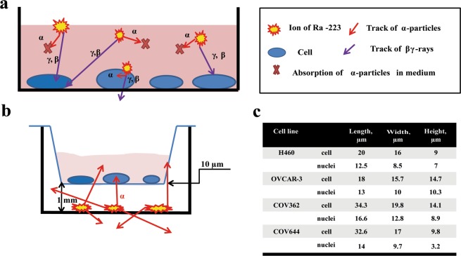
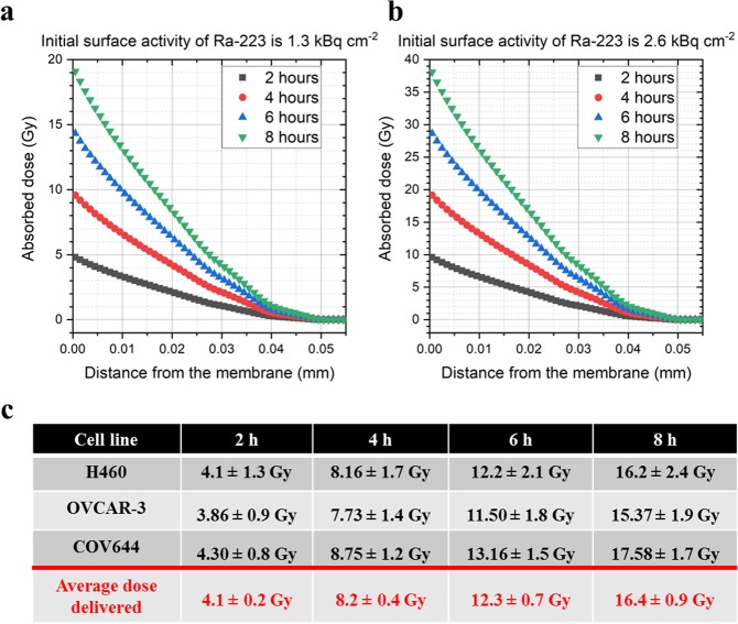
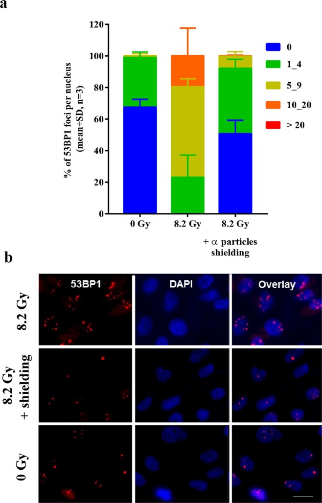
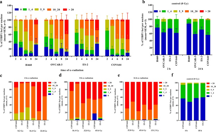
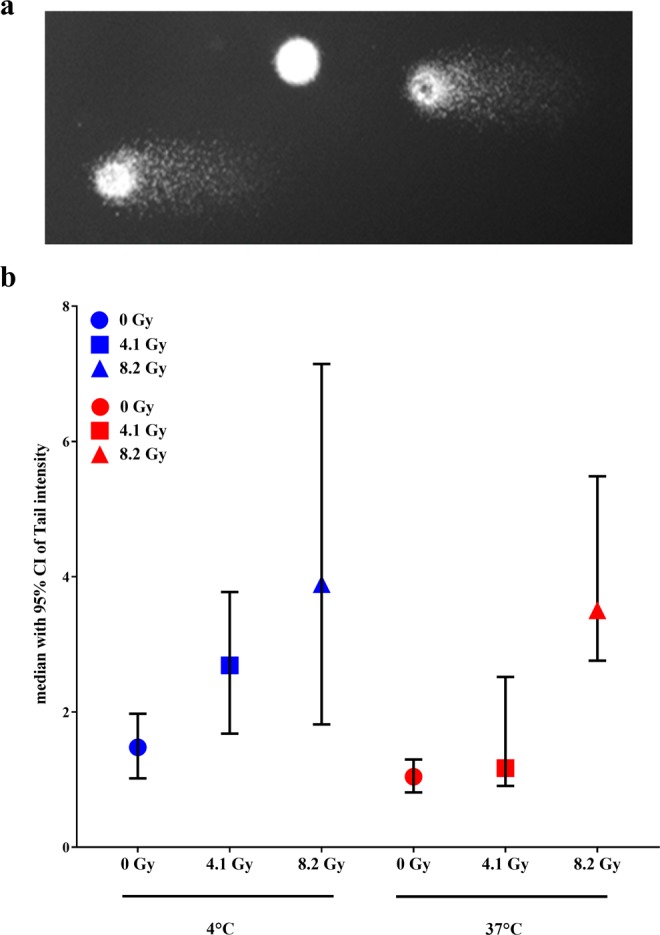
Targeted alpha therapy is an emerging innovative approach for the treatment of advanced cancers, in which targeting agents deliver radionuclides directly to tumors and metastases. The biological effects of α-radiation are still not fully understood - partly due to the lack of sufficiently accurate research methods. The range of α-particles is <100 μm, and therefore, standard in vitro assays may underestimate α-radiation-specific radiation effects. In this report we focus on α-radiation-induced DNA lesions, DNA repair as well as cellular responses to DNA damage. Herein, we used Ra-223 to deliver α-particles to various tumor cells in a Transwell system. We evaluated the time and dose-dependent biological effects of α-radiation on several tumor cell lines by biological endpoints such as clonogenic survival, cell cycle distribution, comet assay, foci analysis for DNA damage, and calculated the absorbed dose by Monte-Carlo simulations. The radiobiological effects of Ra-223 in various tumor cell lines were evaluated using a novel in vitro assay designed to assess α-radiation-mediated effects. The α-radiation induced increasing levels of DNA double-strand breaks (DSBs) as detected by the formation of 53BP1 foci in a time- and dose-dependent manner in tumor cells. Short-term exposure (1-8 h) of different tumor cells to α-radiation was sufficient to double the number of cells in G2/M phase, reduced cell survival to 11-20% and also increased DNA fragmentation measured by tail intensity (from 1.4 to 3.9) dose-dependently. The α-particle component of Ra-223 radiation caused most of the Ra-223 radiation-induced biological effects such as DNA DSBs, cell cycle arrest and micronuclei formation, leading ultimately to cell death. The variable effects of α-radiation onto the different tumor cells demonstrated that tumor cells show diverse sensitivity towards damage caused by α-radiation. If these differences are caused by genetic alterations and if the sensitivity could be modulated by the use of DNA damage repair inhibitors remains a wide field for further investigations.


Conflict of interest statement

Authors #1, 3, 4, 5, 6 and 7 are employees of Bayer A.G. Authors #4, 5 and 6 hold stocks of Bayer A.G. Author #2 has received funding from Bayer A.G. Kristina Bannik is employed by Bayer A.G. Balázs Madas’s work was supported by Bayer A.G. Marco Jarzombek is employed by Bayer A.G. Andreas Sutter owns stock in Bayer A.G. and is employed by Bayer A.G. Gerhard Siemeister owns stock in Bayer A.G. and is employed by Bayer A.G. Dominik Mumberg owns stock in Bayer A.G. and is employed by Bayer A.G. Sabine Zitzmann-Kolbe is employed by Bayer A.G.

Figures

Figure 1
Figure 1
Schematic representation of the irradiation geometry. (a) The absorption of α-particles in medium. (b) The schematic pathway of α-particle from Ra-223. (c) The geometric data of several human cancer cell lines.
Figure 2
Figure 2
Absorbed dose in different layers on the medium/cells as a function of the distance from the mylar membrane with the initial activities of Ra-223 - 1.3 kBq/cm2 (a), 2.6 kBq/cm2 (b). Data points are plotted for the middle of the layers. (c) Table of absorbed dose in various tumor cells (mean ± SD).
Figure 3
Figure 3
Ra-223 induced damage via α-radiation. ES-2 cells were irradiated at 1.3 kBq/cm2 in TWs for 4 h ± α-particles shielding by using 100 μm thick folia. (a) The quantification of 53BP1 foci to indicate DSBs per nucleus in percentage after αβγ- and βγ-radiation at 1.3 kBq/cm2. (b) The immunofluorescence images of 53BP1 (red) and DAPI (blue) after α-radiation ±100 μm thick folia at 4 h. Scale bar is 12 μm.
Figure 4
Figure 4
DNA damage after prolonged α-radiation in tumor cells lines. (a) The quantification of 53BP1 foci per nucleus in percentage at different time points after α-radiation of 1.3 kBq/cm2. (b) Un-irradiated controls. (c,d) DNA damage after α-radiation in ovarian cancer cells lines. The quantification of 53BP1 foci per nucleus in percentage at different time points after α-radiation of 4, 8, 16 hours. (f) un-irradiated controls.
Figure 5
Figure 5
The radiosensitivity of cancer cells after α-radiation from Ra-223. (a) Cell survival after 2, 4, 8 and 24 h α-radiation with the activity of 1.3 and 2.6 kBq/cm2 in H460, n = 2. (b) The table of DNA mutation background in several cancer cell lines. (c) Survival fraction after α-radiation with the activity of 1.3 kBq/cm2 for 2, 4 and 8 hours, n ≥ 2. (d) Survival fraction after α-radiation with the activities of 1.3, 2.6, 5.3 kBq/cm2 for 2 hours, n = 4. Giving absorbed doses of 4.1, 8.2 and 16.4 Gy.
Figure 6
Figure 6
The cell cycle distribution after α-radiation in tumor cell lines.
Figure 7
Figure 7
The detection of DNA damage in H460. (a) The comets after the electrophoresis. (b) The median of tail intensity after α-radiation with absorbed doses of 4.1 and 8.2 Gy for 1 h at 4 °C (blue symbols) and at 37 °C (red symbols).

References

    1. Dekempeneer Y, et al. Targeted alpha therapy using short-lived alpha-particles and the promise of nanobodies as targeting vehicle. Expert Opin. Biol. Ther. 2016;16:1035–1047. doi: 10.1080/14712598.2016.1185412. - DOI - PMC - PubMed
    1. Parker C, et al. Alpha emitter radium-223 and survival in metastatic prostate cancer. N. Engl. J. Med. 2013;369:213–223. doi: 10.1056/NEJMoa1213755. - DOI - PubMed
    1. Carter RJ, et al. Complex DNA Damage Induced by High Linear Energy Transfer Alpha-Particles and Protons Triggers a Specific Cellular DNA Damage Response. Int. J. Radiat. Oncol. Biol. Phys. 2018;100:776–784. doi: 10.1016/j.ijrobp.2017.11.012. - DOI - PMC - PubMed
    1. Asaithamby A, Hu B, Chen DJ. Unrepaired clustered DNA lesions induce chromosome breakage in human cells. Proc. Natl. Acad. Sci. USA. 2011;108:8293–8298. doi: 10.1073/pnas.10160451081016045108. - DOI - PMC - PubMed
    1. Filpula D. Antibody engineering and modification technologies. Biomol. Eng. 2007;24:201–215. doi: 10.1016/j.bioeng.2007.03.004. - DOI - PubMed

Publication types

MeSH terms