. 2020 May;20(5):1431-1438.
doi: 10.1111/ajt.15737. Epub 2019 Dec 27.

B cell clonal expansion within immune infiltrates in human cardiac allograft vasculopathy

Affiliations

B cell clonal expansion within immune infiltrates in human cardiac allograft vasculopathy

Carolina Moore et al. Am J Transplant. 2020 May.

Abstract

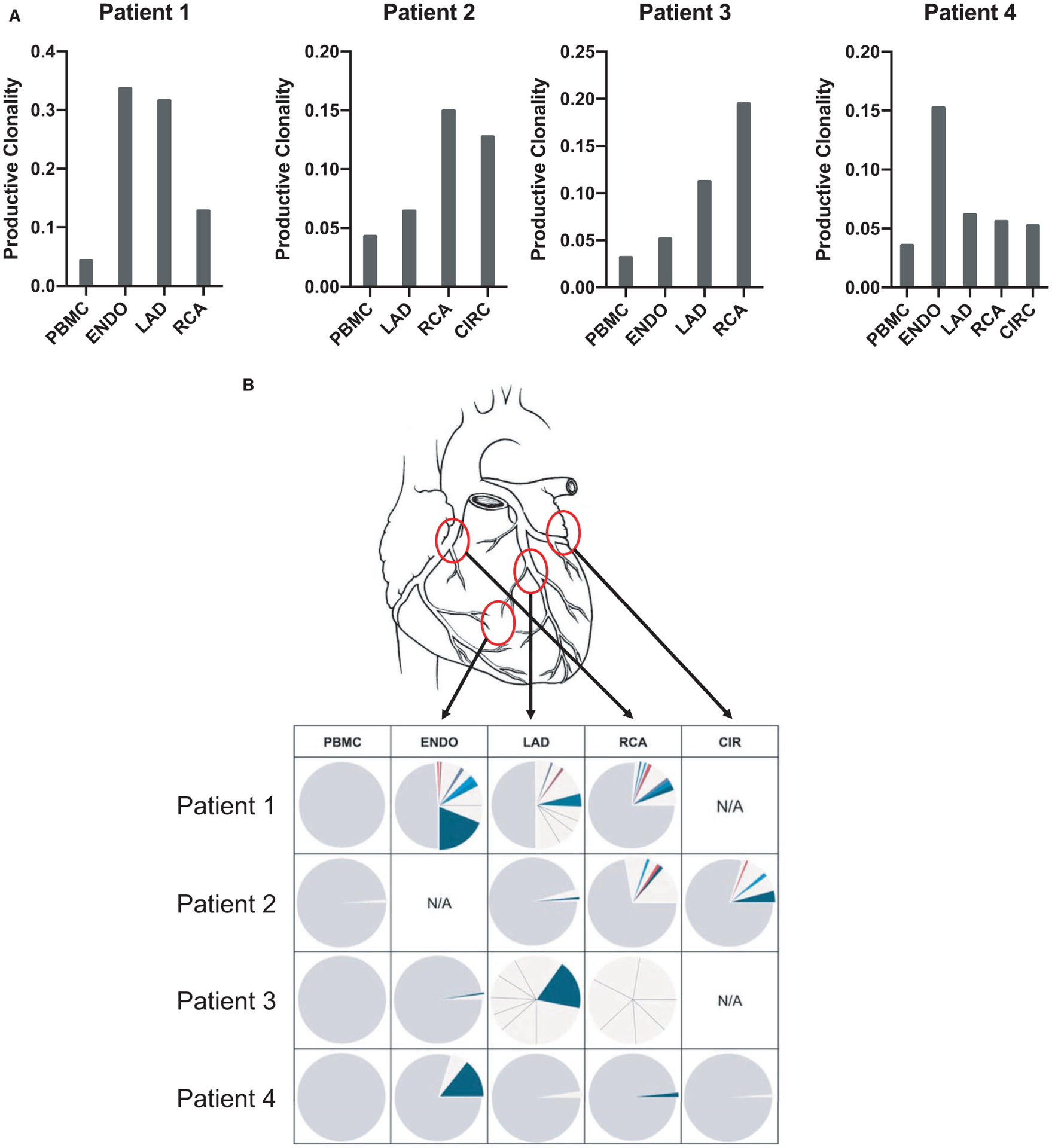
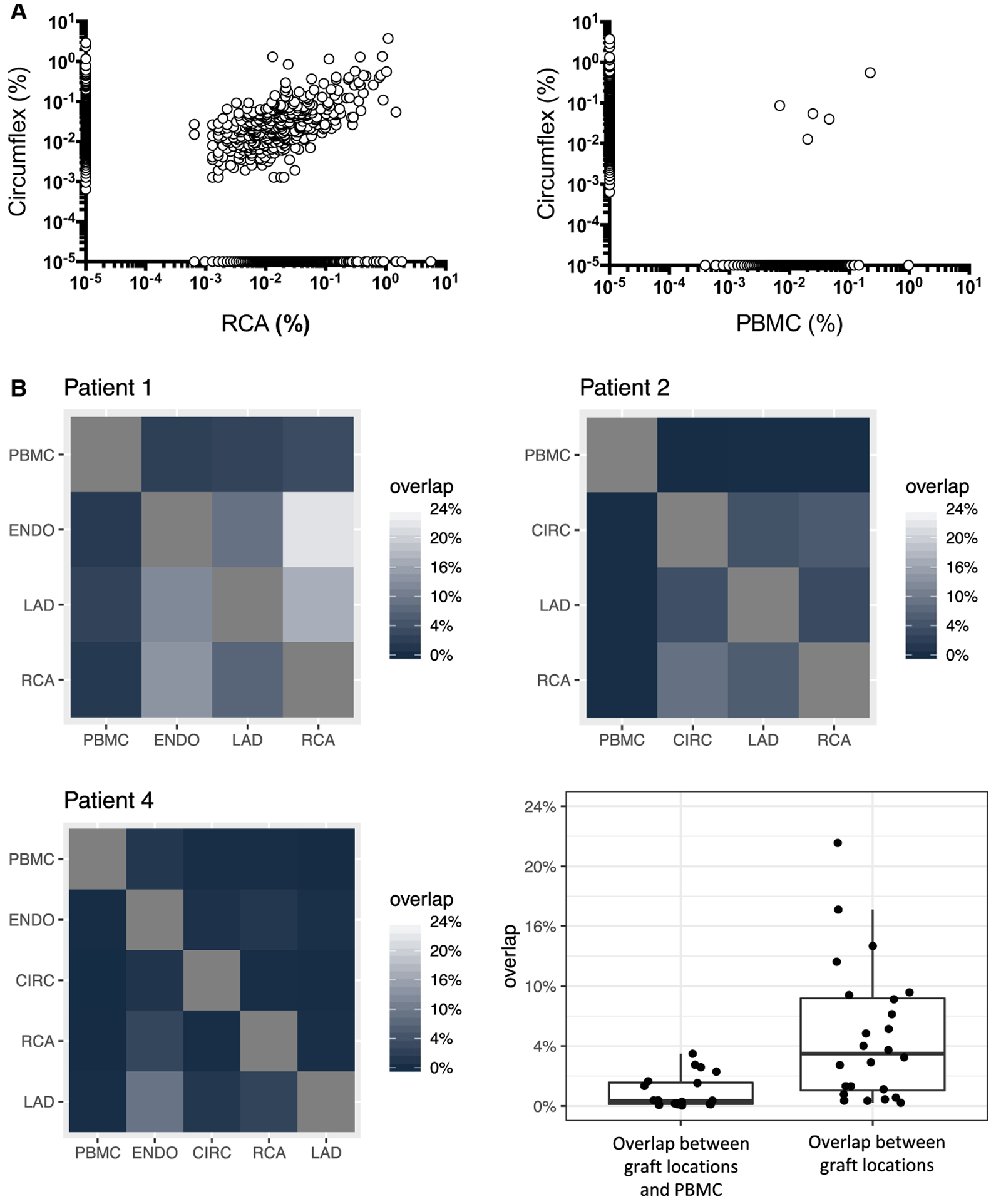
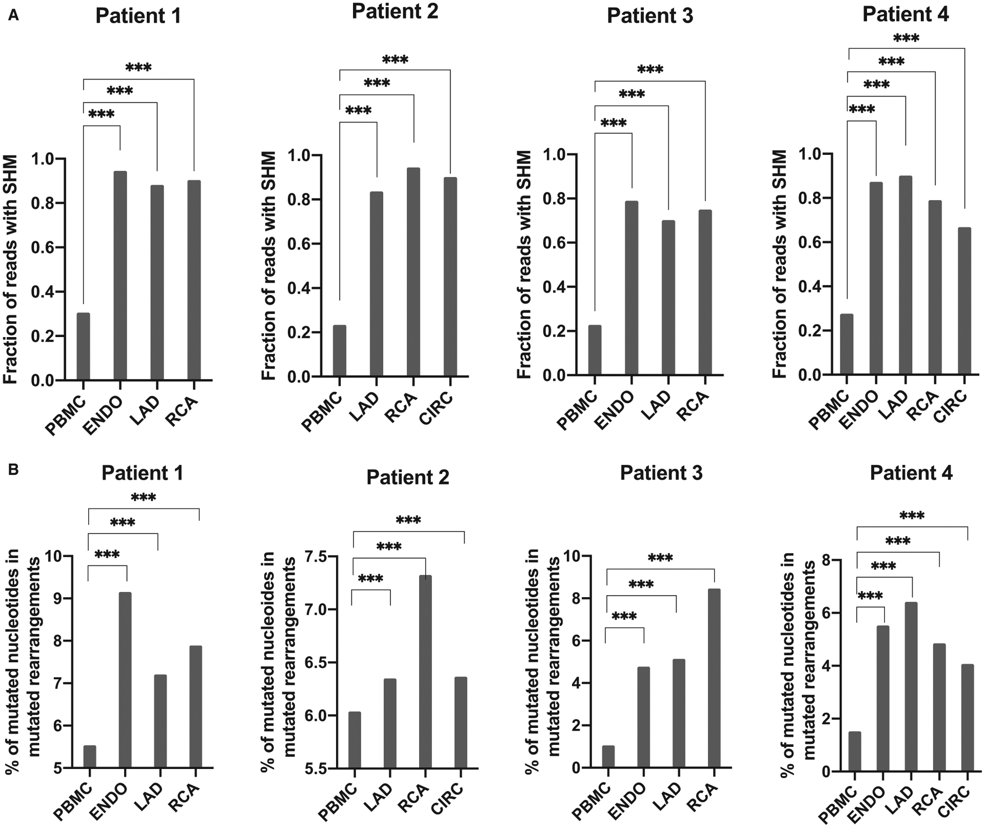
Cardiac allograft vasculopathy (CAV) is associated with intragraft B cell infiltrates. Here, we studied the clonal composition of B cell infiltrates using 4 graft specimens with CAV. Using deep sequencing, we analyzed the immunoglobulin heavy chain variable region repertoire in both graft and blood. Results showed robust B cell clonal expansion in the graft but not in the blood for all cases. Several expanded B cell clones, characterized by their uniquely rearranged complementarity-determining region 3, were detected in different locations in the graft. Sequences from intragraft B cells also showed elevated levels of mutated rearrangements in the graft compared to blood B cells. The number of somatic mutations per rearrangement was also higher in the graft than in the blood, suggesting that B cells continued maturing in situ. Overall, our studies demonstrated B cell clonal expansion in human cardiac allografts with CAV. This local B cell response may contribute to the pathophysiology of CAV through a mechanism that needs to be identified.

Keywords: B cell biology; basic (laboratory) research/science; heart (allograft) function/dysfunction; heart transplantation/cardiology; immunobiology; rejection: chronic.


Conflict of interest statement

DISCLOSURES

The authors of this manuscript have no conflicts to disclose as described by the American Journal of Transplantation.

Figures

FIGURE 1
FIGURE 1
B cell repertoire analysis in graft infiltrates and peripheral blood. A, IGH clonality of graft locations and blood for 4 transplant recipients with active CAV, calculated from productive rearrangements by taking the inverse of the normalized (for sequencing depth) Shannon diversity index. B, Pie chart representation of the frequency of individual CDR3 sequences observed in 4 distinct allograft sites and in the blood for 4 explants. Each pie section corresponds to a unique CDR3 sequence with a frequency above 1%. Sections corresponding to sequences present at less than 1% are merged and depicted as gray. The noncolored sections correspond to clones that were only detected in the indicated location. Colored pie chart sections (blue, red…) correspond to clones that were detected in more than one locale within the graft. CAV, cardiac allograft vasculopathy; ENDO, endocardium; LAD, left anterior descending coronary artery; RCA, right coronary artery; CIR, circumflex coronary artery
FIGURE 2
FIGURE 2
Overlap between B cell repertoires. A, Example of IGHV sequence repertoire overlap between 2 distinct locations within the same graft (RCA and CIR, left panel) and between the graft and PBMC (right panel) for patient 2. Each dot represents a unique sequence. The frequency of each sequence is reported on the x and y axes. Sequences along the x and y axis were detected in only one compartment. B, Summary of the CDR3 productive rearrangements repertoire overlap between the different graft locations and blood for 3 patients. The percentage corresponds to CDR3 sequences from the locations shown on the y-axis that are also found at the location shows on the x-axis. Note that for patient 3, the number of sequences obtained for the coronary arteries was not sufficient to calculate the repertoire overlap. The box-whisker plot shows the distribution of overlaps between graft locations and PBMC (left) or between different graft locations (right). CIR, circumflex coronary artery; IGHV, immunoglobulin heavy chain variable; PBMC, peripheral blood mononuclear cells; RCA, right coronary artery
FIGURE 3
FIGURE 3
Blood and intragraft B cell IGHV mutation rates. A, Overall percentage of productive rearrangements with SHM for all graft locations and blood for the 4 patients. B, Average percentage of mutated nucleotides among productive mutated rearrangements. Differences between locations were assessed using a two-tailed Mann-Whitney test (***P < .001). IGHV, immunoglobulin heavy chain variable; SHM, somatic hypermutations

References

    1. Mehra MR. Contemporary concepts in prevention and treatment of cardiac allograft vasculopathy. Am J Transplant. 2006;6(6):1248–1256. - PubMed
    1. Sipahi I, Starling RC. Cardiac allograft vasculopathy: an update. Heart Fail Clin. 2007;3(1):87–95. - PubMed
    1. Baldwin WM 3rd, Halushka MK, Valujskikh A, Fairchild RL. B cells in cardiac transplants: from clinical questions to experimental models. Semin Immunol. 2012;24(2):122–130. - PMC - PubMed
    1. Gareau A, Hirsch GM, Lee TD, Nashan B. Contribution of B cells and antibody to cardiac allograft vasculopathy. Transplantation. 2009;88(4):470–477. - PubMed
    1. Wehner JR, Fox-Talbot K, Halushka MK, Ellis C, Zachary AA, Baldwin WM 3rd. B cells and plasma cells in coronaries of chronically rejected cardiac transplants. Transplantation. 2010;89(9):1141–1148. - PMC - PubMed

Publication types