

Brain gray matter network organization in psychotic disorders
- PMID: 31812151
- PMCID: PMC7021697
- DOI: 10.1038/s41386-019-0586-2
Brain gray matter network organization in psychotic disorders
Abstract
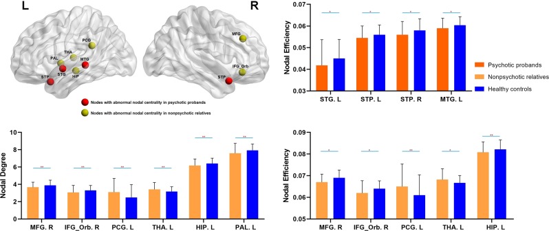
Abnormal neuroanatomic brain networks have been reported in schizophrenia, but their characterization across patients with psychotic disorders, and their potential alterations in nonpsychotic relatives, remain to be clarified. Participants recruited by the Bipolar and Schizophrenia Network for Intermediate Phenotypes consortium included 326 probands with psychotic disorders (107 with schizophrenia (SZ), 87 with schizoaffective disorder (SAD), 132 with psychotic bipolar disorder (BD)), 315 of their nonpsychotic first-degree relatives and 202 healthy controls. Single-subject gray matter graphs were extracted from structural MRI scans, and whole-brain neuroanatomic organization was compared across the participant groups. Compared with healthy controls, psychotic probands showed decreased nodal efficiency mainly in bilateral superior temporal regions. These regions had altered morphological relationships primarily with frontal lobe regions, and their network-level alterations were associated with positive symptoms of psychosis. Nonpsychotic relatives showed lower nodal centrality metrics in the prefrontal cortex and subcortical regions, and higher nodal centrality metrics in the left cingulate cortex and left thalamus. Diagnosis-specific analysis indicated that individuals with SZ had lower nodal efficiency in bilateral superior temporal regions than controls, probands with SAD only exhibited lower nodal efficiency in the left superior and middle temporal gyrus, and individuals with psychotic BD did not show significant differences from healthy controls. Our findings provide novel evidence of clinically relevant disruptions in the anatomic association of the superior temporal lobe with other regions of whole-brain networks in patients with psychotic disorders, but not in their unaffected relatives, suggesting that it is a disease-related trait. Network disorganization primarily involving frontal lobe and subcortical regions in nonpsychotic relatives may be related to familial illness risk.
Figures

Similar articles
-
Neural complexity as a potential translational biomarker for psychosis.J Affect Disord. 2017 Jul;216:89-99. doi: 10.1016/j.jad.2016.10.016. Epub 2016 Oct 26. J Affect Disord. 2017. PMID: 27814962 Free PMC article.
-
Gray matter volume as an intermediate phenotype for psychosis: Bipolar-Schizophrenia Network on Intermediate Phenotypes (B-SNIP).Am J Psychiatry. 2013 Nov;170(11):1285-96. doi: 10.1176/appi.ajp.2013.13010126. Am J Psychiatry. 2013. PMID: 24185241 Free PMC article.
-
Brain structural alterations in pediatric bipolar disorder patients with and without psychotic symptoms.J Affect Disord. 2021 May 1;286:87-93. doi: 10.1016/j.jad.2021.02.077. Epub 2021 Mar 4. J Affect Disord. 2021. PMID: 33714175
-
Brain structural abnormalities as potential markers for detecting individuals with ultra-high risk for psychosis: A systematic review and meta-analysis.Schizophr Res. 2019 Jul;209:22-31. doi: 10.1016/j.schres.2019.05.015. Epub 2019 May 16. Schizophr Res. 2019. PMID: 31104914
-
Structural brain changes in schizophrenia at different stages of the illness: A selective review of longitudinal magnetic resonance imaging studies.Aust N Z J Psychiatry. 2017 May;51(5):500-508. doi: 10.1177/0004867417699473. Epub 2017 Mar 21. Aust N Z J Psychiatry. 2017. PMID: 28415873 Review.
Cited by
-
Aberrant Gray Matter Networks in Non-comorbid Medication-Naive Patients With Major Depressive Disorder and Those With Social Anxiety Disorder.Front Hum Neurosci. 2020 Jun 10;14:172. doi: 10.3389/fnhum.2020.00172. eCollection 2020. Front Hum Neurosci. 2020. PMID: 32587507 Free PMC article.
-
Changes in the structural brain connectome over the course of a nonrandomized clinical trial for acute mania.Neuropsychopharmacology. 2022 Oct;47(11):1961-1968. doi: 10.1038/s41386-022-01328-y. Epub 2022 May 18. Neuropsychopharmacology. 2022. PMID: 35585125 Free PMC article. Clinical Trial.
-
Innovative Neuroimaging Biomarker Distinction of Major Depressive Disorder and Bipolar Disorder through Structural Connectome Analysis and Machine Learning Models.Diagnostics (Basel). 2024 Feb 10;14(4):389. doi: 10.3390/diagnostics14040389. Diagnostics (Basel). 2024. PMID: 38396428 Free PMC article.
-
Graph theoretical analysis and independent component analysis of diabetic optic neuropathy: A resting-state functional magnetic resonance imaging study.CNS Neurosci Ther. 2024 Mar;30(3):e14579. doi: 10.1111/cns.14579. CNS Neurosci Ther. 2024. PMID: 38497532 Free PMC article.
-
Categorical and Dimensional Approaches for Psychiatric Classification and Treatment Targeting: Considerations from Psychosis Biotypes.Adv Neurobiol. 2024;40:685-723. doi: 10.1007/978-3-031-69491-2_23. Adv Neurobiol. 2024. PMID: 39562461 Free PMC article. Review.
References
-
- Arnold SJ, Ivleva EI, Gopal TA, Reddy AP, Jeon-Slaughter H, Sacco CB, et al. Hippocampal volume is reduced in schizophrenia and schizoaffective disorder but not in psychotic bipolar I disorder demonstrated by both manual tracing and automated parcellation (FreeSurfer) Schizophr Bull. 2015;41:233–49. doi: 10.1093/schbul/sbu009. - DOI - PMC - PubMed
Publication types
MeSH terms
Grants and funding
LinkOut - more resources
Full Text Sources
Medical
