

Equivalent L-type channel (CaV1.1) function in adult female and male mouse skeletal muscle fibers
- PMID: 31812241
- PMCID: PMC8019284
- DOI: 10.1016/j.bbrc.2019.11.164
Equivalent L-type channel (CaV1.1) function in adult female and male mouse skeletal muscle fibers
Abstract
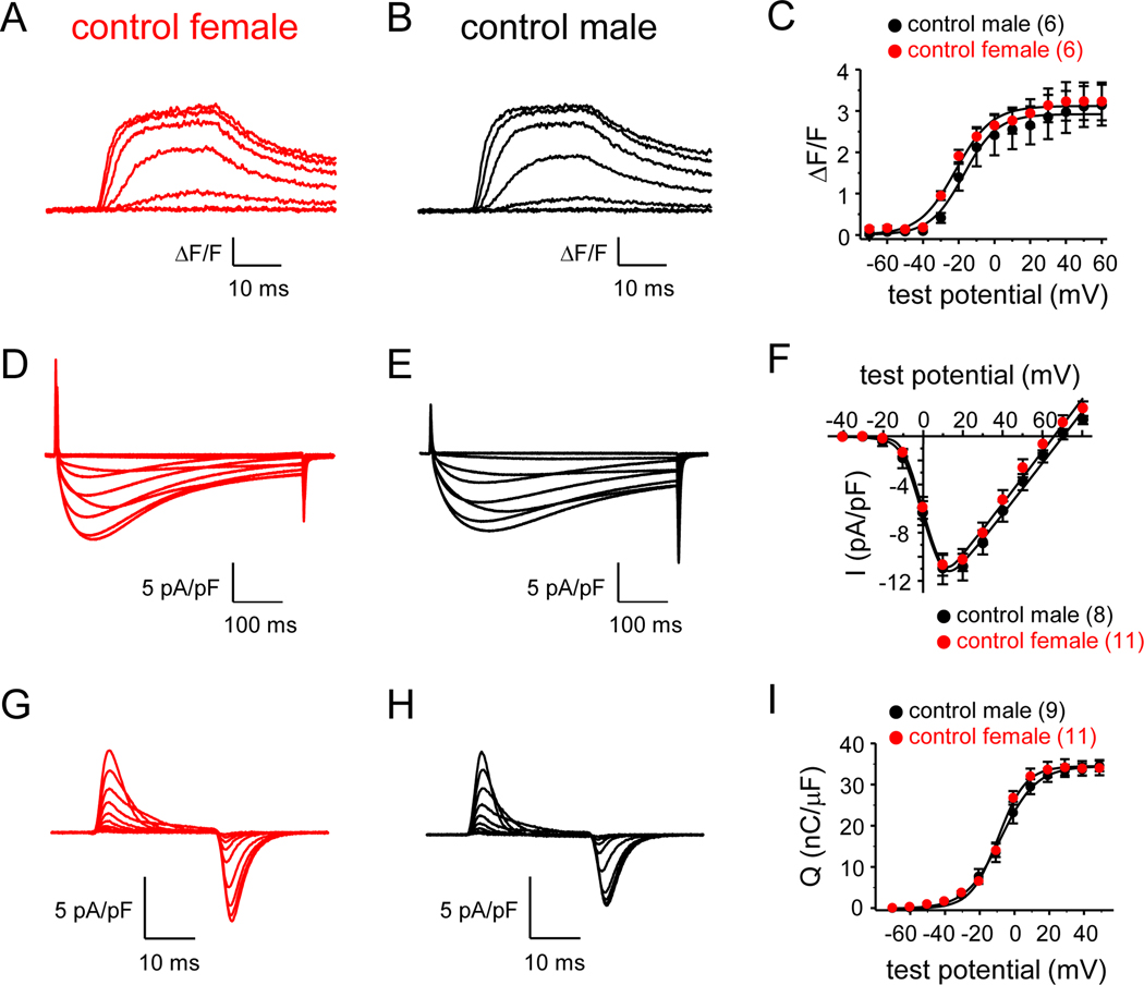
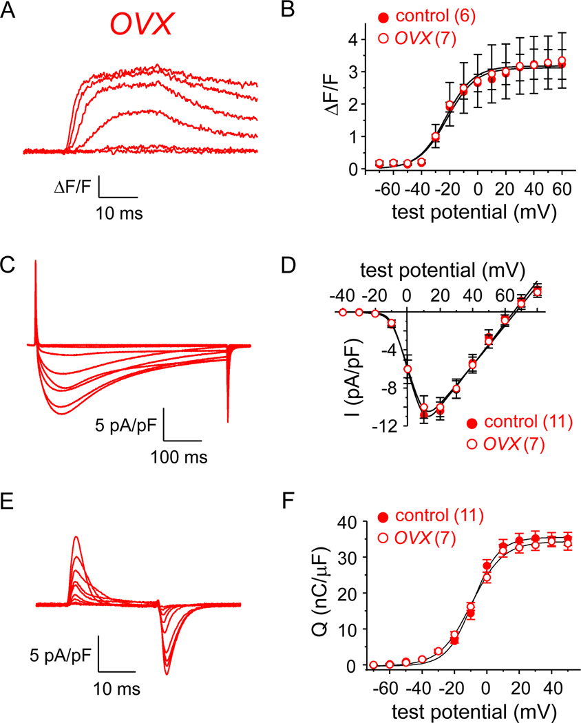
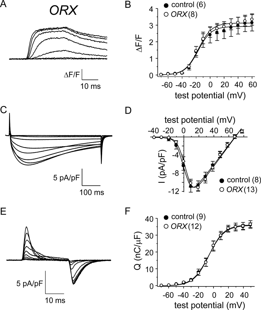
Loss of total muscle force during aging has both atrophic and non-atrophic components. The former deficit is a direct consequence of reduced muscle mass while the latter has been attributed to a depression of excitation-contraction (EC) coupling. It is well established that age-onset reductions in sex hormone production regulate the atrophic component in both males and females. However, it is unknown whether the non-atrophic component is influenced by sex hormones. Since the non-atrophic component has been linked mechanistically to reduced expression of the skeletal muscle L-type Ca2+ channel (CaV1.1), we recorded L-type Ca2+ currents, gating charge movements and depolarization-induced changes in myoplasmic Ca2+ from flexor digitorum brevis (FDB) fibers of naïve and gonadectomized mice of both sexes. Our first set of experiments sought to identify any basal differences in EC coupling or L-type Ca2+ flux between the sexes; no detectable differences in any of the aforementioned parameters were observed between FDB harvested from either naïve males or females. In the latter segments of the study, ovariectomy (OVX) and orchiectomy (ORX) models were used to assess the possible influence of sex hormones on EC coupling and/or L-type Ca2+ flux. In these experiments, FDB fibers harvested from OVX and ORX mice both showed no differences in L-type Ca2+ current, gating charge movement or depolarization-induced changes in Ca2+ release from the sarcoplasmic reticulum. Taken together, our results indicate L-type Ca2+ channel function and EC coupling are: 1) equivalent between the sexes, and 2) not significantly regulated by sex hormones. Since recent NIH review guidelines mandate the consideration of sex differences as a criterion for review, our work indicates the suitability of either sex for the study of the fundamental mechanisms of EC coupling. Thus, our findings may accelerate the research process by conserving animals, labor and financial resources.
Keywords: Ca(V)1.1; Excitation-contraction coupling; L-type; Orchiectomy; Ovariectomy; Sex hormones.
Copyright © 2019 Elsevier Inc. All rights reserved.
Figures
Similar articles
-
Progressive impairment of CaV1.1 function in the skeletal muscle of mice expressing a mutant type 1 Cu/Zn superoxide dismutase (G93A) linked to amyotrophic lateral sclerosis.Skelet Muscle. 2016 Jun 23;6:24. doi: 10.1186/s13395-016-0094-6. eCollection 2016. Skelet Muscle. 2016. PMID: 27340545 Free PMC article.
-
Rem uncouples excitation-contraction coupling in adult skeletal muscle fibers.J Gen Physiol. 2015 Jul;146(1):97-108. doi: 10.1085/jgp.201411314. Epub 2015 Jun 15. J Gen Physiol. 2015. PMID: 26078055 Free PMC article.
-
Stac3 has a direct role in skeletal muscle-type excitation-contraction coupling that is disrupted by a myopathy-causing mutation.Proc Natl Acad Sci U S A. 2016 Sep 27;113(39):10986-91. doi: 10.1073/pnas.1612441113. Epub 2016 Sep 12. Proc Natl Acad Sci U S A. 2016. PMID: 27621462 Free PMC article.
-
Bridging the myoplasmic gap II: more recent advances in skeletal muscle excitation-contraction coupling.J Exp Biol. 2016 Jan;219(Pt 2):175-82. doi: 10.1242/jeb.124123. J Exp Biol. 2016. PMID: 26792328 Review.
-
Functional roles of the gamma subunit of the skeletal muscle DHP-receptor.J Muscle Res Cell Motil. 2006;27(5-7):307-14. doi: 10.1007/s10974-006-9093-2. Epub 2006 Aug 9. J Muscle Res Cell Motil. 2006. PMID: 16897572 Review.
References
-
- Fuller GF, Falls in the elderly. Am. Fam. Physician 61 (2000) 2159–2168. - PubMed
-
- Runge M, Hunter G, Determinants of musculoskeletal frailty and the risk of falls in old age. J Musculoskelet Neuronal Interact. 6 (2006) 167–173. - PubMed
-
- Jasuja R, LeBrasseur NK, Regenerating skeletal muscle in the face of aging and disease. Am. J. Phys. Med. Rehabil 93 (2014) S88–S96. - PubMed
-
- Sipilä S, Taaffe DR, Cheng S, et al., Effects of hormone replacement therapy and high-impact physical exercise on skeletal muscle in post-menopausal women: a randomized placebo-controlled study. Clin. Sci. (Lond) 101 (2001) 147–157. - PubMed
Publication types
MeSH terms
Substances
Grants and funding
LinkOut - more resources
Full Text Sources
Miscellaneous
