

Motor deficit in the mouse ferric chloride-induced distal middle cerebral artery occlusion model of stroke
- PMID: 31812504
- PMCID: PMC6949363
- DOI: 10.1016/j.bbr.2019.112418
Motor deficit in the mouse ferric chloride-induced distal middle cerebral artery occlusion model of stroke
Abstract
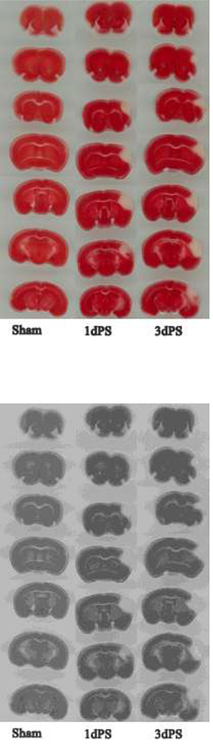
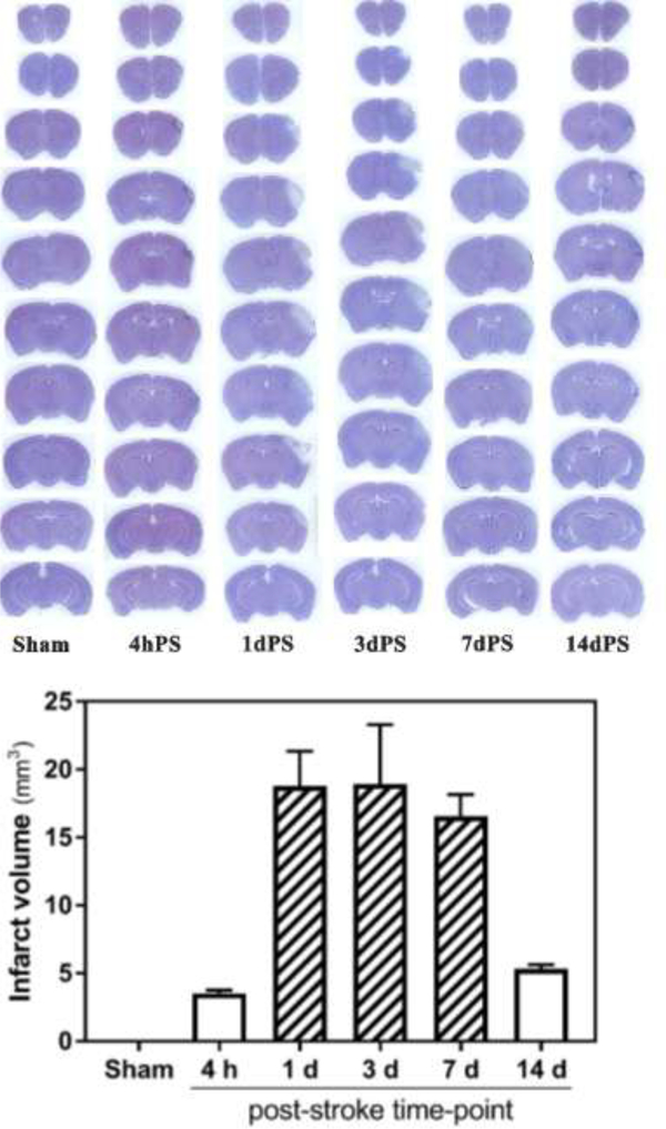
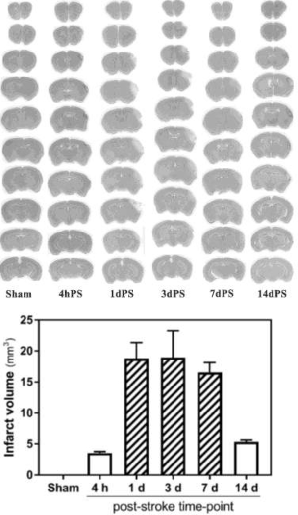
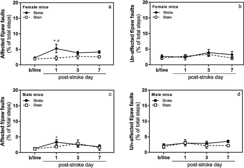
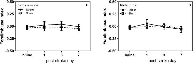
Ferric chloride-induced distal middle cerebral artery occlusion (MCAO) model of stroke was described in mice several years ago, however it lacked in-depth evaluation of the post-stroke functional outcomes in the animals. In this study, we reproduced the recently developed model and expanded its characterization by thorough evaluation of blood supply, cerebral infarction, and motor function in adult male and female mice up to 14 days after stroke. Our observations indicate near complete interruption of blood flow in the distal MCA shortly after application of 20 % ferric chloride over the artery through a cranial window, which remained occluded for at least 4 h. As expected, infarction of the brain tissue, documented by TTC and hematoxylin stains, was restricted to the cerebral cortex. We also systematically evaluated motor impairment of the animals in this model. For this, a series of studies were carried out in male and female mice up to 14 days after stroke, and motor function was assessed in cylinder and grid-walking tests in blinded manner. Contrary to our expectations, the results of both motor tests indicated minor, transient motor deficit in mice after stroke. Based on these observations, we conclude that the mouse ferric chloride-induced distal MCAO model is likely not suitable for proof-of-concept and preclinical studies where motor function is an important outcome measure.
Keywords: Cylinder test; FeCl(3)-induced distal MCAO model of stroke; Grid-walking test; Motor impairment; Mouse stroke model.
Copyright © 2019 Elsevier B.V. All rights reserved.
Figures


Similar articles
-
A novel reproducible model of neonatal stroke in mice: comparison with a hypoxia-ischemia model.Exp Neurol. 2013 Sep;247:218-25. doi: 10.1016/j.expneurol.2013.04.015. Epub 2013 May 4. Exp Neurol. 2013. PMID: 23651512
-
Exposure to female estrous is beneficial for male mice against transient ischemic stroke.Neurol Res. 2019 Jun;41(6):536-543. doi: 10.1080/01616412.2019.1580461. Epub 2019 Feb 27. Neurol Res. 2019. PMID: 30810516
-
Thrombotic distal middle cerebral artery occlusion produced by topical FeCl(3) application: a novel model suitable for intravital microscopy and thrombolysis studies.J Cereb Blood Flow Metab. 2011 Jun;31(6):1452-60. doi: 10.1038/jcbfm.2011.8. Epub 2011 Feb 16. J Cereb Blood Flow Metab. 2011. PMID: 21326267 Free PMC article.
-
Modeling Transient Focal Ischemic Stroke in Rodents by Intraluminal Filament Method of Middle Cerebral Artery Occlusion.Methods Mol Biol. 2018;1717:101-113. doi: 10.1007/978-1-4939-7526-6_9. Methods Mol Biol. 2018. PMID: 29468587 Review.
-
Evaluation of MCAO stroke models in normotensive rats: standardized neocortical infarction by the 3VO technique.Exp Neurol. 2003 Aug;182(2):261-74. doi: 10.1016/s0014-4886(03)00116-x. Exp Neurol. 2003. PMID: 12895438 Review.
Cited by
-
The Effects of Cannabinoids on Ischemic Stroke-Associated Neuroinflammation: A Systematic Review.J Neuroimmune Pharmacol. 2025 Feb 3;20(1):12. doi: 10.1007/s11481-025-10171-z. J Neuroimmune Pharmacol. 2025. PMID: 39899062 Free PMC article.
-
Adenosine receptor A1 enhanced mitochondrial biogenesis and exerted neuroprotection after cerebral ischemia through PGC-1α.Exp Brain Res. 2023 Jun;241(6):1471-1488. doi: 10.1007/s00221-023-06613-w. Epub 2023 Apr 20. Exp Brain Res. 2023. PMID: 37081178
-
Development of a carotid artery thrombolysis stroke model in mice.Blood Adv. 2022 Sep 27;6(18):5449-5462. doi: 10.1182/bloodadvances.2021006008. Blood Adv. 2022. PMID: 35767737 Free PMC article.
-
Functional ultrasound imaging of stroke in awake rats.Elife. 2023 Nov 21;12:RP88919. doi: 10.7554/eLife.88919. Elife. 2023. PMID: 37988288 Free PMC article.
-
The role of peptidase neurolysin in neuroprotection and neural repair after stroke.Neural Regen Res. 2021 Jan;16(1):21-25. doi: 10.4103/1673-5374.284904. Neural Regen Res. 2021. PMID: 32788443 Free PMC article.
References
-
- Durukan A, Tatlisumak T, Acute ischemic stroke: overview of major experimental rodent models, pathophysiology, and therapy of focal cerebral ischemia, Pharmacol Biochem Behav 87(1) (2007) 179–97. - PubMed
-
- Corbett D, Carmichael ST, Murphy TH, Jones TA, Schwab ME, Jolkkonen J, Clarkson AN, Dancause N, Weiloch T, Johansen-Berg H, Nilsson M, McCullough LD, Joy MT, Enhancing the Alignment of the Preclinical and Clinical Stroke Recovery Research Pipeline: Consensus-Based Core Recommendations From the Stroke Recovery and Rehabilitation Roundtable Translational Working Group, Neurorehabil Neural Repair 31(8) (2017) 699–707. - PubMed
Publication types
MeSH terms
Substances
Grants and funding
LinkOut - more resources
Full Text Sources
Medical
Research Materials
