Review
. 2020 Apr 1;32(10):677-700.
doi: 10.1089/ars.2019.7963. Epub 2020 Jan 23.

Redox Regulation via Glutaredoxin-1 and Protein S-Glutathionylation

Affiliations
Review

Redox Regulation via Glutaredoxin-1 and Protein S-Glutathionylation

Reiko Matsui et al. Antioxid Redox Signal. .

Abstract

Significance: Over the past several years, oxidative post-translational modifications of protein cysteines have been recognized for their critical roles in physiology and pathophysiology. Cells have harnessed thiol modifications involving both oxidative and reductive steps for signaling and protein processing. One of these stages requires oxidation of cysteine to sulfenic acid, followed by two reduction reactions. First, glutathione (reduced glutathione [GSH]) forms a S-glutathionylated protein, and second, enzymatic or chemical reduction removes the modification. Under physiological conditions, these steps confer redox signaling and protect cysteines from irreversible oxidation. However, oxidative stress can overwhelm protein S-glutathionylation and irreversibly modify cysteine residues, disrupting redox signaling. Critical Issues: Glutaredoxins mainly catalyze the removal of protein-bound GSH and help maintain protein thiols in a highly reduced state without exerting direct antioxidant properties. Conversely, glutathione S-transferase (GST), peroxiredoxins, and occasionally glutaredoxins can also catalyze protein S-glutathionylation, thus promoting a dynamic redox environment. Recent Advances: The latest studies of glutaredoxin-1 (Glrx) transgenic or knockout mice demonstrate important distinct roles of Glrx in a variety of pathologies. Endogenous Glrx is essential to maintain normal hepatic lipid homeostasis and prevent fatty liver disease. Further, in vivo deletion of Glrx protects lungs from inflammation and bacterial pneumonia-induced damage, attenuates angiotensin II-induced cardiovascular hypertrophy, and improves ischemic limb vascularization. Meanwhile, exogenous Glrx administration can reverse pathological lung fibrosis. Future Directions: Although S-glutathionylation modifies many proteins, these studies suggest that S-glutathionylation and Glrx regulate specific pathways in vivo, and they implicate Glrx as a potential novel therapeutic target to treat diverse disease conditions. Antioxid. Redox Signal. 32, 677-700.

Keywords: NAFLD; NASH; fatty liver disease; hindlimb ischemia; peripheral artery disease.


Conflict of interest statement

The authors have nothing to disclose.

Figures
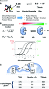
FIG. 1.
FIG. 1.
Reactivity of cysteines. (A) The protonation of a protein cysteine is a major factor that determines the chemical reactivity. A negatively charged thiolate is a much stronger nucleophile than a thiol. Also, the redox potential, the solvent exposure of the cysteine, and the surrounding amino acids influence the cysteine's reactivity. (B) The acid dissociation constant and the pka describe the chemical reactivity of a protein cysteine, with the thiolate being the more reactive form. pH influences the prevalence of a protein cysteine protonation (see qualitative titration curves). (C) Neighboring amino acids stabilize protein thiolates via hydrogen bonds and electrostatic effects and render them sensitive for oxidative post-translational modifications. Color images are available online.
FIG. 2.
FIG. 2.
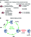
Common oxidative modifications and redox regulation. (A) List of common cysteine modifications involved in or that interfere with redox regulation. (B) Redox-sensitive proteins contain exposed and deprotonated reactive cysteines that exhibit a thiol peroxidase-like activity. These thiolates or “redox sensors” can react with reactive oxygen (OOx) and nitrogen (NOx) species to form labile intermediates such as sulfenic acid (S-OH) and nitrosocysteine (S-NO), respectively ①. These “activated species” quickly react with abundant intracellular glutathione (5–15 mM) ②. The resulting protein GSH adduct is a reversible, oxidative modification that can regulate enzyme activity, localization, protein interactions, and stability. For completion of the redox signaling cycle, Glrx specifically removes GSH adducts ③. Oxidative stress interrupts redox signaling by irreversible oxidation of cysteine thiolates (red arrow). Glrx, glutaredoxin-1; GSH, reduced glutathione; GSSG, oxidized glutathione. Color images are available online.
FIG. 3.
FIG. 3.
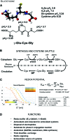
Glutathione. (A) Structure of glutathione (GSH) and pKa values for chemical systems. The pKa of the GSH thiol is lower in the intracellular environment. (B) Glutathione synthesis and distribution through the blood circulation as a source of cysteine. (C) Calculation of the redox potential for glutathione-containing buffers using the Nernst equation. The y-axis depicts the ratio of GSH:GSSG in the redox buffer system. The effects of pH on the redox potential are plotted for pH 7.0, 7.4, and 8.0 and demonstrate that increasing the pH lowers the redox potential of the GSH:GSSG buffer. Glutathione redox buffers are useful tools to investigate redox-sensitive proteins. Redox potentials for thioredoxin, glutaredoxin, glutathione, and NADPH under standard conditions are listed. (D) Biological functions of glutathione. γGCS, γ-glutamylcysteine ligase; DP, dipeptidases; GGT, γ-glutamyl transferase; GS, GSH synthase; NADPH, nicotinamide adenine dinucleotide phosphate. Color images are available online.
FIG. 4.
FIG. 4.
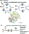
Glrx catalysis. (A) Dithiol and monothiol catalytic mechanisms of Glrx. Glrx was modeled by using the deposited PDB NMR structure 1GRX of Escherichia coli with bound glutathione (arrow) (191) and the Java web plugin Protein workshop (128). The ball-and-stick model depicts glutathione bound to Glrx via its specific GSH-binding groove. (B) The evolutionarily highly conserved function of Glrx3 (cytoplasm) and Glrx5 (mitochondria) in the maintenance and regulation of intracellular iron homeostasis. Glrx3 forms an iron-GSH complex as an intermediate to load specific cytoplasmic proteins with iron-sulfur clusters [2Fe-2S] aided by the highly conserved BolA (DNA-binding transcriptional regulator). The interaction results in various protein-iron-sulfur complexes that may or may not involve BolA or Glrx3. These complexes serve as iron storage depots or mediate cellular stress responses. Color images are available online.
FIG. 5.
FIG. 5.
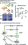
Angiogenesis and ischemic revascularization. (A) Redox regulation is an essential process that is involved in the ischemic neovascularization of the hindlimb. Oxidant sources such as NOX, eNOS, and the thioredoxin system promote angiogenesis. Antioxidant systems, including Glrx, Cat, and Nrf2, suppress angiogenesis. Glrx TG mice have impaired angiogenesis in the ischemic hindlimb after surgical-induced blockage of the femoral artery. Data were obtained by Murdoch et al. (130). *p < 0.05 (B) Angiogenic signaling cascades regulated by Glrx. ?, the direct effect of Glrx was not demonstrated. Cat, catalase; eNOS, endothelial nitric oxide synthase; NOX, NADPH oxidases; Nrf2, nuclear factor (erythroid-derived 2)-like 2; TG, transgenic. Color images are available online.
FIG. 6.
FIG. 6.
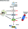
The NF-κB-pathway. The NF-κB inflammatory signaling pathway is regulated via S-glutathionylation at various checkpoints. S-glutathionylation inhibits TRAF6 and IKK-β as well as DNA binding of p50 and p65. Thus, Glrx promotes the inflammatory response. IκBα, an inhibitor of NF-κB; IKK-β, nuclear factor-kappa-β kinase subunit beta; IRAK1/4, interleukin-1 receptor-associated kinase 4; LPS, lipopolysaccharides; MyD88, myeloid differentiation primary response 88; NF-κB, nuclear factor-κB; TLR4, toll-like receptor 4. Color images are available online.
FIG. 7.
FIG. 7.
The HIF-1α pathway. Oxidants cause oxidative modification and S-glutathionylation of Cys533 located in the ODD domain of HIF-1α. S-glutathionylation prevents interaction with vHL, a component of the ubiquitin ligase E3 complex, and proteasomal degradation. Thus, the S-glutathionylation of HIF-1α activates the proangiogenic program, which also includes the expression of VEGF. EC, endothelial cell; HIF, hypoxia-inducible factor; ODD, oxygen-dependent degradation; PTP, protein tyrosine phosphatase; Rac1, Ras-related C3 botulinum toxin substrate 1; SERCA, sarcoplasmic-endoplasmic reticulum calcium ATPase; sVEGFR1 (sFlt), soluble vascular endothelial growth factor receptor 1 (soluble fms-like tyrosine kinase-1s); VEGF, vascular endothelial growth factor; vHL, von Hippel–Lindau. Color images are available online.
FIG. 8.
FIG. 8.
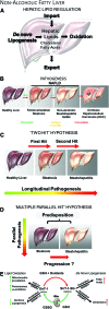
NAFLD and de novo lipogenesis. (A) Major pathways that control hepatic lipid content. Top right: Pathological stages of NAFLD. Liver disease progression adapted from the GB HealthWatch. Major working hypotheses explaining NAFLD disease progression (B) the “two-hit” hypothesis and (C) the “multiple parallel hit” hypothesis. (D) Effects of Glrx on the central metabolic regulator and NAD+-dependent class III histone deacetylase SirT1. (E) S-glutathionylation inhibits SirT1 activity, causing hyperacetylation and transcriptional activation of SREBP. These transcription factors increase expression of the key enzymes—fatty acid synthase and HMG-CoA reductase—for hepatic lipid synthesis. Active SirT1 inhibits de novo lipogenesis and promotes mitochondrial and peroxisomal fatty acid oxidation through the central transcription factors PPARγ and PGC1α. HMG, β-hydroxy β-methylglutaryl; NAFLD, nonalcoholic fatty liver disease; PGC1α, peroxisome proliferator-activated receptor gamma coactivator 1-alpha; PPARγ, peroxisome proliferator-activated receptor gamma; SirT1, sirtuin-1; SREBP, sterol regulatory element-binding protein. Color images are available online.

Similar articles

Cited by

References

    1. Abdelsaid MA and El-Remessy AB. S-glutathionylation of LMW-PTP regulates VEGF-mediated FAK activation and endothelial cell migration. J Cell Sci 125: 4751–4760, 2012 - PubMed
    1. Adachi T, Weisbrod RM, Pimentel DR, Ying J, Sharov VS, Schöneich C, and Cohen RA. S-Glutathiolation by peroxynitrite activates SERCA during arterial relaxation by nitric oxide. Nat Med 10: 1200–1207, 2004 - PubMed
    1. Adluri RS, Thirunavukkarasu M, Zhan L, Dunna NR, Akita Y, Selvaraju V, Otani H, Sanchez JA, Ho Y-S, and Maulik N. Glutaredoxin-1 overexpression enhances neovascularization and diminishes ventricular remodeling in chronic myocardial infarction. PLoS One 7: e34790, 2012 - PMC - PubMed
    1. Aesif SW, Anathy V, Kuipers I, Guala AS, Reiss JN, Ho Y-S, and Janssen-Heininger YMW. Ablation of glutaredoxin-1 attenuates lipopolysaccharide-induced lung inflammation and alveolar macrophage activation. Am J Respir Cell Mol Biol 44: 491–499, 2011 - PMC - PubMed
    1. Aesif SW, Janssen-Heininger YMW, and Reynaert NL. Protocols for the detection of s-glutathionylated and s-nitrosylated proteins in situ. Methods Enzymol 474: 289–296, 2010 - PMC - PubMed

Publication types